

The DOI matches its Crossref record; 6 of the 6 references it cites were found in a registry or an index.
Discovery of a potent HMG-CoA reductase degrader that eliminates statin-induced reductase accumulation and lowers cholesterol
Abstract
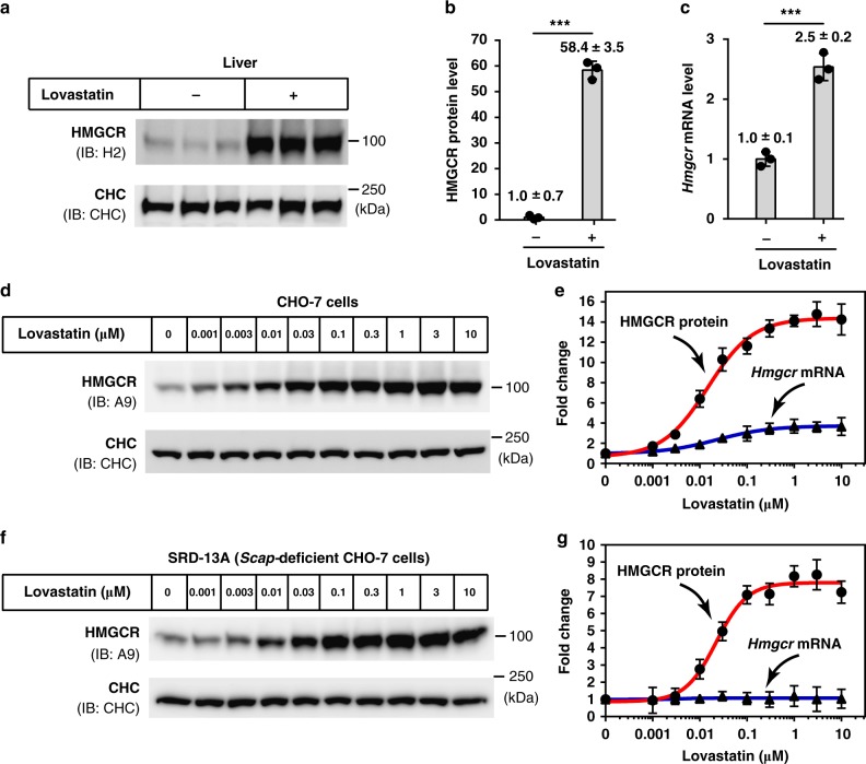
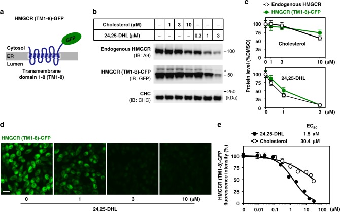
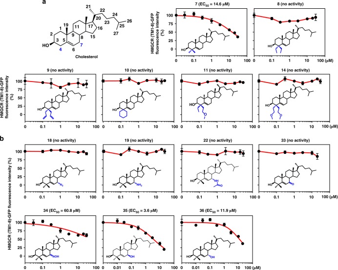
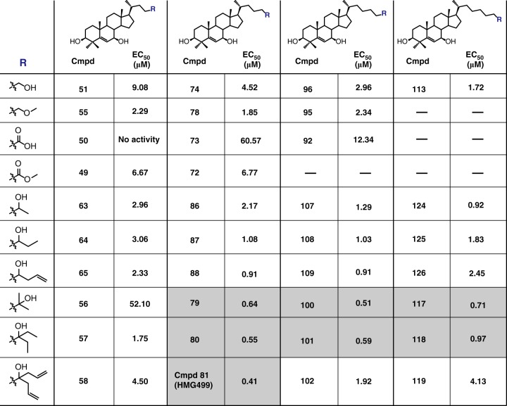
Statins are inhibitors of HMG-CoA reductase, the rate-limiting enzyme of cholesterol biosynthesis, and have been clinically used to treat cardiovascular disease. However, a paradoxical increase of reductase protein following statin treatment may attenuate the effect and increase the side effects. Here we present a previously unexplored strategy to alleviate statin-induced reductase accumulation by inducing its degradation. Inspired by the observations that cholesterol intermediates trigger reductase degradation, we identify a potent degrader, namely Cmpd 81, through structure–activity relationship analysis of sterol analogs. Cmpd 81 stimulates ubiquitination and degradation of reductase in an Insig-dependent manner, thus dramatically reducing protein accumulation induced by various statins. Cmpd 81 can act alone or synergistically with statin to lower cholesterol and reduce atherosclerotic plaques in mice. Collectively, our work suggests that inducing reductase degradation by Cmpd 81 or similar chemicals alone or in combination with statin therapy can be a promising strategy for treating cardiovascular disease.
Overview
The claims and statistics of this paper haven’t been checked yet.
Run the check — free, no account needed. It reads the open-access text and checks the sources it cites.
Integrity signals
Registry
Cites
Statistics
Figures
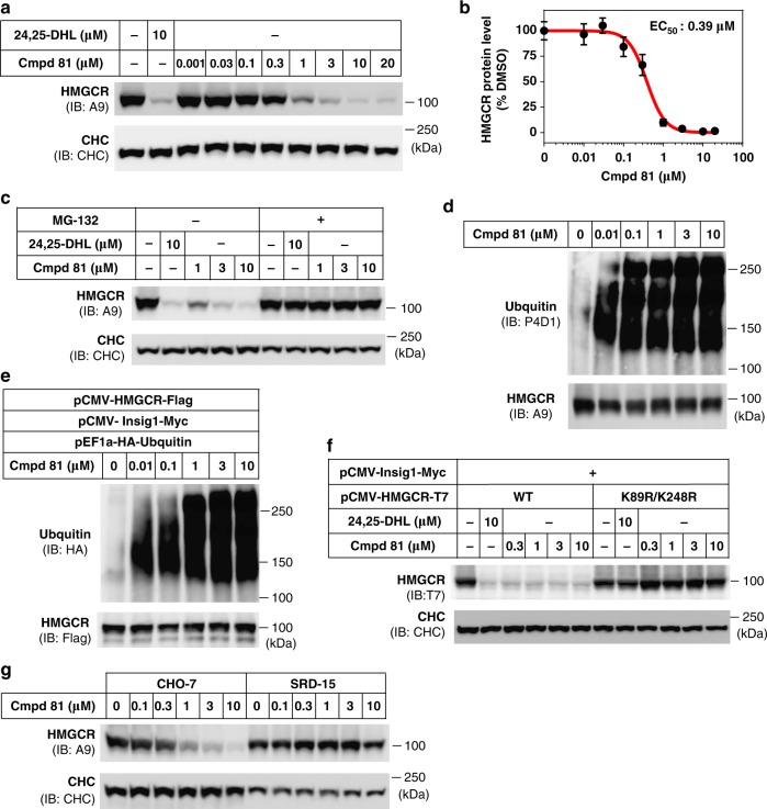
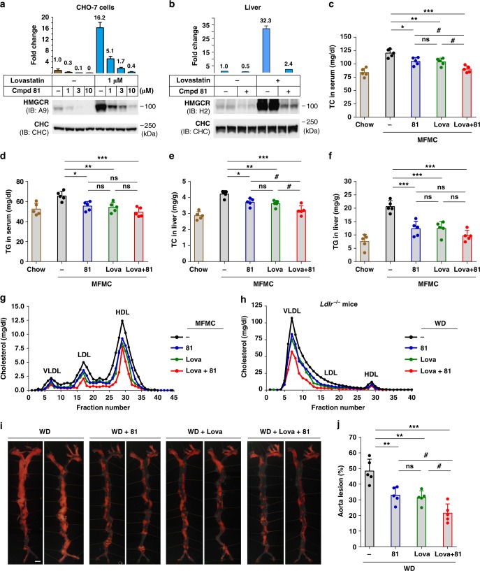
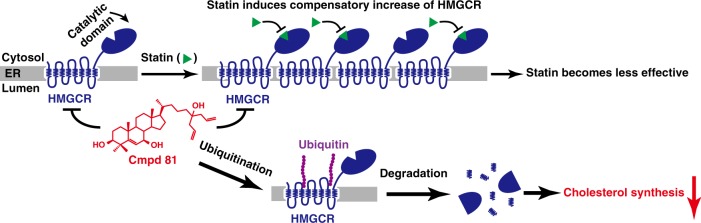
Verification
Claims the paper makes
The claims and statistics of this paper haven’t been checked yet.
Run the check — free, no account needed. It reads the open-access text and checks the sources it cites.
Claims other papers make about this one
Each is checked against this paper’s own text: does it say what the citing paper says it does?
The source backs part of the claim, not all of it — or backs it for a different population, condition or comparison.
In the citing paper
“In addition, in some patients, these treatments can cause a rebound in plasma cholesterol following the withdrawal of the drug(s) to higher than that before treatment [13]”
From Post-translational regulation of the low-density lipoprotein receptor provides new targets for cholesterol regulation (2024) · cites this paper as [13]
Experimental — accuracy not yet measured
Self-check
Percentages, sums and reported differences re-computed from the paper’s own numbers.
The self-check needs the paper’s full text.
Citations
Cites
Cholesterol Treatment Trialists' (CTT) Collaboration (2010) · The Lancet · doi:10.1016/s0140-6736(10)61350-5 · Cited by 6,550
Structural mechanism for statin inhibition of HMG-CoA reductase
Istvan, E. S., & Deisenhofer, J. (2001) · Science · doi:10.1126/science.1059344 · Cited by 1,644
New insights into the pharmacodynamic and pharmacokinetic properties of statins
Corsini, A., Bellosta, S., Baetta, R., Fumagalli, R., Paoletti, R., & Bernini, F. (1999) · Pharmacology & Therapeutics · doi:10.1016/s0163-7258(99)00045-5 · Cited by 796
An assessment by the statin muscle safety task force: 2014 update
Rosenson, R. S., Baker, S. K., Jacobson, T. A., Kopecky, S. L., & Parker, B. A. (2014) · Journal of Clinical Lipidology · doi:10.1016/j.jacl.2014.03.004 · Cited by 485
Statin use and non-alcoholic steatohepatitis in at risk individuals
Dongiovanni, P., Petta, S., Mannisto, V., Mancina, R. M., Pipitone, R., Karja, V., Maggioni, M., Kakela, P., Wiklund, O., Mozzi, E., et al. (2015) · Journal of Hepatology · doi:10.1016/j.jhep.2015.05.006 · Cited by 390
Research works printed but not linked: 60
- Benjamin, E. J. et al. Heart disease and stroke statistics-2017 update: a report from the American Heart Association. Circulation 135, e146–e603 (2017). · DOI · Paper page
- Delahoy, P. J., Magliano, D. J., Webb, K., Grobler, M. & Liew, D. The relationship between reduction in low-density lipoprotein cholesterol by statins and reduction in risk of cardiovascular outcomes: an updated meta-analysis. Clin. Ther. 31, 236–244 (2009). · DOI
- Baigent, C. et al. Efficacy and safety of cholesterol-lowering treatment: prospective meta-analysis of data from 90,056 participants in 14 randomised trials of statins. Lancet 366, 1267–1278 (2005). · DOI · Paper page
- Scandinavian Simvastatin Survival Study Group. Randomised trial of cholesterol lowering in 4444 patients with coronary heart disease: the Scandinavian Simvastatin Survival Study (4S). Lancet 344, 1383–1389 (1994).
- The LIPID Study Group. Long-term effectiveness and safety of pravastatin in 9014 patients with coronary heart disease and average cholesterol concentrations: the LIPID trial follow-up. Lancet 359, 1379–1387 (2002). · DOI · Paper page
- Brown, M. S. & Goldstein, J. L. Multivalent feedback regulation of HMG CoA reductase, a control mechanism coordinating isoprenoid synthesis and cell growth. J. Lipid Res. 21, 505–517 (1980). · DOI
- Goldstein, J. L., DeBose-Boyd, R. A. & Brown, M. S. Protein sensors for membrane sterols. Cell 124, 35–46 (2006). · DOI · Paper page
- Song, B.-L., Javitt, N. B. & DeBose-Boyd, R. A. Insig-mediated degradation of HMG CoA reductase stimulated by lanosterol, an intermediate in the synthesis of cholesterol. Cell Metab. 1, 179–189 (2005). · DOI
- DeBose-Boyd, R. A. Feedback regulation of cholesterol synthesis: sterol-accelerated ubiquitination and degradation of HMG CoA reductase. Cell Res. 18, 609 (2008). · DOI · Paper page
- Sever, N. et al. Insig-dependent ubiquitination and degradation of mammalian 3-hydroxy-3-methylglutaryl-CoA reductase stimulated by sterols and geranylgeraniol. J. Biol. Chem. 278, 52479–52490 (2003). · DOI
- Brown, M. S., Faust, J. R., Goldstein, J. L., Kaneko, I. & Endo, A. Induction of 3-hydroxy-3-methylglutaryl coenzyme A reductase activity in human fibroblasts incubated with compactin (ML-236B), a competitive inhibitor of the reductase. J. Biol. Chem. 253, 1121–1128 (1978). · DOI
- Lee, P. C., Sever, N. & Debose-Boyd, R. A. Isolation of sterol-resistant Chinese hamster ovary cells with genetic deficiencies in both Insig-1 and Insig-2. J. Biol. Chem. 280, 25242–25249 (2005). · DOI
- Orci, L., Brown, M. S., Goldstein, J. L., Garcia-Segura, L. M. & Anderson, R. G. Increase in membrane cholesterol: a possible trigger for degradation of HMG CoA reductase and crystalloid endoplasmic reticulum in UT-1 cells. Cell 36, 835–845 (1984). · DOI
- Kita, T., Brown, M. S. & Goldstein, J. L. Feedback regulation of 3-hydroxy-3-methylglutaryl coenzyme A reductase in livers of mice treated with mevinolin, a competitive inhibitor of the reductase. J. Clin. Invest. 66, 1094–1100 (1980). · DOI
- Schonewille, M. et al. Statins increase hepatic cholesterol synthesis and stimulate fecal cholesterol elimination in mice. J. Lipid Res. 57, 1455–1464 (2016). · DOI
- Hwang, S. et al. Contribution of accelerated degradation to feedback regulation of 3-hydroxy-3-methylglutaryl coenzyme A reductase and cholesterol metabolism in the liver. J. Biol. Chem. 291, 13479–13494 (2016). · DOI
- Ness, G. C., Chambers, C. M. & Lopez, D. Atorvastatin action involves diminished recovery of hepatic HMG-CoA reductase activity. J. Lipid Res. 39, 75–84 (1998). · DOI
- Bilhartz, L. E., Spady, D. K. & Dietschy, J. M. Inappropriate hepatic cholesterol synthesis expands the cellular pool of sterol available for recruitment by bile acids in the rat. J. Clin. Invest. 84, 1181–1187 (1989). · DOI
- Burnett, J. R. et al. The magnitude of decrease in hepatic very low density lipoprotein apolipoprotein B secretion is determined by the extent of 3-hydroxy-3-methylglutaryl coenzyme A reductase inhibition in miniature pigs. Endocrinology 140, 5293–5302 (1999). · DOI
- Reihner, E. et al. Influence of pravastatin, a specific inhibitor of HMG-CoA reductase, on hepatic metabolism of cholesterol. N. Engl. J. Med. 323, 224–228 (1990). · DOI
- Preiss, D. et al. Risk of incident diabetes with intensive-dose compared with moderate-dose statin therapy: a meta-analysis. JAMA 305, 2556–2564 (2011). · DOI · Paper page
- Maki, K. C. et al. An assessment by the Statin Diabetes Safety Task Force: 2014 update. J. Clin. Lipidol. 8, S17–S29 (2014). · DOI · Paper page
- Armitage, J. The safety of statins in clinical practice. Lancet 370, 1781–1790 (2007). · DOI · Paper page
- Buettner, C. et al. Statin use and musculoskeletal pain among adults with and without arthritis. Am. J. Med. 125, 176–182 (2012). · DOI · Paper page
- Pineda, A. & Cubeddu, L. X. Statin rebound or withdrawal syndrome: does it exist? Curr. Atheroscler. Rep. 13, 23–30 (2011). · DOI
- Daskalopoulou, S. S. When statin therapy stops: implications for the patient. Curr. Opin. Cardiol. 24, 454–460 (2009). · DOI
- Liscum, L. et al. Domain structure of 3-hydroxy-3-methylglutaryl coenzyme A reductase, a glycoprotein of the endoplasmic reticulum. J. Biol. Chem. 260, 522–530 (1985). · DOI
- Roitelman, J., Olender, E. H., Bar-Nun, S., Dunn, W. A. Jr. & Simoni, R. D. Immunological evidence for eight spans in the membrane domain of 3-hydroxy-3-methylglutaryl coenzyme A reductase: implications for enzyme degradation in the endoplasmic reticulum. J. Cell Biol. 117, 959–973 (1992). · DOI
- Sever, N., Yang, T., Brown, M. S., Goldstein, J. L. & DeBose-Boyd, R. A. Accelerated degradation of HMG CoA reductase mediated by binding of insig-1 to its sterol-sensing domain. Mol. Cell 11, 25–33 (2003). · DOI
- Song, B. L., Sever, N. & DeBose-Boyd, R. A. Gp78, a membrane-anchored ubiquitin ligase, associates with Insig-1 and couples sterol-regulated ubiquitination to degradation of HMG CoA reductase. Mol. Cell 19, 829–840 (2005). · DOI · Paper page
- Cao, J. et al. Ufd1 is a cofactor of gp78 and plays a key role in cholesterol metabolism by regulating the stability of HMG-CoA reductase. Cell Metab. 6, 115–128 (2007). · DOI
- Jiang, L.-Y. et al. Ring finger protein 145 (RNF145) is a ubiquitin ligase for sterol-induced degradation of HMG-CoA reductase. J. Biol. Chem. 293, 4047–4055 (2018). · DOI
- Ravid, T., Doolman, R., Avner, R., Harats, D. & Roitelman, J. The ubiquitin-proteasome pathway mediates the regulated degradation of mammalian 3-hydroxy-3-methylglutaryl-coenzyme A reductase. J. Biol. Chem. 275, 35840–35847 (2000). · DOI
- Jo, Y. & Debose-Boyd, R. A. Control of cholesterol synthesis through regulated ER-associated degradation of HMG CoA reductase. Crit. Rev. Biochem. Mol. Biol. 45, 185–198 (2010). · DOI
- Lai, A. C. & Crews, C. M. Induced protein degradation: an emerging drug discovery paradigm. Nat. Rev. Drug. Discov. 16, 101–114 (2017). · DOI · Paper page
- Brown, M. S. & Goldstein, J. L. Suppression of 3-hydroxy-3-methylglutaryl coenzyme A reductase activity and inhibition of growth of human fibroblasts by 7-ketocholesterol. J. Biol. Chem. 249, 7306–7314 (1974). · DOI · Paper page
- Bell, J. J., Sargeant, T. E. & Watson, J. A. Inhibition of 3-hydroxy-3-methylglutaryl coenzyme A reductase activity in hepatoma tissue culture cells by pure cholesterol and several cholesterol derivatives. Evidence supporting two distinct mechanisms.20l. J. Biol. Chem. 251, 1745–1758 (1976). · DOI
- Kandutsch, A. A. & Chen, H. W. Regulation of sterol synthesis in cultured cells by oxygenated derivatives of cholesterol. J. Cell Physiol. 85, 415–424 (1975). · DOI
- Lange, Y. et al. Effectors of rapid homeostatic responses of endoplasmic reticulum cholesterol and 3-hydroxy-3-methylglutaryl-CoA reductase. J. Biol. Chem. 283, 1445–1455 (2008). · DOI
- Nguyen, A. D., McDonald, J. G., Bruick, R. K. & DeBose-Boyd, R. A. Hypoxia stimulates degradation of 3-hydroxy-3-methylglutaryl-coenzyme A reductase through accumulation of lanosterol and hypoxia-inducible factor-mediated induction of insigs. J. Biol. Chem. 282, 27436–27446 (2007). · DOI
- Repa, J. J. et al. Regulation of mouse sterol regulatory element-binding protein-1c gene (SREBP-1c) by oxysterol receptors, LXRalpha and LXRbeta. Genes Dev. 14, 2819–2830 (2000). · DOI
- DeBose-Boyd, R. A., Ou, J., Goldstein, J. L. & Brown, M. S. Expression of sterol regulatory element-binding protein 1c (SREBP-1c) mRNA in rat hepatoma cells requires endogenous LXR ligands. Proc. Natl Acad. Sci. USA 98, 1477–1482 (2001). · DOI · Paper page
- Sharpe, L. J. & Brown, A. J. Controlling cholesterol synthesis beyond 3-hydroxy-3-methylglutaryl-CoA reductase (HMGCR). J. Biol. Chem. 288, 18707–18715 (2013). · DOI · Paper page
- Jo, Y., Lee, P. C., Sguigna, P. V. & DeBose-Boyd, R. A. Sterol-induced degradation of HMG CoA reductase depends on interplay of two Insigs and two ubiquitin ligases, gp78 and Trc8. Proc. Natl Acad. Sci. USA 108, 20503–20508 (2011). · DOI
- Pencina, M. J. et al. Application of new cholesterol guidelines to a population-based sample. N. Engl. J. Med. 370, 1422–1431 (2014). · DOI
- Lloyd-Jones, D. M., Goff, D. & Stone, N. J. Statins, risk assessment, and the new American prevention guidelines. Lancet 383, 600–602 (2014). · DOI
- Stone, N. J. et al. 2013 ACC/AHA guideline on the treatment of blood cholesterol to reduce atherosclerotic cardiovascular risk in adults: a report of the American College of Cardiology/American Heart Association Task Force on Practice Guidelines. J. Am. Coll. Cardiol. 63, 2889–2934 (2014). · DOI · Paper page
- Min, H. K. et al. Increased hepatic synthesis and dysregulation of cholesterol metabolism is associated with the severity of nonalcoholic fatty liver disease. Cell. Metab. 15, 665–674 (2012). · DOI
- Ioannou, G. N. The role of cholesterol in the pathogenesis of NASH. Trends Endocrinol. Metab. 27, 84–95 (2016). · DOI · Paper page
- Mullen, P. J., Yu, R., Longo, J., Archer, M. C. & Penn, L. Z. The interplay between cell signalling and the mevalonate pathway in cancer. Nat. Rev. Cancer 16, 718–731 (2016). · DOI
- Zipp, F. et al. Impact of HMG-CoA reductase inhibition on brain pathology. Trends Pharmacol. Sci. 28, 342–349 (2007). · DOI
- McCarthy, C. et al. Statin as a novel pharmacotherapy of pulmonary alveolar proteinosis. Nat. Commun. 9, 3127 (2018). · DOI
- Xia, Y. et al. The mevalonate pathway is a druggable target for vaccine adjuvant discovery. Cell 175, 1059-1073.e21 (2018). · DOI
- Song, B. L. & DeBose-Boyd, R. A. Ubiquitination of 3-hydroxy-3-methylglutaryl-CoA reductase in permeabilized cells mediated by cytosolic E1 and a putative membrane-bound ubiquitin ligase. J. Biol. Chem. 279, 28798–28806 (2004). · DOI
- Fermini, B. & Fossa, A. A. The impact of drug-induced QT interval prolongation on drug discovery and development. Nat. Rev. Drug. Discov. 2, 439–447 (2003). · DOI
- Sanguinetti, M. C. & Tristani-Firouzi, M. hERG potassium channels and cardiac arrhythmia. Nature 440, 463–469 (2006). · DOI
- Goldstein, J. L., Basu, S. K. & Brown, M. S. Receptor-mediated endocytosis of low-density lipoprotein in cultured cells. Methods Enzymol. 98, 241–260 (1983). · DOI
- Hannah, V. C., Ou, J., Luong, A., Goldstein, J. L. & Brown, M. S. Unsaturated fatty acids down-regulate srebp isoforms 1a and 1c by two mechanisms in HEK-293 cells. J. Biol. Chem. 276, 4365–4372 (2001). · DOI · Paper page
- Li, P. S. et al. The clathrin adaptor Numb regulates intestinal cholesterol absorption through dynamic interaction with NPC1L1. Nat. Med. 20, 80–86 (2014). · DOI
- Tang, J. J. et al. Inhibition of SREBP by a small molecule, betulin, improves hyperlipidemia and insulin resistance and reduces atherosclerotic plaques. Cell. Metab. 13, 44–56 (2011). · DOI · Paper page
Cited by
Sorted by citations
Mechanisms and regulation of cholesterol homeostasis
Luo, J., Yang, H., & Song, B.-L. (2020) · Nature Reviews Molecular Cell Biology · doi:10.1038/s41580-019-0190-7 · Cited by 1,980
Self-citationTargeting Small GTPases and Their Prenylation in Diabetes Mellitus
Gendaszewska-Darmach, E., Garstka, M. A., & Błażewska, K. M. (2021) · Journal of Medicinal Chemistry · doi:10.1021/acs.jmedchem.1c00410 · Cited by 35
Ionescu, R.-B., Nicaise, A. M., Reisz, J. A., Williams, E. C., Prasad, P., Willis, C. M., Simões-Abade, M. B. C., Sbarro, L., Dzieciatkowska, M., Stephenson, D., et al. (2024) · Cell Stem Cell · doi:10.1016/j.stem.2024.09.014 · Cited by 34
Aldworth, H., & Hooper, N. M. (2024) · Biochemical Society Transactions · doi:10.1042/bst20230918 · Cited by 11
Metadata
- Authors
- Jiang, S.-Y., Li, H., Tang, J.-J., Wang, J., Luo, J., Liu, B., Wang, J.-K., Shi, X.-J., Cui, H.-W., Tang, J., Yang, F., Qi, W., Qiu, W.-W., & Song, B.-L.
- Venue
- Nature Communications
- Published
- 2018-12-03
- Volume
- 9
- Issue
- 1
- ISSN
- 2041-1723
- PMC
- PMC6277434
- Cited by
- 173
- References
- 6
- Language
- en
About these checks
Registry, retraction, Cites and statistics results are computed, not judged, with one exception: a printed reference that no index lists is read once by a model before it counts as a citation error. Claims are extracted by a model and kept only when their quote appears verbatim in the text; each cited source is then checked, with a model’s help, for whether it says what the claim relies on. Those verdicts can be wrong — read the claim’s quote and the cited source before relying on one.
Statistics and arithmetic are recomputed, not judged.
Confidence bands are shown only for fields calibrated on our annotated test set. None is calibrated yet, so no band is shown.
What counts as a claim ›How we measure confidence ›How we check this ›
Are you an author, or do you see a problem with this page? Request a review
