

The DOI matches its Crossref record; 69 of the 69 references it cites were found in a registry or an index.
The deubiquitylating enzyme USP15 regulates homologous recombination repair and cancer cell response to PARP inhibitors
Open accessAbstract
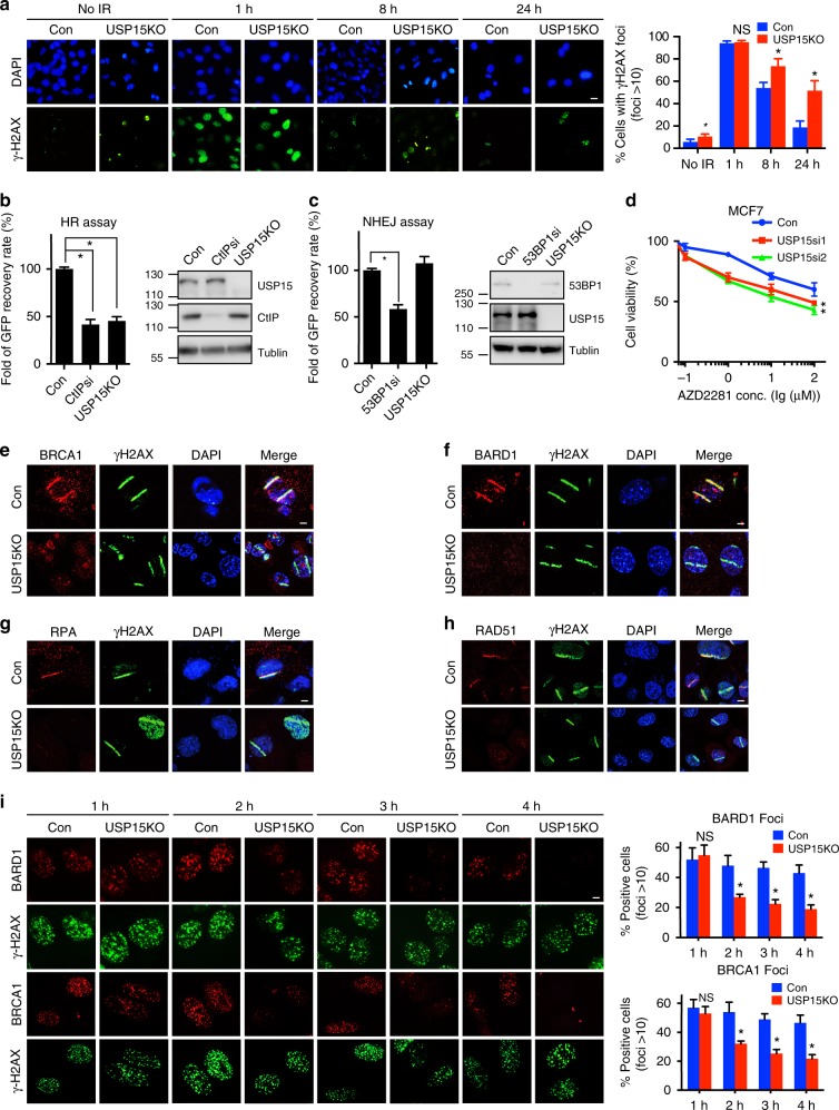
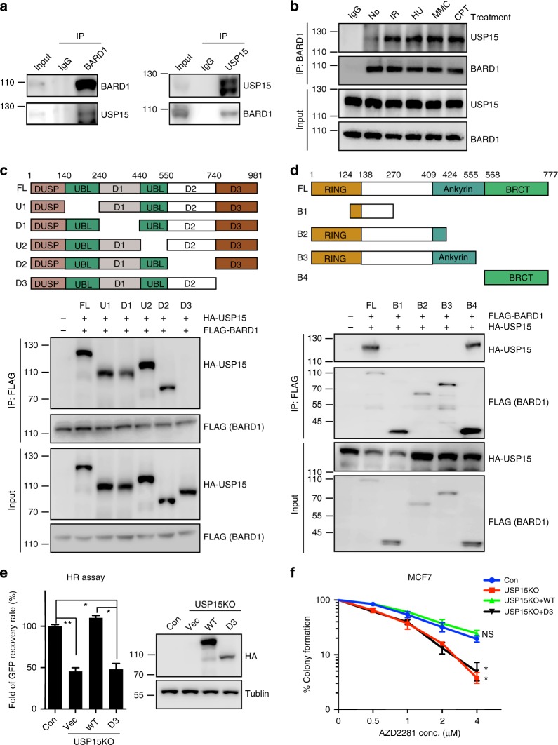
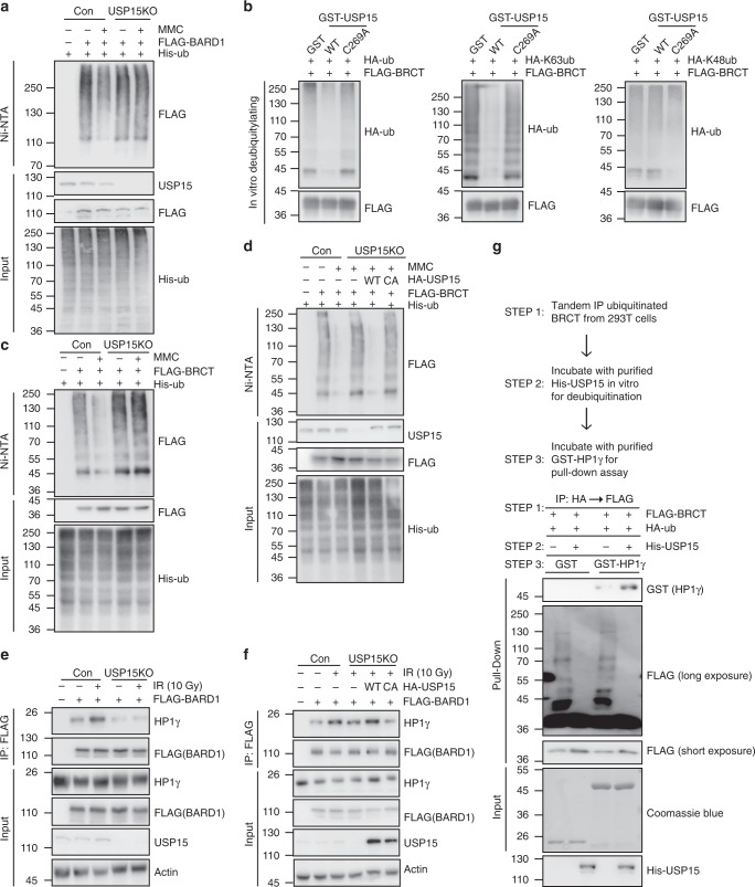
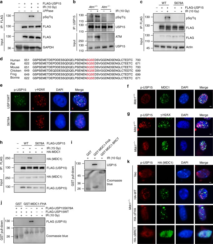
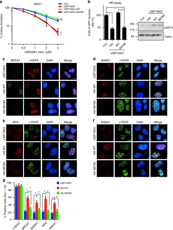
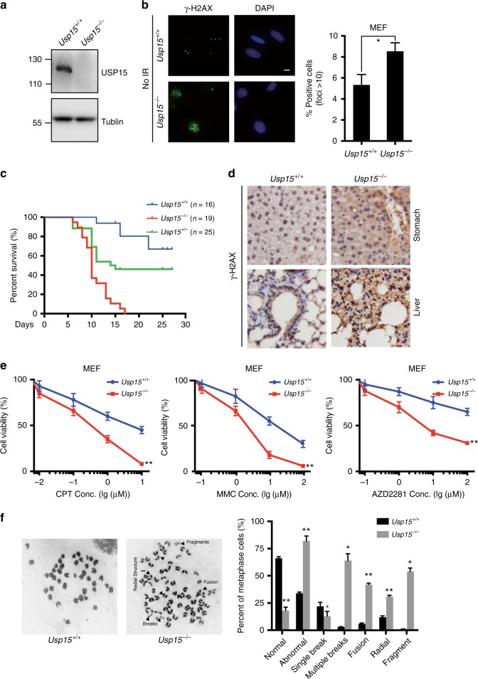
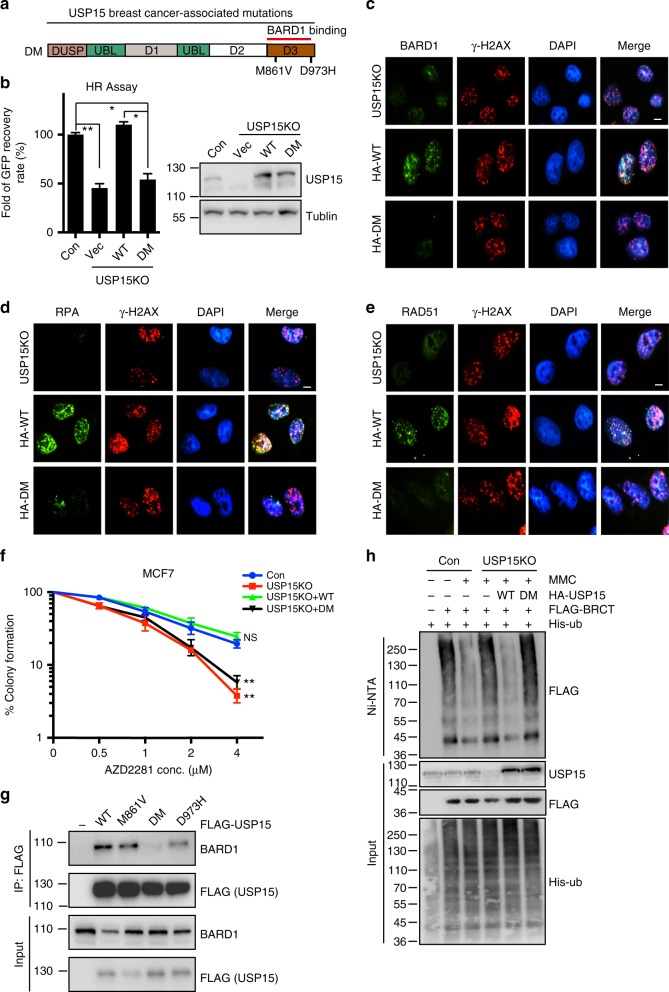
Poly-(ADP-ribose) polymerase inhibitors (PARPi) selectively kill breast and ovarian cancers with defects in homologous recombination (HR) caused by BRCA1/2 mutations. There is also clinical evidence for the utility of PARPi in breast and ovarian cancers without BRCA mutations, but the underlying mechanism is not clear. Here, we report that the deubiquitylating enzyme USP15 affects cancer cell response to PARPi by regulating HR. Mechanistically, USP15 is recruited to DNA double-strand breaks (DSBs) by MDC1, which requires the FHA domain of MDC1 and phosphorylated Ser678 of USP15. Subsequently, USP15 deubiquitinates BARD1 BRCT domain, and promotes BARD1-HP1γ interaction, resulting in BRCA1/BARD1 retention at DSBs. USP15 knockout mice exhibit genomic instability in vivo. Furthermore, cancer-associated USP15 mutations, with decreased USP15-BARD1 interaction, increases PARP inhibitor sensitivity in cancer cells. Thus, our results identify a novel regulator of HR, which is a potential biomarker for therapeutic treatment using PARP inhibitors in cancers.
Overview
The claims and statistics of this paper haven’t been checked yet.
Run the check — free, no account needed. It reads the open-access text and checks the sources it cites.
Integrity signals
Registry
Cites
Statistics
Figures
Verification
Claims the paper makes
The claims and statistics of this paper haven’t been checked yet.
Run the check — free, no account needed. It reads the open-access text and checks the sources it cites.
Claims other papers make about this one
Each is checked against this paper’s own text: does it say what the citing paper says it does?
The source backs part of the claim, not all of it — or backs it for a different population, condition or comparison.
In the citing paper
“Due to the conflicting functions of substrates, USP15 shows dual effects in tumorigenesis. For instance, USP15 copy number is increased in glioblastoma, breast cancer, and ovarian cancer, but decreased in pancreatic cancer40,52,55–58.”
From Proteostatic reactivation of the developmental transcription factor TBX3 drives BRAF/MAPK-mediated tumorigenesis (2024) · cites this paper as [56]
Experimental — accuracy not yet measured
Self-check
Percentages, sums and reported differences re-computed from the paper’s own numbers.
The self-check needs the paper’s full text.
Citations
Cites
1 retracted
RETRACTED ARTICLE: A mechanism for the suppression of homologous recombination in G1 cells
Orthwein, A., Noordermeer, S. M., Wilson, M. D., Landry, S., Enchev, R. I., Sherker, A., Munro, M., Pinder, J., Salsman, J., Dellaire, G., et al. (2015) · Nature · doi:10.1038/nature16142 · Cited by 471
RetractedTargeting the DNA repair defect in BRCA mutant cells as a therapeutic strategy
Farmer, H., McCabe, N., Lord, C. J., Tutt, A. N. J., Johnson, D. A., Richardson, T. B., Santarosa, M., Dillon, K. J., Hickson, I., Knights, C., et al. (2005) · Nature · doi:10.1038/nature03445 · Cited by 6,683
The DNA-damage response in human biology and disease
Jackson, S. P., & Bartek, J. (2009) · Nature · doi:10.1038/nature08467 · Cited by 6,092
The DNA Damage Response: Making It Safe to Play with Knives
Ciccia, A., & Elledge, S. J. (2010) · Molecular Cell · doi:10.1016/j.molcel.2010.09.019 · Cited by 4,368
Sensing DNA Damage Through ATRIP Recognition of RPA-ssDNA Complexes
Zou, L., & Elledge, S. J. (2003) · Science · doi:10.1126/science.1083430 · Cited by 2,693
53BP1 Inhibits Homologous Recombination in Brca1-Deficient Cells by Blocking Resection of DNA Breaks
Bunting, S. F., Callén, E., Wong, N., Chen, H.-T., Polato, F., Gunn, A., Bothmer, A., Feldhahn, N., Fernandez-Capetillo, O., Cao, L., et al. (2010) · Cell · doi:10.1016/j.cell.2010.03.012 · Cited by 1,671
Human CtIP promotes DNA end resection
Sartori, A. A., Lukas, C., Coates, J., Mistrik, M., Fu, S., Bartek, J., Baer, R., Lukas, J., & Jackson, S. P. (2007) · Nature · doi:10.1038/nature06337 · Cited by 1,362
RNF8 Ubiquitylates Histones at DNA Double-Strand Breaks and Promotes Assembly of Repair Proteins
Mailand, N., Bekker‐Jensen, S., Kildegaard, H. F., Melander, F., Bártek, J., Lukas, C., & Lukas, J. (2007) · Cell · doi:10.1016/j.cell.2007.09.040 · Cited by 1,193
Sgs1 Helicase and Two Nucleases Dna2 and Exo1 Resect DNA Double-Strand Break Ends
Zhu, Z., Chung, W.-H., Shim, E. Y., Lee, S. E., & Ira, G. (2008) · Cell · doi:10.1016/j.cell.2008.08.037 · Cited by 1,108
Mechanisms of Deubiquitinase Specificity and Regulation
Mevissen, T. E. T., & Komander, D. (2017) · Annual Review of Biochemistry · doi:10.1146/annurev-biochem-061516-044916 · Cited by 1,069
Sae2, Exo1 and Sgs1 collaborate in DNA double-strand break processing
Mimitou, E. P., & Symington, L. S. (2008) · Nature · doi:10.1038/nature07312 · Cited by 1,060
Doil, C., Mailand, N., Bekker‐Jensen, S., Ménard, P., Larsen, D. H., Pepperkok, R., Ellenberg, J., Panier, S., Durocher, D., Bártek, J., et al. (2009) · Cell · doi:10.1016/j.cell.2008.12.041 · Cited by 982
Orchestration of the DNA-Damage Response by the RNF8 Ubiquitin Ligase
Kolas, N. K., Chapman, J. R., Nakada, S., Ylanko, J., Chahwan, R., Sweeney, F. D., Panier, S., Mendez, M., Wildenhain, J., Thomson, T. M., et al. (2007) · Science · doi:10.1126/science.1150034 · Cited by 932
MDC1 is a mediator of the mammalian DNA damage checkpoint
Stewart, G. S., Wang, B., Bignell, C. R., Taylor, A. M. R., & Elledge, S. J. (2003) · Nature · doi:10.1038/nature01446 · Cited by 874
Homologous Recombination and Human Health: The Roles of BRCA1, BRCA2, and Associated Proteins
Prakash, R., Zhang, Y., Feng, W., & Jasin, M. (2015) · Cold Spring Harbor Perspectives in Biology · doi:10.1101/cshperspect.a016600 · Cited by 869
Role of BRCA2 in Control of the RAD51 Recombination and DNA Repair Protein
Davies, A. A., Masson, J., McIlwraith, M. J., Stasiak, A. Z., Stasiak, A., Venkitaraman, A. R., & West, S. C. (2001) · Molecular Cell · doi:10.1016/s1097-2765(01)00175-7 · Cited by 720
The mechanism of human nonhomologous DNA end joining
Lieber, M. R. (2008) · Journal of Biological Chemistry · doi:10.1074/jbc.r700039200 · Cited by 720
RAP80 Targets BRCA1 to Specific Ubiquitin Structures at DNA Damage Sites
Sobhian, B., Shao, G., Lilli, D. R., Culhane, A. C., Moreau, L. A., Xia, B., Livingston, D. M., & Greenberg, R. A. (2007) · Science · doi:10.1126/science.1139516 · Cited by 697
Abraxas and RAP80 Form a BRCA1 Protein Complex Required for the DNA Damage Response
Wang, B., Matsuoka, S., Ballif, B. A., Zhang, D., Smogorzewska, A., Gygi, S. P., & Elledge, S. J. (2007) · Science · doi:10.1126/science.1139476 · Cited by 695
Cantor, S. B., Bell, D. W., Ganesan, S., Kass, E. M., Drapkin, R., Grossman, S., Wahrer, D. C. R., Sgroi, D. C., Lane, W. S., Haber, D. A., et al. (2001) · Cell · doi:10.1016/s0092-8674(01)00304-x · Cited by 674
CtIP-BRCA1 modulates the choice of DNA double-strand-break repair pathway throughout the cell cycle
Yun, M. H., & Hiom, K. (2009) · Nature · doi:10.1038/nature07955 · Cited by 556
Ubiquitin-Binding Protein RAP80 Mediates BRCA1-Dependent DNA Damage Response
Kim, H., Chen, J., & Yu, X. (2007) · Science · doi:10.1126/science.1139621 · Cited by 540
Loss of 53BP1 Causes PARP Inhibitor Resistance in Brca1 -Mutated Mouse Mammary Tumors
Jaspers, J. E., Kersbergen, A. J., Boon, U., Sol, W., van Deemter, L., Zander, S. A. L., Drost, R., Wientjens, E., Ji, J. J., Aly, A., et al. (2012) · Cancer Discovery · doi:10.1158/2159-8290.cd-12-0049 · Cited by 532
Polak, P., Kim, J., Braunstein, L. Z., Karlic, R., Haradhavala, N. J., Tiao, G., Rosebrock, D., Livitz, D., Kübler, K., Mouw, K. W., et al. (2017) · Nature Genetics · doi:10.1038/ng.3934 · Cited by 531
Structure of a BRCA1-BARD1 heterodimeric RING-RING complex.
Brzović, P. S., Rajagopal, P., Hoyt, D. W., King, M., & Klevit, R. E. (2001) · Nature Structural Biology · doi:10.1038/nsb1001-833 · Cited by 511
Wang, B., & Elledge, S. J. (2007) · Proceedings of the National Academy of Sciences · doi:10.1073/pnas.0710061104 · Cited by 464
DNA end resection by Dna2–Sgs1–RPA and its stimulation by Top3–Rmi1 and Mre11–Rad50–Xrs2
Ćejka, P., Cannavò, E., Polaczek, P., Masuda-Sasa, T., Pokharel, S., Campbell, J. L., & Kowalczykowski, S. C. (2010) · Nature · doi:10.1038/nature09355 · Cited by 442
Moyal, L., Lerenthal, Y., Gana‐Weisz, M., Mass, G., So, S., Wang, S., Eppink, B., Chung, Y. M., Shalev, G., Shema, E., et al. (2011) · Molecular Cell · doi:10.1016/j.molcel.2011.02.015 · Cited by 416
Eichhorn, P. J. A., Rodón, L., Gonzàlez-Juncà, A., Dirac, A., Gili, M., Martínez-Sáez, E., Aura, C., Barba, I., Peg, V., Prat, A., et al. (2012) · Nature Medicine · doi:10.1038/nm.2619 · Cited by 409
BRCA1–BARD1 promotes RAD51-mediated homologous DNA pairing
Zhao, W., Steinfeld, J. B., Liang, F., Chen, X., Maranon, D. G., Jian Ma, C., Kwon, Y., Rao, T., Wang, W., Sheng, C., et al. (2017) · Nature · doi:10.1038/nature24060 · Cited by 393
RPA involvement in the damage-recognition and incision steps of nucleotide excision repair
He, Z., Henricksen, L. A., Wold, M. S., & Ingles, C. J. (1995) · Nature · doi:10.1038/374566a0 · Cited by 389
Gunn, A., & Stark, J. M. (2012) · Methods in molecular biology · doi:10.1007/978-1-61779-998-3_27 · Cited by 363
Regulation of Homologous Recombination by RNF20-Dependent H2B Ubiquitination
Nakamura, K., Kato, A., Kobayashi, J., Yanagihara, H., Sakamoto, S., Oliveira, D. V. N. P., Shimada, M., Tauchi, H., Suzuki, H., Tashiro, S., et al. (2011) · Molecular Cell · doi:10.1016/j.molcel.2011.02.002 · Cited by 361
Function of BRCA1 in the DNA Damage Response Is Mediated by ADP-Ribosylation
Li, M., & Yu, X. (2013) · Cancer Cell · doi:10.1016/j.ccr.2013.03.025 · Cited by 340
Limbo, O., Chahwan, C., Yamada, Y., de Bruin, R. A. M., Wittenberg, C., & Russell, P. R. (2007) · Molecular Cell · doi:10.1016/j.molcel.2007.09.009 · Cited by 322
O'Donovan, P. J., & Livingston, D. M. (2010) · Carcinogenesis · doi:10.1093/carcin/bgq069 · Cited by 307
Sun, C., Yin, J., Fang, Y., Chen, J., Jeong, K. J., Chen, X., Vellano, C. P., Ju, Z., Zhao, W., Zhang, D., et al. (2018) · Cancer Cell · doi:10.1016/j.ccell.2018.01.019 · Cited by 306
USP15 stabilizes MDM2 to mediate cancer-cell survival and inhibit antitumor T cell responses
Zou, Q., Jin, J., Hu, H., Li, H. S., Romano, S., Xiao, Y., Nakaya, M., Zhou, X., Cheng, X., Yang, P., et al. (2014) · Nature Immunology · doi:10.1038/ni.2885 · Cited by 253
Hu, Y., Scully, R., Sobhian, B., Xie, A., Shestakova, E. A., & Livingston, D. M. (2011) · Genes & Development · doi:10.1101/gad.2011011 · Cited by 244
The BRCA1-RAP80 Complex Regulates DNA Repair Mechanism Utilization by Restricting End Resection
Coleman, K. A., & Greenberg, R. A. (2011) · Journal of Biological Chemistry · doi:10.1074/jbc.m110.213728 · Cited by 234
CCDC98 is a BRCA1-BRCT domain-binding protein involved in the DNA damage response
Kim, H., Huang, J., & Chen, J. (2007) · Nature Structural & Molecular Biology · doi:10.1038/nsmb1277 · Cited by 212
USP15 is a deubiquitylating enzyme for receptor-activated SMADs
Inui, M., Manfrin, A., Mamidi, A., Martello, G., Morsut, L., Soligo, S., Enzo, E., Moro, S., Polo, S., Dupont, S., et al. (2011) · Nature Cell Biology · doi:10.1038/ncb2346 · Cited by 193
Fabbro, M., Rodríguez, J. A., Baer, R. J., & Henderson, B. R. (2002) · Journal of Biological Chemistry · doi:10.1074/jbc.m200769200 · Cited by 190
Mu, J., Wáng, Y., Luo, H., Leng, M., Zhang, J., Yang, T., Besusso, D., Jung, S. Y., & Qin, J. (2007) · Journal of Biological Chemistry · doi:10.1074/jbc.c700079200 · Cited by 176
Structures of Human Exonuclease 1 DNA Complexes Suggest a Unified Mechanism for Nuclease Family
Orans, J., McSweeney, E., Iyer, R. R., Hast, M. A., Hellinga, H. W., Modrich, P. L., & Beese, L. S. (2011) · Cell · doi:10.1016/j.cell.2011.03.005 · Cited by 162
CCDC98 targets BRCA1 to DNA damage sites
Liu, Z., Wu, J., & Yu, X. (2007) · Nature Structural & Molecular Biology · doi:10.1038/nsmb1279 · Cited by 148
McCarthy, E. E., Celebi, J. T., Baer, R. J., & Ludwig, T. E. (2003) · Molecular and Cellular Biology · doi:10.1128/mcb.23.14.5056-5063.2003 · Cited by 140
Ctp1/CtIP and the MRN Complex Collaborate in the Initial Steps of Homologous Recombination
Takeda, S., Nakamura, K. M., Taniguchi, Y., & Paull, T. T. (2007) · Molecular Cell · doi:10.1016/j.molcel.2007.10.016 · Cited by 139
Lee, M. S., Edwards, R. A., Thede, G. L., & Glover, J. N. M. (2005) · Journal of Biological Chemistry · doi:10.1074/jbc.c500273200 · Cited by 129
Ubc13/Rnf8ユビキチンリガーゼはDNS損傷に反応してRap80/Abraxas/Brca1/Brca1/Brcc36複合体のフォーカス形成を制御する
Wang, B., & Stephen, J. E. (2007) · Cited by 125
Shakya, R., Szabolcs, M. J., McCarthy, E. E., Ospina, E., Basso, K., Nandula, S. V., Murty, V. V., Baer, R. J., & Ludwig, T. E. (2008) · Proceedings of the National Academy of Sciences · doi:10.1073/pnas.0711032105 · Cited by 125
MRE11-RAD50-NBS1 Complex Dictates DNA Repair Independent of H2AX
Yuan, J., & Chen, J. (2009) · Journal of Biological Chemistry · doi:10.1074/jbc.m109.078436 · Cited by 117
Interaction of BARD1 and HP1 Is Required for BRCA1 Retention at Sites of DNA Damage
Wu, W., Nishikawa, H., Fukuda, T., Vittal, V., Asano, M., Miyoshi, Y., Klevit, R. E., & Ohta, T. (2015) · Cancer Research · doi:10.1158/0008-5472.can-14-2796 · Cited by 109
USP48 restrains resection by site-specific cleavage of the BRCA1 ubiquitin mark from H2A
Uckelmann, M., Densham, R. M., Baas, R., Winterwerp, H. H. K., Fish, A., Sixma, T. K., & Morris, J. R. (2018) · Nature Communications · doi:10.1038/s41467-017-02653-3 · Cited by 105
Cancer predisposing BARD1 mutations in breast–ovarian cancer families
Ratajska, M., Antoszewska, E., Piskorz, A., Brożek, I., Borg, Å., Kusmierek, H., Biernat, W., & Limon, J. (2011) · Breast Cancer Research and Treatment · doi:10.1007/s10549-011-1403-8 · Cited by 96
Structural Basis of BACH1 Phosphopeptide Recognition by BRCA1 Tandem BRCT Domains
Botuyan, M. V., Nominé, Y., Yu, X., Juranić, N., Macura, S. I., Chen, J., & Mer, G. (2004) · Structure · doi:10.1016/j.str.2004.06.002 · Cited by 95
USP4 Auto-Deubiquitylation Promotes Homologous Recombination
Wijnhoven, P. W. G., Konietzny, R., Blackford, A. N., Travers, J., Kessler, B. M., Nishi, R., & Jackson, S. P. (2015) · Molecular Cell · doi:10.1016/j.molcel.2015.09.019 · Cited by 78
Agathanggelou, A., Smith, E., Davies, N. J., Kwok, M., Zlatanou, A., Oldreive, C. E., Mao, J., Da Costa, D., Yadollahi, S., Perry, T., et al. (2017) · Blood · doi:10.1182/blood-2016-12-758219 · Cited by 75
Single-molecule imaging brings Rad51 nucleoprotein filaments into focus
Forget, A. L., & Kowalczykowski, S. C. (2010) · Trends in Cell Biology · doi:10.1016/j.tcb.2010.02.004 · Cited by 74
Coordination of DNA replication and recombination activities in the maintenance of genome stability
Maher, R. L., Branagan, A. M., & Morrical, S. W. (2011) · Journal of Cellular Biochemistry · doi:10.1002/jcb.23211 · Cited by 71
Evolution of the highly networked deubiquitinating enzymes USP4, USP15, and USP11
Vlasschaert, C., Xia, X., Coulombe, J., & Gray, D. A. (2015) · BMC Evolutionary Biology · doi:10.1186/s12862-015-0511-1 · Cited by 70
Rothenberg, M., Kohli, J., & Ludin, K. (2009) · PLoS Genetics · doi:10.1371/journal.pgen.1000722 · Cited by 69
The Deubiquitylating Enzyme USP4 Cooperates with CtIP in DNA Double-Strand Break End Resection
Liu, H., Zhang, H., Wang, X., Tian, Q., Hu, Z., Peng, C., Jiang, P., Wang, T., Guo, W., Chen, Y., et al. (2015) · Cell Reports · doi:10.1016/j.celrep.2015.08.056 · Cited by 62
Self-citationUbiquitin-specific protease 21 stabilizes BRCA2 to control DNA repair and tumor growth
Liu, J., Kruswick, A., Dang, H. T., Tran, A. D., Kwon, S. M., Wang, X. W., & Oberdoerffer, P. (2017) · Nature Communications · doi:10.1038/s41467-017-00206-2 · Cited by 60
Zou, Q., Jin, J., Xiao, Y., Zhou, X., Hu, H., Cheng, X., Kazimi, N., Ullrich, S. E., & Sun, S. (2015) · Cell Reports · doi:10.1016/j.celrep.2015.11.046 · Cited by 49
Wu, J., Liu, C., Chen, J., & Yu, X. (2012) · Journal of Biological Chemistry · doi:10.1074/jbc.m112.351007 · Cited by 48
Self-citationEstablishment of a new human cell line (EN) with TP53 mutation derived from endometrial carcinoma
Isaka, K., Nishi, H., Sagawa, Y., Nakada, T., Osakabe, Y., Serizawa, H., Ebihara, Y., & Takayama, M. (2003) · Cancer Genetics and Cytogenetics · doi:10.1016/s0165-4608(02)00673-8 · Cited by 19
Structural Delineation of MDC1-FHA Domain Binding with CHK2-pThr68
Wu, H.-H., Wu, P., Huang, K.-F., Kao, Y.-Y., & Tsai, M. (2012) · Biochemistry · doi:10.1021/bi201709w · Cited by 13
The BARD1/HP1 interaction: Another clue to heterochromatin involvement in homologous recombination
Fukuda, T., Tsuruga, T., Kuroda, T., Takeuchi, J., Wu, W., & Ohta, T. (2015) · Molecular & Cellular Oncology · doi:10.1080/23723556.2015.1030535 · Cited by 7
Cited by
Sorted by citations
PARP inhibitor resistance: the underlying mechanisms and clinical implications
Li, H., Liu, Z., Wu, N., Chen, Y., Cheng, Q., & Wang, J. (2020) · Molecular Cancer · doi:10.1186/s12943-020-01227-0 · Cited by 578
Self-citationMolecular mechanisms of tumor resistance to radiotherapy
Wu, Y., Song, Y., Wang, R., & Wang, T. (2023) · Molecular Cancer · doi:10.1186/s12943-023-01801-2 · Cited by 527
Targeting replication stress in cancer therapy
da Costa, A. A. B. A., Chowdhury, D., Shapiro, G. I., D’Andrea, A. D., & Konstantinopoulos, P. A. (2022) · Nature Reviews Drug Discovery · doi:10.1038/s41573-022-00558-5 · Cited by 380
Dewson, G., Eichhorn, P. J. A., & Komander, D. (2023) · Nature reviews. Cancer · doi:10.1038/s41568-023-00633-y · Cited by 353
Deubiquitylating Enzymes in Cancer and Immunity
Ren, J., Yu, P., Liu, S., Li, R., Niu, X., Chen, Y., Zhang, Z., Zhou, F., & Zhang, L. (2023) · Advanced Science · doi:10.1002/advs.202303807 · Cited by 118
Self-citation
Metadata
- Authors
- Peng, Y., Liao, Q., Tan, W., Peng, C., Hu, Z., Chen, Y., Li, Z., Li, J., Zhen, B., Zhu, W., Li, X., Yao, Y., Song, Q., Liu, C., Qi, X., He, F., & Pei, H.
- Affiliations
- George Washington University (US)
- Tianjin University of Science and Technology (CN)
- Beijing Proteome Research Center (CN)
- Renmin Hospital of Wuhan University (CN)
- Corresponding author
- Qi, Xiangdong
- He, Fuchu
- Pei, Huadong
- ORCID
- Venue
- Nature Communications
- Published
- 2019-03-15
- Volume
- 10
- Issue
- 1
- Pages
- 1224-1224
- ISSN
- 2041-1723 (ISSN-L)
- PMC
- PMC6420636
- OpenAlex
- W2920931417
- Cited by
- 104
- References
- 69
- Citations by year
- 2026: 11 · 2025: 21 · 2024: 17 · 2023: 15 · 2022: 13 · 2021: 10 · 2020: 11 · 2019: 6
- FWCI
- 4.18
- Type
- journal article
- Language
- en
- Open access
- gold
- doi.org
- Locations
- Nature Communications (publishedVersion, cc-by)
- Europe PMC (PubMed Central) (publishedVersion, cc-by)
- PubMed Central (publishedVersion, cc-by)
- PubMed (publishedVersion)
- RePEc: Research Papers in Economics (submittedVersion)
- DOAJ (DOAJ: Directory of Open Access Journals) (submittedVersion)
- DOAJ (DOAJ: Directory of Open Access Journals) (submittedVersion, cc-by-sa)
- Funders
- National Natural Science Foundation of China (31571463, 2015CB910601, 31371433, 81572740)
- National Key Research and Development Program of China (2015CB910601, 2018YFC0910303)
- National Cancer Institute (R01CA184717, R01CA177898)
- Topics
- Health Sciences › Medicine › Oncology › PARP inhibition in cancer therapy
- Life Sciences › Biochemistry, Genetics and Molecular Biology › Molecular Biology › DNA Repair Mechanisms
- Life Sciences › Biochemistry, Genetics and Molecular Biology › Molecular Biology › Ubiquitin and proteasome pathways
- Keywords
- USP15, deubiquitinase, homologous recombination repair, PARP inhibitors, BARD1, BRCA1, MDC1, DNA double-strand breaks, deubiquitination, genomic instability, breast cancer, ovarian cancer, biomarkers
- MeSH terms
- Poly(ADP-ribose) Polymerase Inhibitors, Animals, Humans, Mice, Inbred C57BL, Mutation, Neoplasms, Neoplasms, Experimental, Nuclear Proteins, Whole-Body Irradiation, Trans-Activators, Treatment Outcome, Mice, Knockout, Cell Cycle Proteins, Drug Resistance, Neoplasm, Tumor Suppressor Proteins, RNA, Small Interfering, Ubiquitin-Protein Ligases, Intracellular Signaling Peptides and Proteins, Adaptor Proteins, Signal Transducing, Mice, Kaplan-Meier Estimate, DNA Breaks, Double-Stranded, HEK293 Cells, Recombinational DNA Repair, MCF-7 Cells, Ubiquitin-Specific Proteases
About these checks
Registry, retraction, Cites and statistics results are computed, not judged, with one exception: a printed reference that no index lists is read once by a model before it counts as a citation error. Claims are extracted by a model and kept only when their quote appears verbatim in the text; each cited source is then checked, with a model’s help, for whether it says what the claim relies on. Those verdicts can be wrong — read the claim’s quote and the cited source before relying on one.
Statistics and arithmetic are recomputed, not judged.
Confidence bands are shown only for fields calibrated on our annotated test set. None is calibrated yet, so no band is shown.
What counts as a claim ›How we measure confidence ›How we check this ›
Are you an author, or do you see a problem with this page? Request a review
