

The DOI matches its Crossref record; 97 of the 97 references it cites were found in a registry or an index.
Tau activates microglia via the PQBP1-cGAS-STING pathway to promote brain inflammation
Open accessAbstract
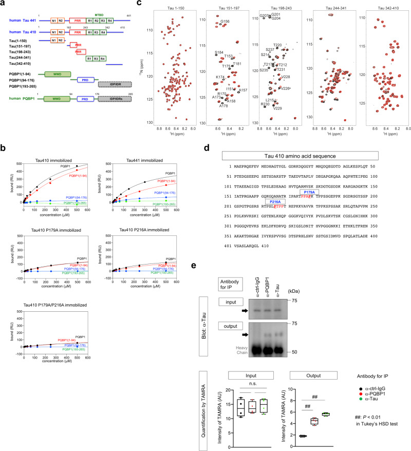
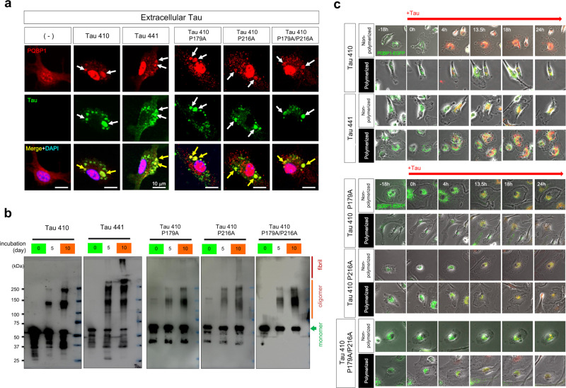
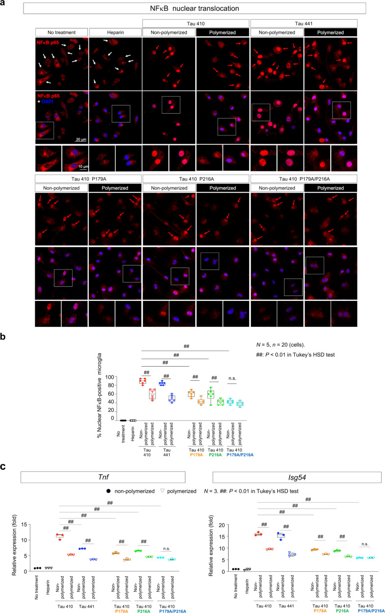
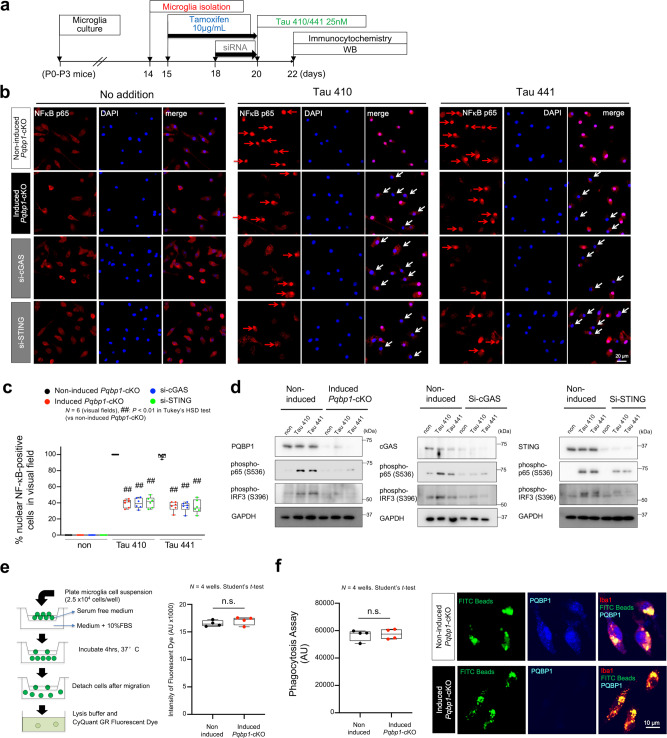
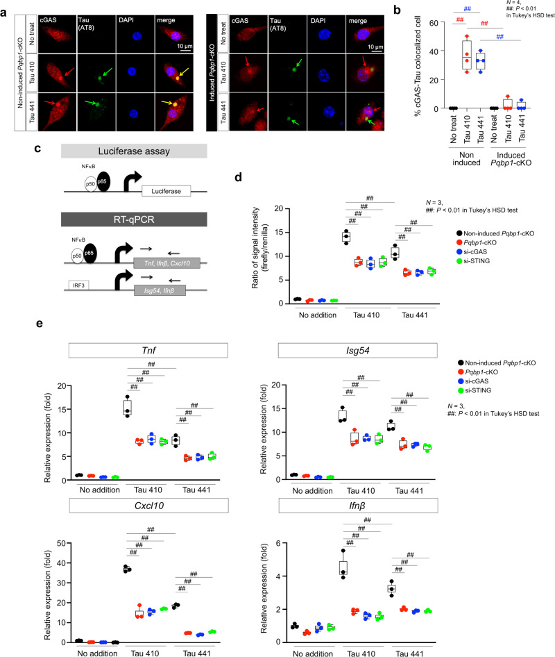
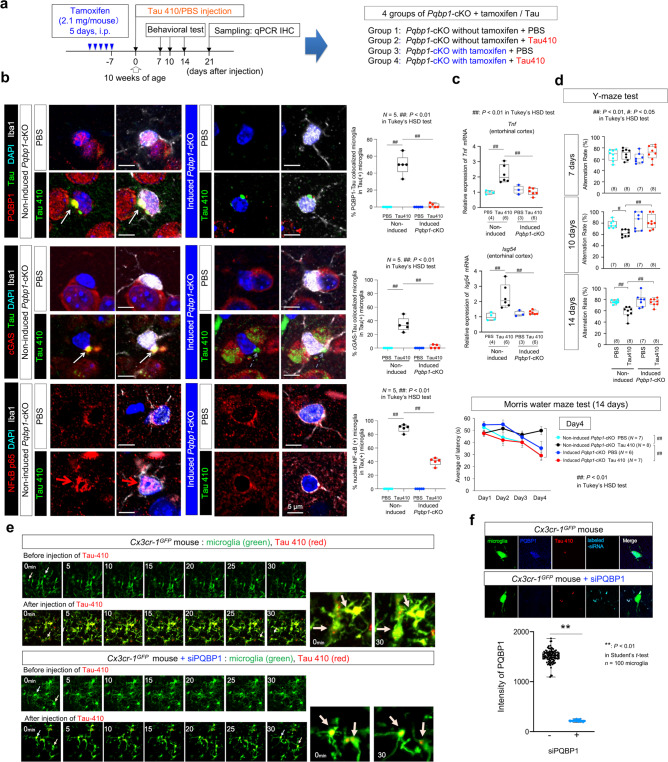
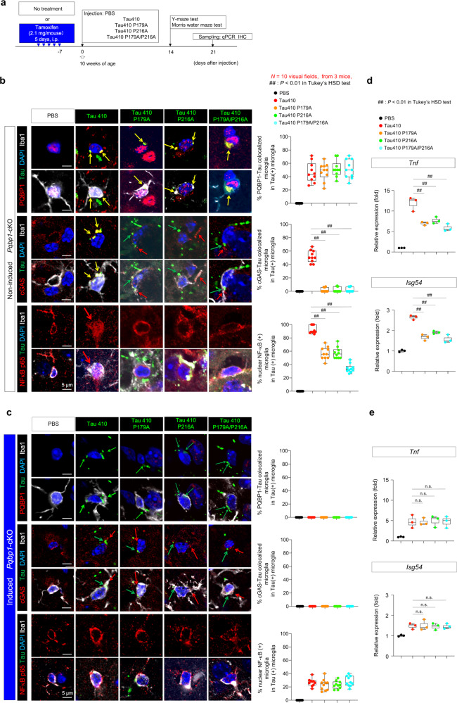
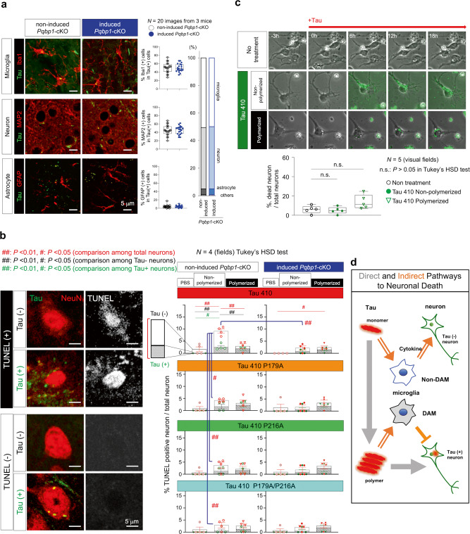
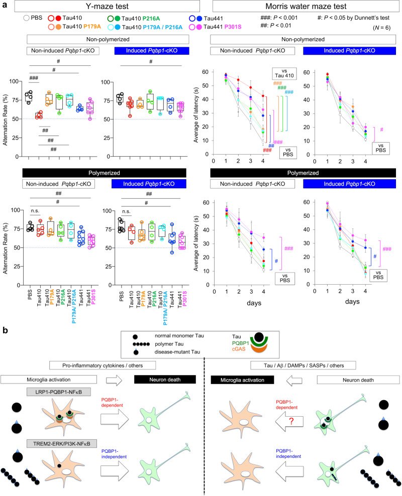
Brain inflammation generally accompanies and accelerates neurodegeneration. Here we report a microglial mechanism in which polyglutamine binding protein 1 (PQBP1) senses extrinsic tau 3R/4R proteins by direct interaction and triggers an innate immune response by activating a cyclic GMP-AMP synthase (cGAS)-Stimulator of interferon genes (STING) pathway. Tamoxifen-inducible and microglia-specific depletion of PQBP1 in primary culture in vitro and mouse brain in vivo shows that PQBP1 is essential for sensing-tau to induce nuclear translocation of nuclear factor κB (NFκB), NFκB-dependent transcription of inflammation genes, brain inflammation in vivo, and eventually mouse cognitive impairment. Collectively, PQBP1 is an intracellular receptor in the cGAS-STING pathway not only for cDNA of human immunodeficiency virus (HIV) but also for the transmissible neurodegenerative disease protein tau. This study characterises a mechanism of brain inflammation that is common to virus infection and neurodegenerative disorders.
Overview
The claims and statistics of this paper haven’t been checked yet.
Run the check — free, no account needed. It reads the open-access text and checks the sources it cites.
Integrity signals
Registry
Cites
Statistics
Figures
Verification
Claims the paper makes
The claims and statistics of this paper haven’t been checked yet.
Run the check — free, no account needed. It reads the open-access text and checks the sources it cites.
Claims other papers make about this one
Each is checked against this paper’s own text: does it say what the citing paper says it does?
The cited source says what the claim attributes to it.
In the citing paper
“Jin et al. reported an interesting finding that AD pathogenic protein tau can bind to polyglutamine binding protein 1 (PQBP1) and trigger inflammation in the CNS by activating cGAS-STING pathway.405”
From Role of neuroinflammation in neurodegeneration development (2023) · cites this paper as [405]
The cited source says what the claim attributes to it.
In the citing paper
“While it is currently unknown whether tau-induced retrotransposons form eccDNA, recent work from Gan's group (Udeochu et al. 2023) suggests that the cGAS-STING pathway is robustly activated in brains of tau transgenic mice, consistent with previous work in primary microglia treated with tau aggregates (Jin et al. 2021).”
From The Role of Retrotransposons and Endogenous Retroviruses in Age-Dependent Neurodegenerative Disorders (2024) · cites this paper as Jin et al. 2021
In the citing paper
“Numerous studies have reported that misfolded proteins and protein aggregates, including tau,136–139 Aβ,138,140 α-synuclein,141–144 mutant huntingtin,145 TDP-43,132,146 superoxide dismutase 1 (SOD1),147–149 and fused in sarcoma (FUS)150 induce microglial activation and neuroinflammation.”
From Microbiota–gut–brain axis and its therapeutic applications in neurodegenerative diseases (2024) · cites this paper as [136]
In the citing paper
“In addition, several studies have shown that tau seeds taken up by microglia can activate microglia, further exacerbating tau propagation and pathology [186, 192, 193].”
From Tau and neuroinflammation in Alzheimer’s disease: interplay mechanisms and clinical translation (2023) · cites this paper as [192]
In the citing paper
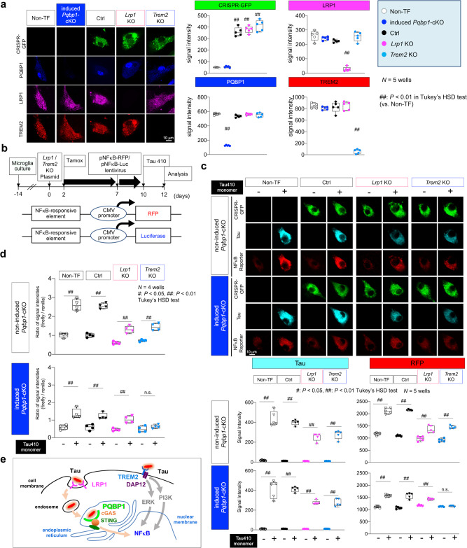
“Jin et al. reported that microglia uptake exogenous monomeric tau in parallel via two surface receptors, LRP1 and TREM2, and then induce nuclear factor NF-κB activation in microglia through two different pathways, LRP1/polyglutamine binding protein 1 (PQBP1)/cyclic GMP–AMP synthase (cGAS)/Stimulator of interferon genes (STING) and TREM2/extracellular signal-regulated kinase (ERK)/Phosphoinositide”
From Tau and neuroinflammation in Alzheimer’s disease: interplay mechanisms and clinical translation (2023) · cites this paper as [192]
Self-check
Percentages, sums and reported differences re-computed from the paper’s own numbers.
The self-check needs the paper’s full text.
Citations
Cites
Amyloid oligomers exacerbate tau pathology in a mouse model of tauopathy
Selenica, M.-L. B., Brownlow, M., Jimenez, J. P., Lee, D. C., Pena, G., Dickey, C. A., Gordon, M. N., & Morgan, D. (2013) · Neurodegenerative Diseases · doi:10.1159/000337230 · Cited by 10,359
Alzheimer's Disease: Genes, Proteins, and Therapy
Selkoe, D. J. (2001) · Physiological Reviews · doi:10.1152/physrev.2001.81.2.741 · Cited by 6,063
A Unique Microglia Type Associated with Restricting Development of Alzheimer’s Disease
Keren-Shaul, H., Spinrad, A., Weiner, A., Matcovitch-Natan, O., Dvir-Szternfeld, R., Ulland, T. K., David, E., Baruch, K., Lara-Astaiso, D., Toth, B., et al. (2017) · Cell · doi:10.1016/j.cell.2017.05.018 · Cited by 5,493
Release of chromatin protein HMGB1 by necrotic cells triggers inflammation
Scaffidi, P., Misteli, T., & Bianchi, M. E. (2002) · Nature · doi:10.1038/nature00858 · Cited by 4,221 · cited 1×
“1a) and analyzed the interaction of these fragments with PQBP1(1-94) (Fig.”
BackgroundMechanisms underlying inflammation in neurodegeneration
Glass, C. K., Saijo, K., Winner, B., Marchetto, M. C., & Gage, F. H. (2010) · Cell · doi:10.1016/j.cell.2010.02.016 · Cited by 3,580
Association of missense and 5′-splice-site mutations in tau with the inherited dementia FTDP-17
Hutton, M., Lendon, C. L., Rizzu, P., Baker, M., Froelich, S., Houlden, H., Pickering-Brown, S., Chakraverty, S., Isaacs, A., Grover, A., et al. (1998) · Nature · doi:10.1038/31508 · Cited by 3,498
TREM2 Variants in Alzheimer's Disease
Guerreiro, R., Wojtas, A., Bras, J., Carrasquillo, M., Rogaeva, E., Majounie, E., Cruchaga, C., Sassi, C., Kauwe, J. S. K., Younkin, S., et al. (2013) · New England Journal of Medicine · doi:10.1056/nejmoa1211851 · Cited by 3,020
Variant of TREM2 Associated with the Risk of Alzheimer's Disease
Jonsson, T., Stefansson, H., Steinberg, S., Jonsdottir, I., Jonsson, P. V., Snaedal, J., Bjornsson, S., Huttenlocher, J., Levey, A. I., Lah, J. J., et al. (2013) · New England Journal of Medicine · doi:10.1056/nejmoa1211103 · Cited by 2,558
Regulation and function of the cGAS–STING pathway of cytosolic DNA sensing
Chen, Q., Sun, L., & Chen, Z. J. (2016) · Nature Immunology · doi:10.1038/ni.3558 · Cited by 2,333
Immune attack: the role of inflammation in Alzheimer disease
Heppner, F. L., Ransohoff, R. M., & Becher, B. (2015) · Nature Reviews Neuroscience · doi:10.1038/nrn3880 · Cited by 2,134
Cited by
Sorted by citations
Items 201–224 of 224
Zhu, X., Liu, W., Tang, X., Chen, Y., Ge, X., Ke, Q., Liang, X., Gan, Y., Zheng, Y., Zou, M., et al. (2024) · Figshare · doi:10.6084/m9.figshare.26590775
Bao, H., Yan, J., Huang, J., Deng, W., Zhang, C., Liu, C., Huang, A., Zhang, Q., Xiong, Y., Wang, Q., et al. (2024) · Figshare · doi:10.6084/m9.figshare.26568898
Wang, Z., Liu, J., Han, J., Zhang, T., Li, S., Hou, Y., Su, H., Han, F., & Zhang, C. (2024) · Figshare · doi:10.6084/m9.figshare.26751308
Disrupting PQBP1–eEF2 protein-protein interaction: From synaptic translation to immunity and cancer
Kamel, E. M., Khadrawy, S. M., Ali, M. A. M., Ahmed, N. A., Yassin, N. Y. S., Alkhedhairi, S., Alkhayl, F. F. A., & Lamsabhi, A. M. (2026) · Neurochemistry International · doi:10.1016/j.neuint.2026.106154
Zhang, Y., Zhang, X., Huang, J., & Sun, Z. (2026) · Brain Research Bulletin · doi:10.1016/j.brainresbull.2026.112014
Mirjalili, F. S., Jambarsang, S., Yazd, E. F., Dianatinasab, M., Eussen, S. J. P. M., Schram, M. T., Koster, A., Salehi‐Abargouei, A., Yu, E. Y.-W., Zeegers, M. P., et al. (2026) · American Journal of Clinical Nutrition · doi:10.1016/j.ajcnut.2026.101415
PQBP1 couples HIV-1 capsid recognition to cGAS recruitment through conformational remodelingPreprint
Allen, D., Manna, A. K., Beusch, C. M., Zhang, T.-H., Ganser‐Pornillos, B. K., Piacentini, J., Oh, S., Prabhu, S., Mehta, A., Gordon, D. E., et al. (2026) · bioRxiv (Cold Spring Harbor Laboratory) · doi:10.64898/2026.07.07.737097
Ahmad, A., & Khan, M. M. (2026) · Molecular Neurobiology · doi:10.1007/s12035-026-06061-x
Wu, Y., Zhang, X., Gao, C., Zhang, Z., Liao, X., & Kang, Y. M. (2026) · Cellular and Molecular Neurobiology · doi:10.1007/s10571-026-01786-8
Xu, N., Ma, W., Dong, W., & Yú, L. (2026) · NeuroMolecular Medicine · doi:10.1007/s12017-026-08938-5
Chu, M., Tan, M., Gan, X., Cui, S., & Shi, L. (2026) · Molecular Neurobiology · doi:10.1007/s12035-026-06091-5
Shi, C., Liu, Y., & Zhu, Y. (2026) · Molecular Neurobiology · doi:10.1007/s12035-026-06081-7
Xinyuan, Y. U., Song, Y. A. O., Xu, P., & Wang, A. (2026) · Journal of Zhejiang University (Medical Sciences) · doi:10.3724/zdxbyxb-2026-0036
Xu, L., Zhang, Y., Jiang, L., Xing, J. F., Hu, J., Lan, T., & Ta, G. (2026) · Frontiers in Cellular Neuroscience · doi:10.3389/fncel.2026.1902577
Ferroptosis and Alzheimer’s disease: Neuroimmune crosstalk and pharmacological opportunities
Marakhovskaya, Y. A., Churov, A. V., Arbatsky, M. S., & Sergeeva, S. P. (2026) · Brain Research · doi:10.1016/j.brainres.2026.150514
The cGAS-STING pathway in inflammaging and neuroinflammation
Zhang, C., Liu, C., Zhao, Y., Wang, H., Fang, Z., He, Z., Mao, Y., & Yang, H. (2026) · Science China Life Sciences · doi:10.1007/s11427-026-3414-8
Therapeutic targeting of tauopathies: From druggable biology to precision intervention
Zhang, C., Ma, J., Wu, X., Wang, X., Huang, X., Zheng, H., & Zhao, Y. (2026) · Pharmacological Research · doi:10.1016/j.phrs.2026.108432
陈, 旸., & Li, Y. (2026) · Scientia Sinica Chimica · doi:10.1360/ssc-2026-0161
Höfs, L., Luksch, H., Schulze, F., Rösen‐Wolff, A., & Falkenburger, B. H. (2026) · Scientific Reports · doi:10.1038/s41598-026-70399-4
Disrupted PQBP1-HNRNPU-LINE-1 axis underlies aberrant neurodevelopment in renpenning syndrome
Zhang, J., Qu, W., Cheng, X., Li, L., Li, Y., Li, S., Ye, L., Yu, L., Li, Y., Jiang, P., et al. (2026) · Molecular Psychiatry · doi:10.1038/s41380-026-03908-6
Thupil, V. A., & Satchidanand, N. (2026) · Cureus · doi:10.7759/cureus.117175
Shao, J., & Lei, Z. (2026) · Frontiers in Aging Neuroscience · doi:10.3389/fnagi.2026.1983962
Zhang, C., & Li, Q. (2026) · Frontiers in Neurology · doi:10.3389/fneur.2026.1979957
Chen, R., Zhang, X., 宋恩杰, Chen, X., Wang, M., Xu, S., Fu, F., He, J., & Wan, S. (2026) · Reviews in the Neurosciences · doi:10.1515/revneuro-2026-0161
Metadata
- Authors
- Jin, M., Shiwaku, H., Tanaka, H., Obita, T., Ohuchi, S., Yoshioka, Y., Jin, X., Kondo, K., Fujita, K., Homma, H., Nakajima, K., Mizuguchi, M., & Okazawa, H.
- Affiliations
- Tokyo Medical and Dental University (JP)
- University of Toyama (JP)
- Soka University (JP)
- Corresponding author
- Okazawa, Hitoshi
- ORCID
- Venue
- Nature Communications
- Published
- 2021-11-15
- Volume
- 12
- Issue
- 1
- Pages
- 6565-6565
- ISSN
- 2041-1723 (ISSN-L)
- PMC
- PMC8592984
- OpenAlex
- W3214109787
- Cited by
- 223
- References
- 97
- Citations by year
- 2026: 43 · 2025: 49 · 2024: 66 · 2023: 37 · 2022: 28
- FWCI
- 13.21
- Type
- journal article
- Language
- en
- Open access
- gold
- doi.org
- Locations
- Nature Communications (publishedVersion, cc-by)
- PubMed Central (publishedVersion, cc-by)
- PubMed (publishedVersion)
- RePEc: Research Papers in Economics (submittedVersion)
- DOAJ (DOAJ: Directory of Open Access Journals) (submittedVersion, cc-by-sa)
- Funders
- MEXT | Japan Society for the Promotion of Science (16H02655, 19H01042)
- Ministry of Education, Culture, Sports, Science and Technology (22110001, 22110002)
- Topics
- Life Sciences › Neuroscience › Neurology › Neuroinflammation and Neurodegeneration Mechanisms
- Life Sciences › Neuroscience › Cellular and Molecular Neuroscience › Alzheimer's disease research and treatments
- Life Sciences › Immunology and Microbiology › Immunology › interferon and immune responses
- Keywords
- tau protein, microglia, PQBP1, cGAS-STING pathway, neuroinflammation, neurodegeneration, NF-κB, innate immune response, HIV, cognitive impairment, intracellular receptors
- MeSH terms
- STING Protein, Cyclic Guanosine Monophosphate-Adenosine Monophosphate Synthase, Animals, Brain, DNA-Binding Proteins, Encephalitis, Female, HIV, Humans, Immunity, Innate, Male, Membrane Glycoproteins, Membrane Proteins, Mice, Inbred C57BL, Nucleotidyltransferases, Tamoxifen, NF-kappa B, Microglia, Mice, Knockout, Neurodegenerative Diseases, Mice
About these checks
Registry, retraction, Cites and statistics results are computed, not judged, with one exception: a printed reference that no index lists is read once by a model before it counts as a citation error. Claims are extracted by a model and kept only when their quote appears verbatim in the text; each cited source is then checked, with a model’s help, for whether it says what the claim relies on. Those verdicts can be wrong — read the claim’s quote and the cited source before relying on one.
Statistics and arithmetic are recomputed, not judged.
Confidence bands are shown only for fields calibrated on our annotated test set. None is calibrated yet, so no band is shown.
What counts as a claim ›How we measure confidence ›How we check this ›
Are you an author, or do you see a problem with this page? Request a review
