

The DOI matches its Crossref record; 97 of the 97 references it cites were found in a registry or an index.
Tau activates microglia via the PQBP1-cGAS-STING pathway to promote brain inflammation
Open accessAbstract
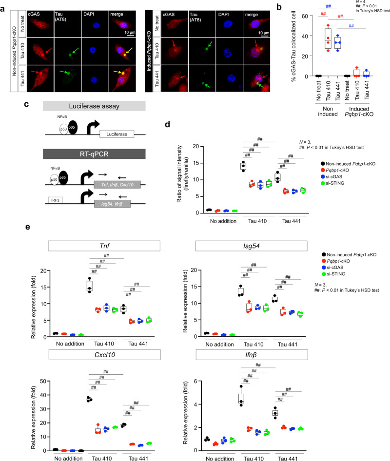
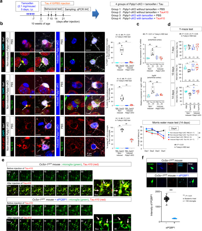
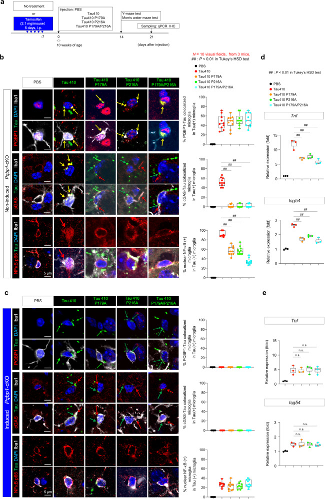
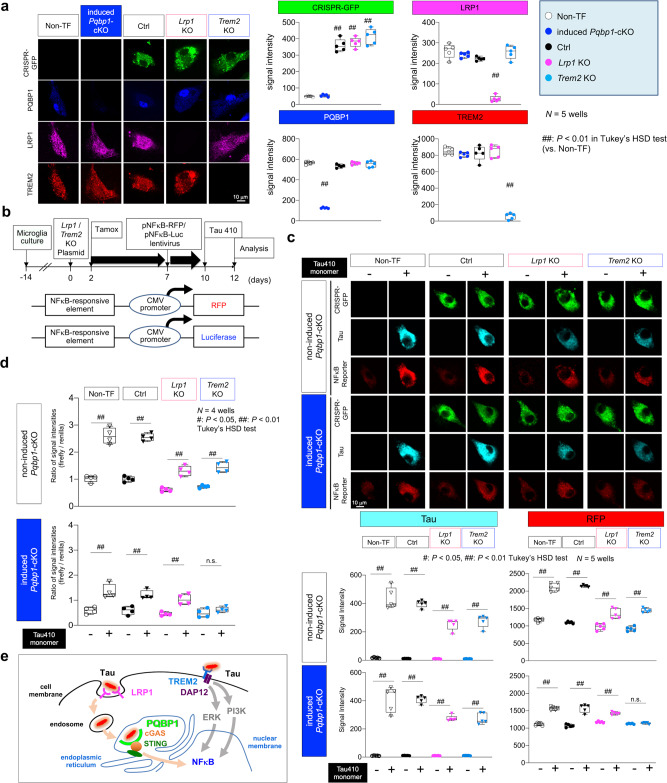
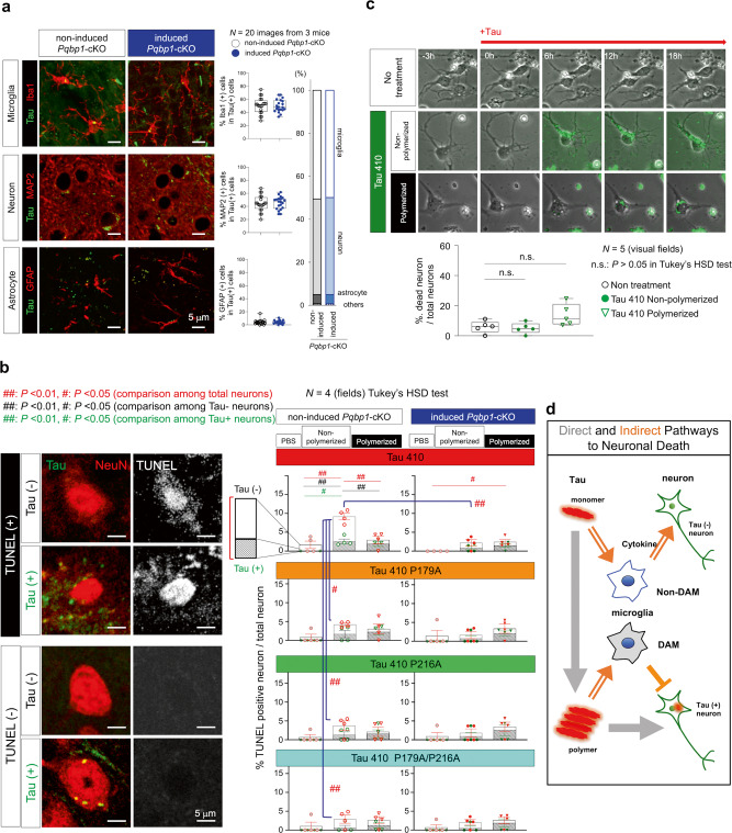
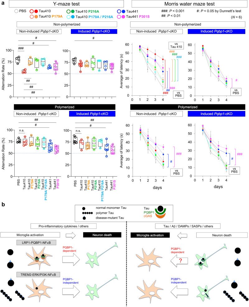
Brain inflammation generally accompanies and accelerates neurodegeneration. Here we report a microglial mechanism in which polyglutamine binding protein 1 (PQBP1) senses extrinsic tau 3R/4R proteins by direct interaction and triggers an innate immune response by activating a cyclic GMP-AMP synthase (cGAS)-Stimulator of interferon genes (STING) pathway. Tamoxifen-inducible and microglia-specific depletion of PQBP1 in primary culture in vitro and mouse brain in vivo shows that PQBP1 is essential for sensing-tau to induce nuclear translocation of nuclear factor κB (NFκB), NFκB-dependent transcription of inflammation genes, brain inflammation in vivo, and eventually mouse cognitive impairment. Collectively, PQBP1 is an intracellular receptor in the cGAS-STING pathway not only for cDNA of human immunodeficiency virus (HIV) but also for the transmissible neurodegenerative disease protein tau. This study characterises a mechanism of brain inflammation that is common to virus infection and neurodegenerative disorders.
Overview
The claims and statistics of this paper haven’t been checked yet.
Run the check — free, no account needed. It reads the open-access text and checks the sources it cites.
Integrity signals
Registry
Cites
Statistics
Figures
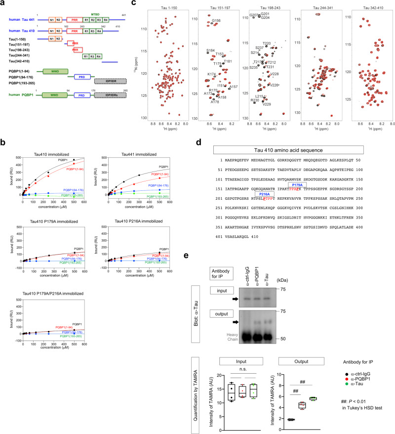
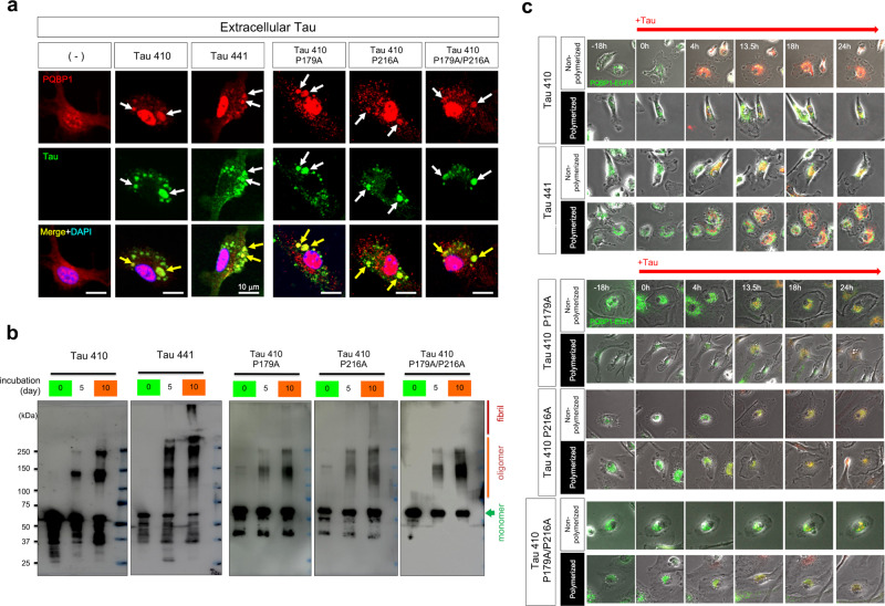
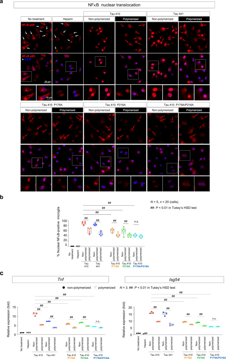
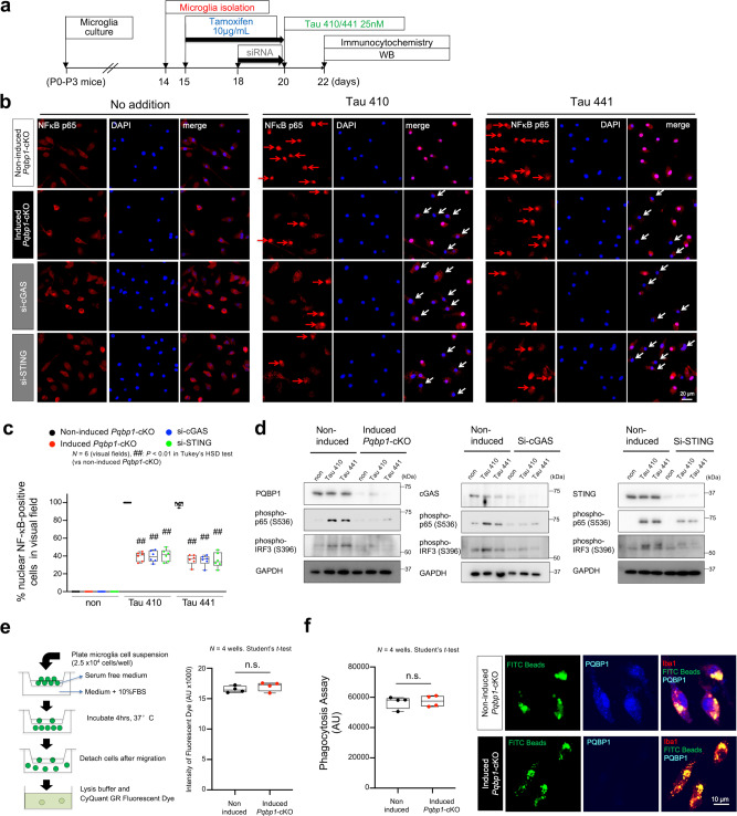
Verification
Claims the paper makes
The claims and statistics of this paper haven’t been checked yet.
Run the check — free, no account needed. It reads the open-access text and checks the sources it cites.
Claims other papers make about this one
Each is checked against this paper’s own text: does it say what the citing paper says it does?
The cited source says what the claim attributes to it.
In the citing paper
“Jin et al. reported an interesting finding that AD pathogenic protein tau can bind to polyglutamine binding protein 1 (PQBP1) and trigger inflammation in the CNS by activating cGAS-STING pathway.405”
From Role of neuroinflammation in neurodegeneration development (2023) · cites this paper as [405]
The cited source says what the claim attributes to it.
In the citing paper
“While it is currently unknown whether tau-induced retrotransposons form eccDNA, recent work from Gan's group (Udeochu et al. 2023) suggests that the cGAS-STING pathway is robustly activated in brains of tau transgenic mice, consistent with previous work in primary microglia treated with tau aggregates (Jin et al. 2021).”
From The Role of Retrotransposons and Endogenous Retroviruses in Age-Dependent Neurodegenerative Disorders (2024) · cites this paper as Jin et al. 2021
In the citing paper
“Numerous studies have reported that misfolded proteins and protein aggregates, including tau,136–139 Aβ,138,140 α-synuclein,141–144 mutant huntingtin,145 TDP-43,132,146 superoxide dismutase 1 (SOD1),147–149 and fused in sarcoma (FUS)150 induce microglial activation and neuroinflammation.”
From Microbiota–gut–brain axis and its therapeutic applications in neurodegenerative diseases (2024) · cites this paper as [136]
In the citing paper
“In addition, several studies have shown that tau seeds taken up by microglia can activate microglia, further exacerbating tau propagation and pathology [186, 192, 193].”
From Tau and neuroinflammation in Alzheimer’s disease: interplay mechanisms and clinical translation (2023) · cites this paper as [192]
In the citing paper
“Jin et al. reported that microglia uptake exogenous monomeric tau in parallel via two surface receptors, LRP1 and TREM2, and then induce nuclear factor NF-κB activation in microglia through two different pathways, LRP1/polyglutamine binding protein 1 (PQBP1)/cyclic GMP–AMP synthase (cGAS)/Stimulator of interferon genes (STING) and TREM2/extracellular signal-regulated kinase (ERK)/Phosphoinositide”
From Tau and neuroinflammation in Alzheimer’s disease: interplay mechanisms and clinical translation (2023) · cites this paper as [192]
Self-check
Percentages, sums and reported differences re-computed from the paper’s own numbers.
The self-check needs the paper’s full text.
Citations
Cites
Amyloid oligomers exacerbate tau pathology in a mouse model of tauopathy
Selenica, M.-L. B., Brownlow, M., Jimenez, J. P., Lee, D. C., Pena, G., Dickey, C. A., Gordon, M. N., & Morgan, D. (2013) · Neurodegenerative Diseases · doi:10.1159/000337230 · Cited by 10,359
Alzheimer's Disease: Genes, Proteins, and Therapy
Selkoe, D. J. (2001) · Physiological Reviews · doi:10.1152/physrev.2001.81.2.741 · Cited by 6,063
A Unique Microglia Type Associated with Restricting Development of Alzheimer’s Disease
Keren-Shaul, H., Spinrad, A., Weiner, A., Matcovitch-Natan, O., Dvir-Szternfeld, R., Ulland, T. K., David, E., Baruch, K., Lara-Astaiso, D., Toth, B., et al. (2017) · Cell · doi:10.1016/j.cell.2017.05.018 · Cited by 5,493
Release of chromatin protein HMGB1 by necrotic cells triggers inflammation
Scaffidi, P., Misteli, T., & Bianchi, M. E. (2002) · Nature · doi:10.1038/nature00858 · Cited by 4,221 · cited 1×
“1a) and analyzed the interaction of these fragments with PQBP1(1-94) (Fig.”
BackgroundMechanisms underlying inflammation in neurodegeneration
Glass, C. K., Saijo, K., Winner, B., Marchetto, M. C., & Gage, F. H. (2010) · Cell · doi:10.1016/j.cell.2010.02.016 · Cited by 3,580
Association of missense and 5′-splice-site mutations in tau with the inherited dementia FTDP-17
Hutton, M., Lendon, C. L., Rizzu, P., Baker, M., Froelich, S., Houlden, H., Pickering-Brown, S., Chakraverty, S., Isaacs, A., Grover, A., et al. (1998) · Nature · doi:10.1038/31508 · Cited by 3,498
TREM2 Variants in Alzheimer's Disease
Guerreiro, R., Wojtas, A., Bras, J., Carrasquillo, M., Rogaeva, E., Majounie, E., Cruchaga, C., Sassi, C., Kauwe, J. S. K., Younkin, S., et al. (2013) · New England Journal of Medicine · doi:10.1056/nejmoa1211851 · Cited by 3,020
Variant of TREM2 Associated with the Risk of Alzheimer's Disease
Jonsson, T., Stefansson, H., Steinberg, S., Jonsdottir, I., Jonsson, P. V., Snaedal, J., Bjornsson, S., Huttenlocher, J., Levey, A. I., Lah, J. J., et al. (2013) · New England Journal of Medicine · doi:10.1056/nejmoa1211103 · Cited by 2,558
Regulation and function of the cGAS–STING pathway of cytosolic DNA sensing
Chen, Q., Sun, L., & Chen, Z. J. (2016) · Nature Immunology · doi:10.1038/ni.3558 · Cited by 2,333
Immune attack: the role of inflammation in Alzheimer disease
Heppner, F. L., Ransohoff, R. M., & Becher, B. (2015) · Nature Reviews Neuroscience · doi:10.1038/nrn3880 · Cited by 2,134
Cited by
Sorted by citations
Role of neuroinflammation in neurodegeneration development
Zhang, W., Xiao, D., Mao, Q., & Xia, H. (2023) · Signal Transduction and Targeted Therapy · doi:10.1038/s41392-023-01486-5 · Cited by 1,668
“reported an interesting finding that AD pathogenic protein tau can bind to polyglutamine binding protein 1 (PQBP1) and trigger inflammation in the CNS by activating cGASSTING pathway.(405) Recently, Xie et al.”
BackgroundMicrobiota–gut–brain axis and its therapeutic applications in neurodegenerative diseases
Loh, J. S., Mak, W. Q., Tan, L. K. S., Ng, C. X., Chan, H. H., Yeow, S. H., Foo, J. B., Ong, Y. S., How, C. W., & Khaw, K. Y. (2024) · Signal Transduction and Targeted Therapy · doi:10.1038/s41392-024-01743-1 · Cited by 1,193
Neuroinflammation in Alzheimer disease
Heneka, M. T., van der Flier, W. M., Jessen, F., Hoozemanns, J., Thal, D. R., Boche, D., Brosseron, F., Teunissen, C., Zetterberg, H., Jacobs, A. H., et al. (2025) · Nature Reviews Immunology · doi:10.1038/s41577-024-01104-7 · Cited by 638
Tau and neuroinflammation in Alzheimer’s disease: interplay mechanisms and clinical translation
Chen, Y., & Yu, Y. (2023) · Journal of Neuroinflammation · doi:10.1186/s12974-023-02853-3 · Cited by 516
Role of the cGAS–STING pathway in systemic and organ-specific diseases
Skopelja‐Gardner, S., An, J., & Elkon, K. B. (2022) · Nature Reviews Nephrology · doi:10.1038/s41581-022-00589-6 · Cited by 318
The effects of microglia-associated neuroinflammation on Alzheimer’s disease
Wang, C., Zong, S., Cui, X., Wang, X., Wu, S., Wang, L., Liu, Y., & Lu, Z. (2023) · Frontiers in Immunology · doi:10.3389/fimmu.2023.1117172 · Cited by 331
Activation of innate immune cGAS-STING pathway contributes to Alzheimer’s pathogenesis in 5×FAD mice
Xie, X., Ma, G., Li, X., Zhao, J., Zhao, Z., & Zeng, J. (2023) · Nature Aging · doi:10.1038/s43587-022-00337-2 · Cited by 292
Tau Activation of Microglial cGAS–IFN Reduces MEF2C‐mediated Cognitive Resilience
Udeochu, J. C., Amin, S., Huang, Y., Fan, L., Torres, E. R. S., Carling, G. K., Liu, B., McGurran, H., Coronas-Samano, G., Kauwe, G., et al. (2023) · Nature Neuroscience · doi:10.1038/s41593-023-01315-6 · Cited by 289
Regulation of cGAS–STING signalling and its diversity of cellular outcomes
Zhang, Z., & Zhang, C. (2025) · Nature Reviews Immunology · doi:10.1038/s41577-024-01112-7 · Cited by 287
Cellular and pathological functions of tau
Bravo, C. P., Naguib, S. A., & Gan, L. (2024) · Nature Reviews Molecular Cell Biology · doi:10.1038/s41580-024-00753-9 · Cited by 255
STING orchestrates the neuronal inflammatory stress response in multiple sclerosis
Woo, M. S., Mayer, C., Binkle-Ladisch, L., Sonner, J. K., Rosenkranz, S. C., Shaposhnykov, A., Rothammer, N., Tsvilovskyy, V., Lorenz, S., Raich, L., et al. (2024) · Cell · doi:10.1016/j.cell.2024.05.031 · Cited by 197
Lau, V. Z., Ramer, L. M., & Tremblay, M. (2023) · Nature Communications · doi:10.1038/s41467-023-37304-3 · Cited by 173
Innate immune activation in neurodegenerative diseases
Castro‐Gomez, S., & Heneka, M. T. (2024) · Immunity · doi:10.1016/j.immuni.2024.03.010 · Cited by 171
Mechanism and therapeutic potential of targeting cGAS-STING signaling in neurological disorders
Huang, Y., Liu, B., Sinha, S. C., Amin, S., & Gan, L. (2023) · Molecular Neurodegeneration · doi:10.1186/s13024-023-00672-x · Cited by 164
Molecular Mechanisms of Neuroinflammation in Aging and Alzheimer’s Disease Progression
Andronie-Cioară, F. L., Ardelean, A. I., Nistor-Cseppento, C. D., Jurcău, A., Jurcău, M. C., Pașcalău, N., & Marcu, F. M. (2023) · International Journal of Molecular Sciences · doi:10.3390/ijms24031869 · Cited by 168
Mitophagy and cGAS–STING crosstalk in neuroinflammation
Zhou, X., Wang, J., Yu, L., Qiao, G., Qin, D., Yuen-Kwan Law, B., Ren, F., Wu, J., & Wu, A. (2024) · Acta Pharmaceutica Sinica B · doi:10.1016/j.apsb.2024.05.012 · Cited by 131
Microglial CMPK2 promotes neuroinflammation and brain injury after ischemic stroke
Guan, X., Zhu, S., Song, J.-Q., Liu, K., Liu, M., Xie, L., Wang, Y., Wu, J. S., Xu, X., & Pang, T. (2024) · Cell Reports Medicine · doi:10.1016/j.xcrm.2024.101522 · Cited by 114
Hou, Y., Wang, Z., Liu, P., Wei, X., Zhang, Z., Fan, S., Zhang, L., Han, F., Song, Y., Chu, L., et al. (2023) · Immunity · doi:10.1016/j.immuni.2023.10.001 · Cited by 112
Li, T., Lu, L., Pember, E., Li, X., Zhang, B., & Zhu, Z. (2022) · Cells · doi:10.3390/cells11121925 · Cited by 109
Chung, S., Jeong, J.-H., Park, J.-C., Han, J. W., Lee, Y., Kim, J.-I., & Mook-Jung, I. (2024) · Experimental & Molecular Medicine · doi:10.1038/s12276-024-01295-y · Cited by 103
“In addition, A β can induce pathological neuritic plaque-dependent tau conversion 31 , and microglia-engulfed tau has been reported to induce PQBP1-dependent cGAS activation 32 .”
Quan, S., Fu, X., Cai, H., Ren, Z., Xu, Y., & Jia, L. (2025) · Molecular Neurodegeneration · doi:10.1186/s13024-025-00815-2 · Cited by 110
Chronic effects of inflammation on tauopathies
Langworth-Green, C., Patel, S., Jaunmuktane, Z., Jabbari, E., Morris, H. R., Thom, M., Lees, A. J., Hardy, J., Zandi, M. S., & Duff, K. (2023) · The Lancet Neurology · doi:10.1016/s1474-4422(23)00038-8 · Cited by 81
Wu, Y., Bogale, T. A., Koıstınaho, J., Pizzi, M., Rõlova, T., & Bellucci, A. (2024) · Acta Neuropathologica · doi:10.1007/s00401-024-02696-z · Cited by 82
Zhang, Y., Zou, M., Wu, H., Zhu, J., & Jin, T. (2024) · Neurobiology of Disease · doi:10.1016/j.nbd.2024.106710 · Cited by 73
Lei, W., Cheng, Y., Liu, X., Gao, J., Zhu, Z., Ding, W., Xu, X., Li, Y., Ling, Z., Jiang, R., et al. (2025) · Frontiers in Immunology · doi:10.3389/fimmu.2025.1582119 · Cited by 68
Boza-Serrano, A., Vrillon, A., Minta, K., Paulus, A., Camprubí-Ferrer, L., Garcia, M., Andreasson, U., Antonell, A., Wennström, M., Gouras, G., et al. (2022) · Acta Neuropathologica · doi:10.1007/s00401-022-02469-6 · Cited by 66
“This microglial inflammatory response is dependent on NFκB-dependent transcription of inflammatory genes, leading to brain inflammation [27].”
Targeting tau in Alzheimer's disease: from mechanisms to clinical therapy
Ye, J., Wan, H., Chen, S., & Liu, G. (2023) · Neural Regeneration Research · doi:10.4103/1673-5374.385847 · Cited by 70
STING signaling in the brain: Molecular threats, signaling activities, and therapeutic challenges
Yang, K., Tang, Z., Xing, C., & Yan, N. (2023) · Neuron · doi:10.1016/j.neuron.2023.10.014 · Cited by 63
Li, H., Cai, R., Zhou, Y., Jiang, Y., & Tan, S. (2025) · Journal of Neuroinflammation · doi:10.1186/s12974-025-03563-8 · Cited by 72
Wang, B., Wang, Y., Qiu, J., Gao, S., Yu, S., Sun, D., & Lou, H. (2023) · International Immunopharmacology · doi:10.1016/j.intimp.2023.110827 · Cited by 64
Relationship between the cGAS−STING and NF-κB pathways-role in neurotoxicity
Zhong, S., Zhou, Q., Yang, J., Zhang, Z., Zhang, X., Liu, J., Chang, X., & Wang, H. (2024) · Biomedicine & Pharmacotherapy · doi:10.1016/j.biopha.2024.116698 · Cited by 62
Wang, Z., Liu, J., Han, J., Zhang, T., Li, S., Hou, Y., Su, H., Han, F., & Zhang, C. (2024) · Journal of Neuroinflammation · doi:10.1186/s12974-024-03166-9 · Cited by 60
Microglial Priming in Infections and Its Risk to Neurodegenerative Diseases
Lima, M. N., Barbosa‐Silva, M. C., & Maron‐Gutierrez, T. (2022) · Frontiers in Cellular Neuroscience · doi:10.3389/fncel.2022.878987 · Cited by 62
Anti-herpetic tau preserves neurons via the cGAS-STING-TBK1 pathway in Alzheimer’s disease
Hyde, V. R., Zhou, C., y Fernández, J. R. M., Chatterjee, K., Ramakrishna, P., Lin, A., Fisher, G. W., Celiker, O. T., Caldwell, J., Bender, O., et al. (2025) · Cell Reports · doi:10.1016/j.celrep.2024.115109 · Cited by 61
cGAS-STING and neurodegenerative diseases: A molecular crosstalk and therapeutic perspective
Dhapola, R., Paidlewar, M., Kumari, S., Sharma, P., Vellingiri, B., Medhi, B., & HariKrishnaReddy, D. (2025) · International Immunopharmacology · doi:10.1016/j.intimp.2025.114902 · Cited by 60
Wu, C., Zhang, S., Sun, H., Li, A., Hou, F., Qi, L., & Liao, H. (2024) · Journal of Neuroinflammation · doi:10.1186/s12974-024-03086-8 · Cited by 54
Role of cGAS–Sting Signaling in Alzheimer’s Disease
Govindarajulu, M., Ramesh, S., Beasley, M., Lynn, G., Wallace, C., Labeau, S., Pathak, S., Nadar, R. M., Moore, T., & Dhanasekaran, M. (2023) · International Journal of Molecular Sciences · doi:10.3390/ijms24098151 · Cited by 59
DNA Double-Strand Breaks as Pathogenic Lesions in Neurological Disorders
Provasek, V. E., Mitra, J., Malojirao, V. H., & Hegde, M. L. (2022) · International Journal of Molecular Sciences · doi:10.3390/ijms23094653 · Cited by 54
Control of innate immunity by the cGAS‐STING pathway
Mosallanejad, K., & Kagan, J. C. (2022) · Immunology and Cell Biology · doi:10.1111/imcb.12555 · Cited by 52
Aβ and Tau Regulate Microglia Metabolism via Exosomes in Alzheimer’s Disease
Zhao, Y., Liu, B., Wang, J., Xu, L., Yu, S., Fu, J., Yan, X., & Su, J. (2022) · Biomedicines · doi:10.3390/biomedicines10081800 · Cited by 47
Cancer immunotherapy strategies that target the cGAS-STING pathway
Tian, Z., Zeng, Y., Peng, Y., Liu, J., & Wu, F. (2022) · Frontiers in Immunology · doi:10.3389/fimmu.2022.996663 · Cited by 47
Abdolmaleky, H. M., & Zhou, J. (2023) · Nutrients · doi:10.3390/nu15153456 · Cited by 47
Frost, B., & Dubnau, J. (2024) · Annual Review of Neuroscience · doi:10.1146/annurev-neuro-082823-020615 · Cited by 45
Zhu, X., Liu, W., Tang, X., Chen, Y.-L., Ge, X., Ke, Q., Liang, X., Gan, Y., Zheng, Y., Zou, M., et al. (2023) · Journal of Neuroinflammation · doi:10.1186/s12974-023-02804-y · Cited by 46
Wang, R., Sun, H., Cao, Y., Zhang, Z., Chen, Y., Wang, X., Liu, L., Wu, J., Xu, H., Wu, D., et al. (2024) · Science Signaling · doi:10.1126/scisignal.adk8249 · Cited by 44
Type-I Interferons in Alzheimer's Disease and Other Tauopathies
Sanford, S., & McEwan, W. A. (2022) · Frontiers in Cellular Neuroscience · doi:10.3389/fncel.2022.949340 · Cited by 42
cGAS/STING and innate brain inflammation following acute high-fat feeding
Elzinga, S. E., Henn, R. E., Murdock, B. J., Kim, B., Hayes, J. M., Mendelson, F. E., Webber‐Davis, I. F., Teener, S. J., Pacut, C. M., Lentz, S. I., et al. (2022) · Frontiers in Immunology · doi:10.3389/fimmu.2022.1012594 · Cited by 39
Maimaiti, M., Li, C., Cheng, M., Zhong, Z., Hu, J., Yang, L., Zhang, L., Hong, Z., Song, J., Pan, M., et al. (2024) · Med · doi:10.1016/j.medj.2024.03.018 · Cited by 39
PQBP5/NOL10 maintains and anchors the nucleolus under physiological and osmotic stress conditions
Jin, X., Tanaka, H., Jin, M., Fujita, K., Homma, H., Inotsume, M., Yong, H., Umeda, K., Kodera, N., Ando, T., et al. (2023) · Nature Communications · doi:10.1038/s41467-022-35602-w · Cited by 39
Self-citationTargeting STING: From antiviral immunity to treat osteoporosis
Gao, Z., Gao, Z., Zhang, H., Hou, S., Zhou, Y., & Liu, X. (2023) · Frontiers in Immunology · doi:10.3389/fimmu.2022.1095577 · Cited by 38
Immunotherapies in chronic pain through modulation of neuroimmune interactions
Zhao, J., Huh, Y., Bortsov, A. V., Diatchenko, L. B., & Ji, R. (2023) · Pharmacology & Therapeutics · doi:10.1016/j.pharmthera.2023.108476 · Cited by 40
Mechanistic insights into the role of herpes simplex virus 1 in Alzheimer’s disease
Feng, S., Liu, Y., Zhou, Y., Shu, Z., Cheng, Z., Brenner, C., & Feng, P. (2023) · Frontiers in Aging Neuroscience · doi:10.3389/fnagi.2023.1245904 · Cited by 36
Donison, N. S., Palik, J., Volkening, K., & Strong, M. J. (2025) · Molecular Neurodegeneration · doi:10.1186/s13024-025-00842-z · Cited by 39
Španić, E., Horvat, L. L., Ilić, K., Hof, P. R., & Šimić, G. (2022) · Cells · doi:10.3390/cells11142223 · Cited by 35
Role of microglia autophagy and mitophagy in age-related neurodegenerative diseases
Lin, M., Yu, H., Xie, Q., Xu, Z., & Shang, P. (2023) · Frontiers in Aging Neuroscience · doi:10.3389/fnagi.2022.1100133 · Cited by 37
Hu, J., Sha, W., Yuan, S., Wu, J., & Huang, Y. (2023) · International Journal of Molecular Sciences · doi:10.3390/ijms241915023 · Cited by 40
Nanomedicines for Alzheimer's disease: Therapies based on pathological mechanisms
Cheng, G., Xie, A., Yan, Z., Zhu, X., Song, Y., & Chen, T. (2023) · Brain‐X · doi:10.1002/brx2.27 · Cited by 35
Luo, H., Lv, J., Wen, P., Zhang, S., Ma, W., & Yáng, Z. (2025) · Nature Communications · doi:10.1038/s41467-025-57718-5 · Cited by 35
Microglial autophagy in Alzheimer’s disease and Parkinson’s disease
Wang, Z., Wang, Q., Li, S., Li, X.-J., Yang, W., & He, D. (2023) · Frontiers in Aging Neuroscience · doi:10.3389/fnagi.2022.1065183 · Cited by 35
Wang, L., Zhao, H., Han, M. H., Yang, H., Lei, M., Wang, W., Li, K., Li, Y., Sang, Y., Xin, T., et al. (2024) · Advanced Science · doi:10.1002/advs.202307527 · Cited by 31
Regulation of cGAS Activity and Downstream Signaling
Joshi, B., Joshi, J. C., & Mehta, D. (2022) · Cells · doi:10.3390/cells11182812 · Cited by 32
Gao, S., Shen, R., Li, J., Jiang, Y., Sun, H., Wu, X., Li, X., Miao, C., He, M., Wang, J., et al. (2024) · Proceedings of the National Academy of Sciences · doi:10.1073/pnas.2410564121 · Cited by 31
Ju, C., Wang, Y., Zang, C., Liu, H., Yuan, F., Ning, J., Shang, M., Ma, J., Li, G., Yang, Y., et al. (2022) · Inflammation · doi:10.1007/s10753-022-01699-w · Cited by 31
Microglial Dysfunction in Neurodegenerative Diseases via RIPK1 and ROS
Wu, Q., & Zou, C. (2022) · Antioxidants · doi:10.3390/antiox11112201 · Cited by 30
Gut microbiota, pathogenic proteins and neurodegenerative diseases
Wei, W., Wang, S., Xu, C., Zhou, X., Lian, X., He, L., & Li, K. (2022) · Frontiers in Microbiology · doi:10.3389/fmicb.2022.959856 · Cited by 30
Pathophysiological evaluation of the LRRK2 G2385R risk variant for Parkinson’s disease
Tezuka, T., Taniguchi, D., Sano, M., Shimada, T., Oji, Y., Tsunemi, T., Ikeda, A., Li, Y., Yoshino, H., Ogata, J., et al. (2022) · npj Parkinson s Disease · doi:10.1038/s41531-022-00367-y · Cited by 27
Bao, H., Yan, J., Huang, J., Deng, W., Zhang, C., Liu, C., Huang, A., Zhang, Q., Xiong, Y., Wang, Q., et al. (2023) · Journal of Neuroinflammation · doi:10.1186/s12974-023-02724-x · Cited by 28
Zhou, Q., Luo, J., Chai, X., Yang, J., Zhong, S., Zhang, Z., Chang, X., & Wang, H. (2025) · International Immunopharmacology · doi:10.1016/j.intimp.2025.114679 · Cited by 31
Bogár, F., Fülöp, L., & Penke, B. (2022) · Biomolecules · doi:10.3390/biom12030363 · Cited by 27
Chidambaram, H., & Chinnathambi, S. (2025) · Brain Network Disorders · doi:10.1016/j.bnd.2024.12.005 · Cited by 25
Role of Microglial Mitophagy in Alleviating Postoperative Cognitive Dysfunction: a Mechanistic Study
Zhang, L., Li, J., Li, C., Wu, Y., Liu, S., Li, Q., & Qi, S. (2024) · Molecular Neurobiology · doi:10.1007/s12035-024-04405-z · Cited by 25
Zhang, X., Li, X., Wang, W., Zhang, Y., Gong, Z., Peng, Y., Wu, J., & You, X. (2022) · Cancers · doi:10.3390/cancers14215188 · Cited by 25
Qu, W., Canoll, P., & Hargus, G. (2023) · Neuroscience · doi:10.1016/j.neuroscience.2022.05.006 · Cited by 24
Is Drp1 a link between mitochondrial dysfunction and inflammation in Alzheimer’s disease?
Sbai, O., Bazzani, V., Tapaswi, S., McHale, J., Vascotto, C., & Perrone, L. (2023) · Frontiers in Molecular Neuroscience · doi:10.3389/fnmol.2023.1166879 · Cited by 24
Microglia internalize tau monomers and fibrils using distinct receptors but similar mechanisms
Falkon, K. F., Danford, L., Kuri, E. G., Esquinca‐Moreno, P., Señeriz, Y. L. P., Smith, S. A., Wickline, J. L., Louwrier, A., McPhail, J. A., Sayre, N. L., et al. (2024) · Alzheimer s & Dementia · doi:10.1002/alz.14418 · Cited by 25
Asveda, T., Talwar, P., & Ravanan, P. (2023) · Life Sciences · doi:10.1016/j.lfs.2023.121920 · Cited by 23
Non–Interferon-Dependent Role of STING Signaling in Pulmonary Hypertension
Pham, A. T., de Oliveira, A. C., Fu, C., Alves, M. D., Dupee, Z., Mukhsinova, L., Ebrahimi, E., Patel, H., Patel, R., Nguyen, A., et al. (2023) · Arteriosclerosis Thrombosis and Vascular Biology · doi:10.1161/atvbaha.123.320121 · Cited by 21
Cui, Z., Yuan, X., Wang, Y., Liu, Z., Fei, X., Chen, K., Shen, H., Wu, Y., & Xia, D. (2024) · The Science of The Total Environment · doi:10.1016/j.scitotenv.2024.176786 · Cited by 22
Guzman‐Hernandez, R. A., & Fossati, S. (2025) · Alzheimer s & Dementia · doi:10.1002/alz.70077 · Cited by 23
Joshi, R., Brezáni, V., Mey, G. M., Guixé‐Muntet, S., Ortega‐Ribera, M., Zhuang, Y., Zivny, A., Werneburg, S., Gracia‐Sancho, J., & Szabó, G. (2024) · Journal of Neuroinflammation · doi:10.1186/s12974-024-03203-7 · Cited by 22
Type I interferon signalling and interferon‐responsive microglia in health and disease
López‐Atalaya, J. P., & Bhojwani-Cabrera, A. M. (2025) · FEBS Journal · doi:10.1111/febs.70126 · Cited by 21
Lau, S., Fu, A. K. Y., & Ip, N. Y. (2023) · Journal of Neurochemistry · doi:10.1111/jnc.15933 · Cited by 22
Kaur, A., & Aran, K. R. (2025) · Neuroscience · doi:10.1016/j.neuroscience.2025.04.001 · Cited by 23
Dapsone is an anticatalysis for Alzheimer’s disease exacerbation
Lee, J., Kanwar, B. A., Lee, C. J., Sergi, C. M., & Coleman, M. D. (2022) · iScience · doi:10.1016/j.isci.2022.104274 · Cited by 19
Xu, L., Li, B., Li, F., Li, Y., Qin, W., Zhao, Y., & Jia, J. (2022) · Journal of Alzheimer s Disease · doi:10.3233/jad-220747 · Cited by 20
Necrosis Links Neurodegeneration and Neuroinflammation in Neurodegenerative Disease
Homma, H., Tanaka, H., Fujita, K., & Okazawa, H. (2024) · International Journal of Molecular Sciences · doi:10.3390/ijms25073636 · Cited by 20
Self-citationcGAS-STING signaling in Alzheimer's disease: Microglial mechanisms and therapeutic opportunities
Fazal, F., Dar, N. J., Ahamad, S., Khan, S., Bano, N., Saha, S., Nazir, A., & Bhat, S. A. (2026) · Molecular Aspects of Medicine · doi:10.1016/j.mam.2025.101444 · Cited by 21
Xie, J., Cheng, J., Ko, H., & Tang, Y. (2024) · EMBO Molecular Medicine · doi:10.1038/s44321-024-00046-w · Cited by 17
Emerging roles for innate and adaptive immunity in tauopathies
Johnson, A. M., & Lukens, J. R. (2025) · Cell Reports · doi:10.1016/j.celrep.2025.116232 · Cited by 19
Chemistry and Function of Glycosaminoglycans in the Nervous System
Schwartz, N. B., & Domowicz, M. S. (2022) · Advances in neurobiology · doi:10.1007/978-3-031-12390-0_5 · Cited by 17
Human stem cell transplantation models of Alzheimer’s disease
Ifediora, N., Canoll, P., & Hargus, G. (2024) · Frontiers in Aging Neuroscience · doi:10.3389/fnagi.2024.1354164 · Cited by 18
Yang, G., Kantapan, J., Mazhar, M., Hu, Q., Bai, X. H., Zou, Y., Wang, H., Yang, S., Wang, L., & Dechsupa, N. (2024) · Journal of Inflammation Research · doi:10.2147/jir.s449579 · Cited by 16
PQBP1: The Key to Intellectual Disability, Neurodegenerative Diseases, and Innate Immunity
Tanaka, H., & Okazawa, H. (2022) · International Journal of Molecular Sciences · doi:10.3390/ijms23116227 · Cited by 18
Self-citationType I interferons in health and disease: molecular aspects and clinical implications
Mavragani, C. P., & Crow, M. K. (2025) · Physiological Reviews · doi:10.1152/physrev.00047.2024 · Cited by 16
STING-mediated neuroinflammation: a therapeutic target in neurodegenerative diseases
Zhang, H., He, Z., Yin, C., Yang, S., Li, J., Lin, H. R., Hu, G., Wu, A., Qin, D., Yu, L., et al. (2025) · Frontiers in Aging Neuroscience · doi:10.3389/fnagi.2025.1659216 · Cited by 16
Wang, W., Wang, L., Zhong, X., Dong, L., Zhang, L., & Hu, J. X. (2025) · Cell Biochemistry and Function · doi:10.1002/cbf.70146 · Cited by 15
Cytosolic DNA sensors and glial responses to endogenous DNA
Suptela, A. J., & Marriott, I. (2023) · Frontiers in Immunology · doi:10.3389/fimmu.2023.1130172 · Cited by 15
Amyloid solutions: lecanemab, gantenerumab, and donanemab in the treatment of Alzheimer's disease
Ameen, T. B., Ali, U., Salma, O., Samee, M. A., Abbas, S. M. I., Kashif, S. N., Arifi, M. A., Ali, M., Khowaja, M., Ali, S. M. S., et al. (2025) · The Egyptian Journal of Neurology Psychiatry and Neurosurgery · doi:10.1186/s41983-025-00968-3 · Cited by 18
cGAS–STING signaling pathway as a therapeutic target in human diseases
Xiao, Y., Ma, Y., Zhao, J., Zhang, X., Gan, C., Gao, J., & Huang, Z. (2025) · Chinese Medical Journal · doi:10.1097/cm9.0000000000003904 · Cited by 15
Complement receptor 4 mediates the clearance of extracellular tau fibrils by microglia
Yoo, C. J., Choi, Y., Bok, E., Lin, Y., Cheon, M., Lee, Y., & Kim, J. (2024) · FEBS Journal · doi:10.1111/febs.17150 · Cited by 16
Metadata
- Authors
- Jin, M., Shiwaku, H., Tanaka, H., Obita, T., Ohuchi, S., Yoshioka, Y., Jin, X., Kondo, K., Fujita, K., Homma, H., Nakajima, K., Mizuguchi, M., & Okazawa, H.
- Affiliations
- Tokyo Medical and Dental University (JP)
- University of Toyama (JP)
- Soka University (JP)
- Corresponding author
- Okazawa, Hitoshi
- ORCID
- Venue
- Nature Communications
- Published
- 2021-11-15
- Volume
- 12
- Issue
- 1
- Pages
- 6565-6565
- ISSN
- 2041-1723 (ISSN-L)
- PMC
- PMC8592984
- OpenAlex
- W3214109787
- Cited by
- 223
- References
- 97
- Citations by year
- 2026: 43 · 2025: 49 · 2024: 66 · 2023: 37 · 2022: 28
- FWCI
- 13.21
- Type
- journal article
- Language
- en
- Open access
- gold
- doi.org
- Locations
- Nature Communications (publishedVersion, cc-by)
- PubMed Central (publishedVersion, cc-by)
- PubMed (publishedVersion)
- RePEc: Research Papers in Economics (submittedVersion)
- DOAJ (DOAJ: Directory of Open Access Journals) (submittedVersion, cc-by-sa)
- Funders
- MEXT | Japan Society for the Promotion of Science (16H02655, 19H01042)
- Ministry of Education, Culture, Sports, Science and Technology (22110001, 22110002)
- Topics
- Life Sciences › Neuroscience › Neurology › Neuroinflammation and Neurodegeneration Mechanisms
- Life Sciences › Neuroscience › Cellular and Molecular Neuroscience › Alzheimer's disease research and treatments
- Life Sciences › Immunology and Microbiology › Immunology › interferon and immune responses
- Keywords
- tau protein, microglia, PQBP1, cGAS-STING pathway, neuroinflammation, neurodegeneration, NF-κB, innate immune response, HIV, cognitive impairment, intracellular receptors
- MeSH terms
- STING Protein, Cyclic Guanosine Monophosphate-Adenosine Monophosphate Synthase, Animals, Brain, DNA-Binding Proteins, Encephalitis, Female, HIV, Humans, Immunity, Innate, Male, Membrane Glycoproteins, Membrane Proteins, Mice, Inbred C57BL, Nucleotidyltransferases, Tamoxifen, NF-kappa B, Microglia, Mice, Knockout, Neurodegenerative Diseases, Mice
About these checks
Registry, retraction, Cites and statistics results are computed, not judged, with one exception: a printed reference that no index lists is read once by a model before it counts as a citation error. Claims are extracted by a model and kept only when their quote appears verbatim in the text; each cited source is then checked, with a model’s help, for whether it says what the claim relies on. Those verdicts can be wrong — read the claim’s quote and the cited source before relying on one.
Statistics and arithmetic are recomputed, not judged.
Confidence bands are shown only for fields calibrated on our annotated test set. None is calibrated yet, so no band is shown.
What counts as a claim ›How we measure confidence ›How we check this ›
Are you an author, or do you see a problem with this page? Request a review
