

The DOI matches its Crossref record; 13 of the 13 references it cites were found in a registry or an index.
Control of SARS-CoV-2 infection by MT1-MMP-mediated shedding of ACE2
Abstract
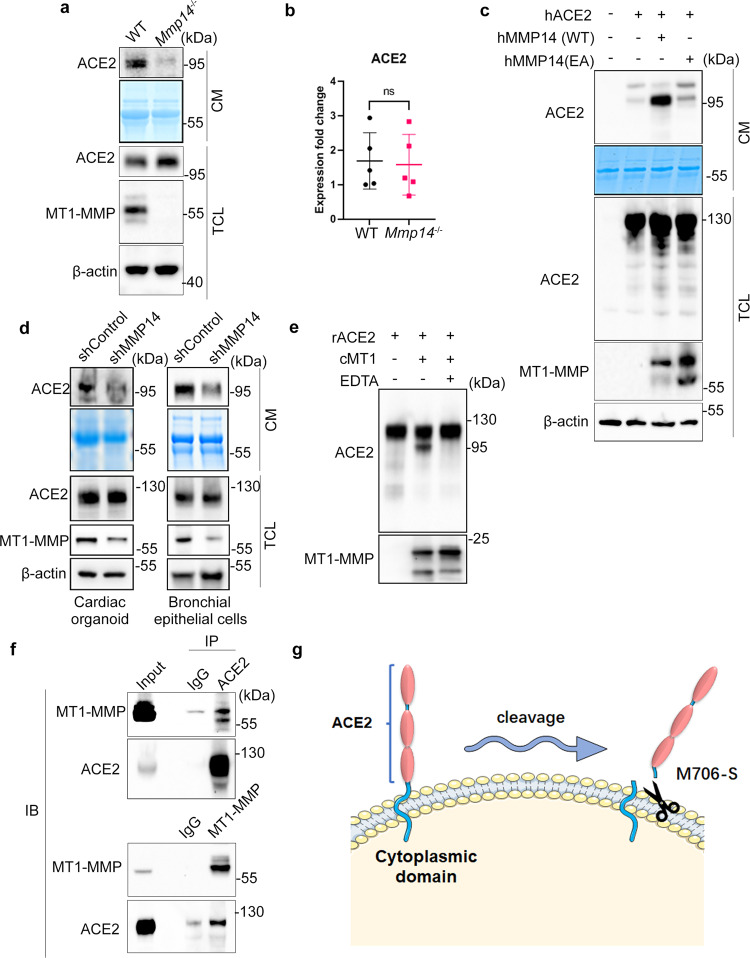
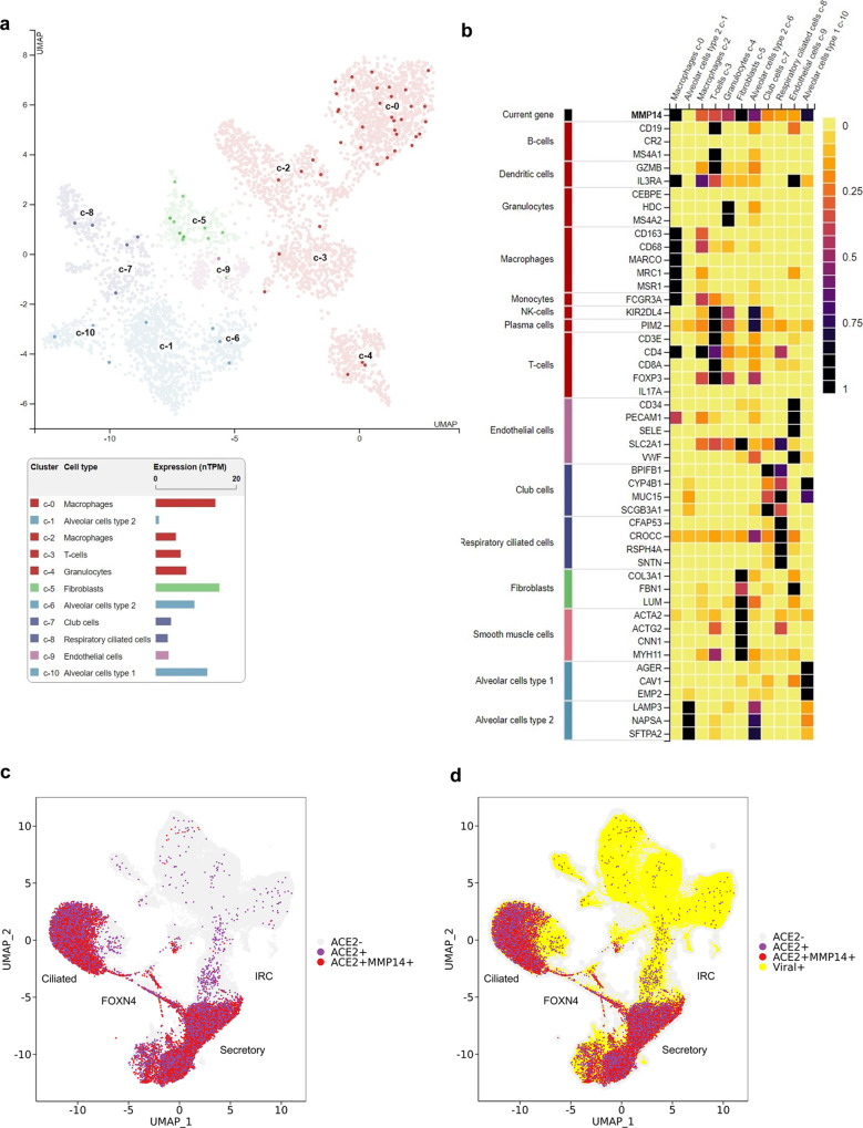
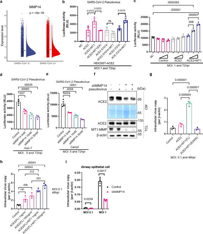
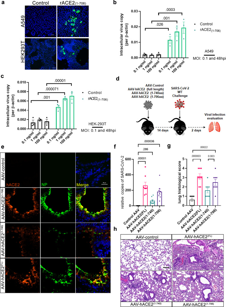
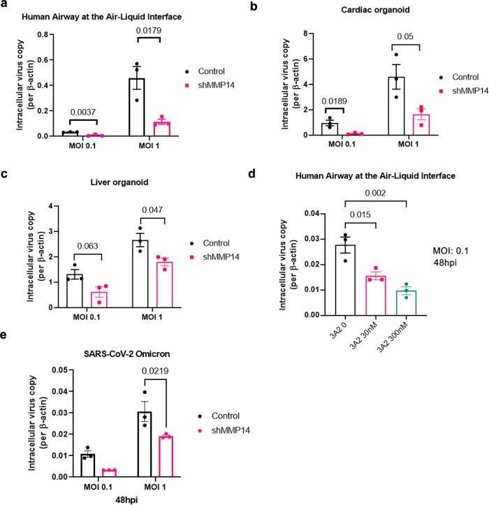
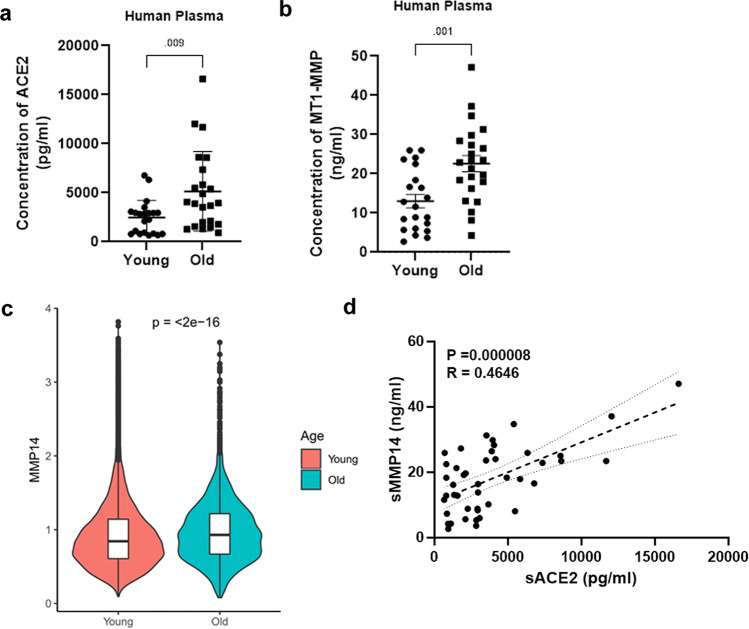
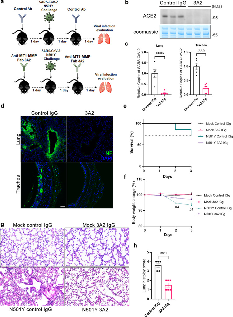
Severe acute respiratory syndrome coronavirus 2 (SARS-CoV-2) has caused a global pandemic. Angiotensin-converting enzyme 2 (ACE2) is an entry receptor for SARS-CoV-2. The full-length membrane form of ACE2 (memACE2) undergoes ectodomain shedding to generate a shed soluble form (solACE2) that mediates SARS-CoV-2 entry via receptor-mediated endocytosis. Currently, it is not known how the physiological regulation of ACE2 shedding contributes to the etiology of COVID-19 in vivo. The present study identifies Membrane-type 1 Matrix Metalloproteinase (MT1-MMP) as a critical host protease for solACE2-mediated SARS-CoV-2 infection. SARS-CoV-2 infection leads to increased activation of MT1-MMP that is colocalized with ACE2 in human lung epithelium. Mechanistically, MT1-MMP directly cleaves memACE2 at M706-S to release solACE2 18-706 that binds to the SARS-CoV-2 spike proteins (S), thus facilitating cell entry of SARS-CoV-2. Human solACE2 18-706 enables SARS-CoV-2 infection in both non-permissive cells and naturally insusceptible C57BL/6 mice. Inhibition of MT1-MMP activities suppresses solACE2-directed entry of SARS-CoV-2 in human organoids and aged mice. Both solACE2 and circulating MT1-MMP are positively correlated in plasma of aged mice and humans. Our findings provide in vivo evidence demonstrating the contribution of ACE2 shedding to the etiology of COVID-19.
Overview
The claims and statistics of this paper haven’t been checked yet.
Run the check — free, no account needed. It reads the open-access text and checks the sources it cites.
Integrity signals
Registry
Cites
Statistics
Figures
Verification
Claims the paper makes
The claims and statistics of this paper haven’t been checked yet.
Run the check — free, no account needed. It reads the open-access text and checks the sources it cites.
Claims other papers make about this one
Each is checked against this paper’s own text: does it say what the citing paper says it does?
The check wasn’t confident either way, so it gives no verdict — never disputed.
In the citing paper
“preliminary evidence suggests that MT1-MMP inhibition would not result in developmental defects under physiological conditions, as Mmp14 heterozygous mice do not exhibit any gross phenotypes during aging.”
From MT1-MMP inhibition rejuvenates ageing brain and rescues cognitive deficits in obesity (2025) · cites this paper as [16]
Experimental — accuracy not yet measuredIn the citing paper
“Our recent findings, along with those of other studies, demonstrate that MT1-MMP is a key regulator of energy homeostasis and that a series of proteolytic events mediated by MT1-MMP contribute to the pathogenesis of multiple age-associated metabolic disorders, including obesity, diabetes, and atherosclerosis15–17.”
From MT1-MMP inhibition rejuvenates ageing brain and rescues cognitive deficits in obesity (2025) · cites this paper as [16]
In the citing paper
“Our previous studies revealed a significant increase in sMT1-MMP levels with increasing age in mice, cynomolgus macaques, and humans16,17.”
From MT1-MMP inhibition rejuvenates ageing brain and rescues cognitive deficits in obesity (2025) · cites this paper as [16]
In the citing paper
“The specificity of this blocking antibody has been well-characterized in our previous studies, in which we showed that blocking the activity of MT1-MMP with this antibody effectively promotes body weight loss in obesity, improves metabolic functions in diabetes, and promotes viral clearance in SARS-CoV-2-infected mice13,15–17.”
From MT1-MMP inhibition rejuvenates ageing brain and rescues cognitive deficits in obesity (2025) · cites this paper as [16]
In the citing paper
“In fact, our previous studies, along with the current findings, demonstrate that genetic ablation or pharmacological inhibition of MT1-MMP confers protection against multiple age-associated disorders, including cognitive decline, obesity, and diabetes, without adverse effects15–17.”
From MT1-MMP inhibition rejuvenates ageing brain and rescues cognitive deficits in obesity (2025) · cites this paper as [16]
Self-check
Percentages, sums and reported differences re-computed from the paper’s own numbers.
The self-check needs the paper’s full text.
Citations
Cites
Cell entry mechanisms of SARS-CoV-2
Shang, J., Wan, Y., Luo, C., Ye, G., Geng, Q., Auerbach, A., & Li, F. (2020) · Proceedings of the National Academy of Sciences · doi:10.1073/pnas.2003138117 · Cited by 3,142
Structural and Functional Basis of SARS-CoV-2 Entry by Using Human ACE2
Wang, Q., Zhang, Y., Wu, L., Niu, S., Song, C., Zhang, Z., Lu, G., Qiao, C., Hu, Y., Yuen, K.-Y., et al. (2020) · Cell · doi:10.1016/j.cell.2020.03.045 · Cited by 2,892
Self-citationHCA Lung Biological Network, Sungnak, W., Huang, N., Bécavin, C., Berg, M., Queen, R., Litvinukova, M., Talavera-López, C., Maatz, H., Reichart, D., et al. (2020) · Nature Medicine · doi:10.1038/s41591-020-0868-6 · Cited by 2,501
Ziegler, C. G. K., Allon, S. J., Nyquist, S. K., Mbano, I. M., Miao, V. N., Tzouanas, C. N., Cao, Y., Yousif, A. S., Bals, J., Hauser, B. M., et al. (2020) · Cell · doi:10.1016/j.cell.2020.04.035 · Cited by 2,284
Omicron escapes the majority of existing SARS-CoV-2 neutralizing antibodies
Cao, Y., Wang, J., Jian, F., Xiao, T., Song, W., Yisimayi, A., Huang, W., Li, Q., Wang, P., An, R., et al. (2022) · Nature · doi:10.1038/s41586-021-04385-3 · Cited by 2,006
Hoffmann, M., Kleine-Weber, H., & Pöhlmann, S. (2020) · Molecular Cell · doi:10.1016/j.molcel.2020.04.022 · Cited by 1,852
Multiorgan and Renal Tropism of SARS-CoV-2
Puelles, V. G., Lütgehetmann, M., Lindenmeyer, M. T., Sperhake, J. P., Wong, M. N., Allweiss, L., Chilla, S., Heinemann, A., Wanner, N., Liu, S., et al. (2020) · New England Journal of Medicine · doi:10.1056/nejmc2011400 · Cited by 1,775
An atlas of the protein-coding genes in the human, pig, and mouse brain
Sjöstedt, E., Zhong, W., Fagerberg, L., Karlsson, M., Mitsios, N., Adori, C., Oksvold, P., Edfors, F., Limiszewska, A., Hikmet, F., et al. (2020) · Science · doi:10.1126/science.aay5947 · Cited by 1,151
Self-citationBroutier, L., Andersson-Rolf, A., Hindley, C. J., Boj, S. F., Clevers, H., Koo, B.-K., & Huch, M. (2016) · Nature Protocols · doi:10.1038/nprot.2016.097 · Cited by 834
Matsuyama, S., Nagata, N., Shirato, K., Kawase, M., Takeda, M., & Taguchi, F. (2010) · Journal of Virology · doi:10.1128/jvi.01542-10 · Cited by 778
SARS-CoV-2 infects cells after viral entry via clathrin-mediated endocytosis
Bayati, A., Kumar, R., Francis, V., & McPherson, P. S. (2021) · Journal of Biological Chemistry · doi:10.1016/j.jbc.2021.100306 · Cited by 499
Li, R. A., Keung, W., Cashman, T. J., Backeris, P. C., Johnson, B. V., Bardot, E. S., Wong, A. O. T., Chan, P. K. W., Chan, C. W. Y., & Costa, K. D. (2018) · Biomaterials · doi:10.1016/j.biomaterials.2018.02.024 · Cited by 175
Genetic Link Between Obesity and MMP14-Dependent Adipogenic Collagen Turnover
Chun, T.-H., Inoue, M., Morisaki, H., Yamanaka, I., Miyamoto, Y., Okamura, T., Sato-Kusubata, K., & Weiss, S. J. (2010) · Diabetes · doi:10.2337/db10-0073 · Cited by 97
Research works printed but not linked: 58
- Yan, R. et al. Structural basis for the recognition of SARS-CoV-2 by full-length human ACE2. Science 367, 1444–1448 (2020). · DOI
- Shang, J. et al. Structural basis of receptor recognition by SARS-CoV-2. Nature 581, 221–224 (2020). · DOI · Paper page
- Lan, J. et al. Structure of the SARS-CoV-2 spike receptor-binding domain bound to the ACE2 receptor. Nature 581, 215–220 (2020). · DOI · Paper page
- Glowacka, I. et al. Evidence that TMPRSS2 activates the severe acute respiratory syndrome coronavirus spike protein for membrane fusion and reduces viral control by the humoral immune response. J. Virol. 85, 4122–4134 (2011). · DOI
- Shulla, A. et al. A transmembrane serine protease is linked to the severe acute respiratory syndrome coronavirus receptor and activates virus entry. J. Virol. 85, 873–882 (2011). · DOI
- Huang, I. C. et al. SARS coronavirus, but not human coronavirus NL63, utilizes cathepsin L to infect ACE2-expressing cells. J. Biol. Chem. 281, 3198–3203 (2006). · DOI
- Simmons, G. et al. Inhibitors of cathepsin L prevent severe acute respiratory syndrome coronavirus entry. Proc. Natl Acad. Sci. 102, 11876–11881 (2005). · DOI
- Hoffmann, M. et al. SARS-CoV-2 Cell Entry Depends on ACE2 and TMPRSS2 and Is Blocked by a Clinically Proven Protease Inhibitor. Cell 181, 271–280.e278 (2020). · DOI · Paper page
- Inoue, Y. et al. Clathrin-dependent entry of severe acute respiratory syndrome coronavirus into target cells expressing ACE2 with the cytoplasmic tail deleted. J. Virol. 81, 8722–8729 (2007). · DOI · Paper page
- Schoeman, D. & Fielding, B. C. Coronavirus envelope protein: current knowledge. Virol. J. 16, 69 (2019). · DOI
- Yeung, M. L. et al. Soluble ACE2-mediated cell entry of SARS-CoV-2 via interaction with proteins related to the renin-angiotensin system. Cell 184, 2212–2228.e2212 (2021). · DOI
- Haga, S. et al. Modulation of TNF-alpha-converting enzyme by the spike protein of SARS-CoV and ACE2 induces TNF-alpha production and facilitates viral entry. Proc. Natl Acad. Sci. 105, 7809–7814 (2008). · DOI
- Hassler, L. et al. A novel soluble ACE2 protein totally protects from lethal disease caused by SARS-CoV-2 infection. bioRxiv, https://doi.org/10.1101/2021.03.12.435191 (2021). · DOI
- Monteil, V. et al. Inhibition of SARS-CoV-2 Infections in Engineered Human Tissues Using Clinical-Grade Soluble Human ACE2. Cell 181, 905–913.e907 (2020). · DOI · Paper page
- Wysocki, J. et al. A Novel Soluble ACE2 Variant with Prolonged Duration of Action Neutralizes SARS-CoV-2 Infection in Human Kidney Organoids. J. Am. Soc. Nephrol. https://doi.org/10.1681/ASN.2020101537 (2021). · DOI
- Zoufaly, A. et al. Human recombinant soluble ACE2 in severe COVID-19. Lancet Respir. Med 8, 1154–1158 (2020). · DOI
- Holmbeck, K. et al. MT1-MMP-deficient mice develop dwarfism, osteopenia, arthritis, and connective tissue disease due to inadequate collagen turnover. Cell 99, 81–92 (1999). · DOI · Paper page
- Zhou, Z. et al. Impaired endochondral ossification and angiogenesis in mice deficient in membrane-type matrix metalloproteinase I. Proc. Natl Acad. Sci. 97, 4052–4057 (2000). · DOI · Paper page
- Chow, C. F. W. et al. Body weight regulation via MT1-MMP-mediated cleavage of GFRAL. Nat. Metab. 4, 203–212 (2022). · DOI · Paper page
- Turunen, S. P., Tatti-Bugaeva, O. & Lehti, K. Membrane-type matrix metalloproteases as diverse effectors of cancer progression. Biochim Biophys. Acta Mol. Cell Res. 1864, 1974–1988 (2017). · DOI
- Chan, K. M. et al. MT1-MMP inactivates ADAM9 to regulate FGFR2 signaling and calvarial osteogenesis. Dev. Cell 22, 1176–1190 (2012). · DOI · Paper page
- Jin, G. et al. MT1-MMP cleaves Dll1 to negatively regulate Notch signalling to maintain normal B-cell development. EMBO J. 30, 2281–2293 (2011). · DOI · Paper page
- Wong, H. L. et al. MT1-MMP sheds LYVE-1 on lymphatic endothelial cells and suppresses VEGF-C production to inhibit lymphangiogenesis. Nat. Commun. 7, 10824 (2016). · DOI
- Kumar, S., Ratnikov, B. I., Kazanov, M. D., Smith, J. W. & Cieplak, P. CleavPredict: A Platform for Reasoning about Matrix Metalloproteinases Proteolytic Events. PLoS One. 10, e0127877 (2015). · DOI · Paper page
- Chua, R. L. et al. COVID-19 severity correlates with airway epithelium-immune cell interactions identified by single-cell analysis. Nat. Biotechnol. 38, 970–979 (2020). · DOI · Paper page
- Trump, S. et al. Hypertension delays viral clearance and exacerbates airway hyperinflammation in patients with COVID-19. Nat. Biotechnol. 39, 705–716 (2021). · DOI
- Loske, J. et al. Pre-activated antiviral innate immunity in the upper airways controls early SARS-CoV-2 infection in children. Nat Biotechnol, https://doi.org/10.1038/s41587-021-01037-9 (2021). · DOI
- Zou, X. et al. Single-cell RNA-seq data analysis on the receptor ACE2 expression reveals the potential risk of different human organs vulnerable to 2019-nCoV infection. Front Med. 14, 185–192 (2020). · DOI · Paper page
- Schuler, B. A. et al. Age-determined expression of priming protease TMPRSS2 and localization of SARS-CoV-2 in lung epithelium. J Clin Invest. 131, https://doi.org/10.1172/JCI140766 (2021). · DOI
- Mills, R. J. et al. BET inhibition blocks inflammation-induced cardiac dysfunction and SARS-CoV-2 infection. Cell 184, 2167–2182 e2122 (2021). · DOI
- Yang, L. et al. A Human Pluripotent Stem Cell-based Platform to Study SARS-CoV-2 Tropism and Model Virus Infection in Human Cells and Organoids. Cell Stem Cell. 27, 125–136.e127 (2020). · DOI
- Lamers, M. M. et al. An organoid-derived bronchioalveolar model for SARS-CoV-2 infection of human alveolar type II-like cells. EMBO J. 40, e105912 (2021). · DOI · Paper page
- Remacle, A. G. et al. Selective function-blocking monoclonal human antibody highlights the important role of membrane type-1 matrix metalloproteinase (MT1-MMP) in metastasis. Oncotarget 8, 2781–2799 (2017). · DOI
- Nam, D. H., Lee, K. B., Kruchowy, E., Pham, H. & Ge, X. Protease Inhibition Mechanism of Camelid-like Synthetic Human Antibodies. Biochemistry 59, 3802–3812 (2020). · DOI
- Nam, D. H., Rodriguez, C., Remacle, A. G., Strongin, A. Y. & Ge, X. Active-site MMP-selective antibody inhibitors discovered from convex paratope synthetic libraries. Proc. Natl Acad. Sci. 113, 14970–14975 (2016). · DOI
- Chakraborty, C., Sharma, A. R., Sharma, G., Bhattacharya, M. & Lee, S. S. SARS-CoV-2 causing pneumonia-associated respiratory disorder (COVID-19): diagnostic and proposed therapeutic options. Eur. Rev. Med Pharm. 24, 4016–4026 (2020).
- Zhou, F., Yu, T. & Du, R. Clinical course and risk factors for mortality of adult inpatients with COVID-19 in Wuhan, China: a retrospective cohort study (395, 1054, 2020). Lancet 395, 1038–1038 (2020). · DOI · Paper page
- Toth, M. et al. Complex pattern of membrane type 1 matrix metalloproteinase shedding. Regulation by autocatalytic cells surface inactivation of active enzyme. J. Biol. Chem. 277, 26340–26350 (2002). · DOI · Paper page
- Maisi, P. et al. Soluble membrane-type 1 matrix metalloproteinase (MT1-MMP) and gelatinase A (MMP-2) in induced sputum and bronchoalveolar lavage fluid of human bronchial asthma and bronchiectasis. APMIS 110, 771–782 (2002). · DOI · Paper page
- Li, H. et al. Immunological characterization of cell-surface and soluble forms of membrane type 1 matrix metalloproteinase in human breast cancer cells and in fibroblasts. Mol. Carcinog. 22, 84–94 (1998). · DOI
- Lukassen, S. et al. SARS-CoV-2 receptor ACE2 and TMPRSS2 are primarily expressed in bronchial transient secretory cells. EMBO J. 39, e105114 (2020). · DOI
- Guo, X. et al. Regulation of age-associated insulin resistance by MT1-MMP-mediated cleavage of insulin receptor. Nat. Commun. 13, 3749 (2022). · DOI · Paper page
- Yang, Z. et al. Genetic Landscape of the ACE2 Coronavirus Receptor. Circulation, https://doi.org/10.1161/CIRCULATIONAHA.121.057888 (2022). · DOI
- Kornilov, S. A. et al. Plasma levels of soluble ACE2are associated with sex, Metabolic Syndrome, and its biomarkers in a large cohort, pointing to a possible mechanism for increased severity in COVID-19. Crit. Care. 24, 452 (2020). · DOI
- Wallentin, L. et al. Angiotensin-converting enzyme 2 (ACE2) levels in relation to risk factors for COVID-19 in two large cohorts of patients with atrial fibrillation. Eur. Heart J. 41, 4037–4046 (2020). · DOI
- Donoghue, M. et al. A novel angiotensin-converting enzyme-related carboxypeptidase (ACE2) converts angiotensin I to angiotensin 1-9. Circ. Res. 87, E1–E9 (2000). · DOI · Paper page
- Jia, H. P. et al. Ectodomain shedding of angiotensin converting enzyme 2 in human airway epithelia. Am. J. Physiol. Lung Cell Mol. Physiol. 297, L84–L96 (2009). · DOI
- Lambert, D. W. et al. Tumor necrosis factor-alpha convertase (ADAM17) mediates regulated ectodomain shedding of the severe-acute respiratory syndrome-coronavirus (SARS-CoV) receptor, angiotensin-converting enzyme-2 (ACE2). J. Biol. Chem. 280, 30113–30119 (2005). · DOI
- Wong, H. L. et al. When MT1-MMP meets ADAMs. Cell Cycle 11, 2793–2798 (2012). · DOI
- Benton, D. J. et al. Receptor binding and priming of the spike protein of SARS-CoV-2 for membrane fusion. Nature 588, 327–330 (2020). · DOI
- Yamamoto, M. et al. Metalloproteinase-Dependent and TMPRSS2-Independent Cell Surface Entry Pathway of SARS-CoV-2 Requires the Furin Cleavage Site and the S2 Domain of Spike Protein. mBio 13, e0051922 (2022). · DOI
- Benlarbi, M. et al. Identification and differential usage of a host metalloproteinase entry pathway by SARS-CoV-2 Delta and Omicron. iScience, 105316, https://doi.org/10.1016/j.isci.2022.105316 (2022). · DOI
- Zhang, X. et al. SARS-CoV-2 Omicron strain exhibits potent capabilities for immune evasion and viral entrance. Signal Transduct. Target Ther. 6, 430 (2021). · DOI
- Cele, S. et al. Omicron extensively but incompletely escapes Pfizer BNT162b2 neutralization. Nature 602, 654–656 (2022). · DOI · Paper page
- Planas, D. et al. Considerable escape of SARS-CoV-2 Omicron to antibody neutralization. Nature 602, 671–675 (2022). · DOI · Paper page
- Kormann, R. et al. Periostin Promotes Cell Proliferation and Macrophage Polarization to Drive Repair after AKI. J. Am. Soc. Nephrol. 31, 85–100 (2020). · DOI
- Park, E. J. et al. The Spike Glycoprotein of SARS-CoV-2 Binds to beta1 Integrins Expressed on the Surface of Lung Epithelial Cells. Viruses 13, https://doi.org/10.3390/v13040645 (2021). · DOI
- Yuan, S. et al. Viruses harness YxxO motif to interact with host AP2M1 for replication: A vulnerable broad-spectrum antiviral target. Sci. Adv. 6, eaba7910 (2020). · DOI
Cited by
Sorted by citations
MT1-MMP inhibition rejuvenates ageing brain and rescues cognitive deficits in obesity
Asthana, P., Li, L., Lu, L., Wu, J., Zhang, S., Li, N., Wong, S. K. K., Gurung, S., Zhang, Y., Lin, Y., et al. (2025) · Cell Discovery · doi:10.1038/s41421-025-00825-w · Cited by 4
Self-citation
Metadata
- Authors
- Guo, X., Cao, J., Cai, J.-P., Wu, J., Huang, J., Asthana, P., Wong, S. K. K., Ye, Z.-W., Gurung, S., Zhang, Y., Wang, S., Wang, Z., Ge, X., Kwan, H. Y., Lyu, A., Chan, K. M., Wong, N., Huang, J., Zhou, Z., Bian, Z.-X., Yuan, S., & Wong, H. L. X.
- ORCID
- Venue
- Nature Communications
- Published
- 2022-12-23
- Volume
- 13
- Issue
- 1
- ISSN
- 2041-1723
- PMC
- PMC9780620
- Cited by
- 25
- References
- 13
- Language
- en
- Funders
- Research Grants Council, University Grants Committee (R4017-18, 12101019, 12102020)
- Food and Health Bureau of the Government of the Hong Kong Special Administrative Region | Health and Medical Research Fund (06170056, 08793626)
- Natural Science Foundation of Guangdong Province (2021A1515011128)
About these checks
Registry, retraction, Cites and statistics results are computed, not judged, with one exception: a printed reference that no index lists is read once by a model before it counts as a citation error. Claims are extracted by a model and kept only when their quote appears verbatim in the text; each cited source is then checked, with a model’s help, for whether it says what the claim relies on. Those verdicts can be wrong — read the claim’s quote and the cited source before relying on one.
Statistics and arithmetic are recomputed, not judged.
Confidence bands are shown only for fields calibrated on our annotated test set. None is calibrated yet, so no band is shown.
What counts as a claim ›How we measure confidence ›How we check this ›
Are you an author, or do you see a problem with this page? Request a review
