

The DOI matches its Crossref record; 75 of the 77 references it cites were found in a registry or an index.
BANKSY unifies cell typing and tissue domain segmentation for scalable spatial omics data analysis
Open accessPublished version of a preprint
Abstract
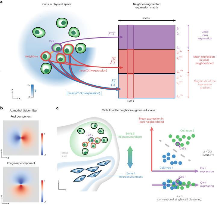
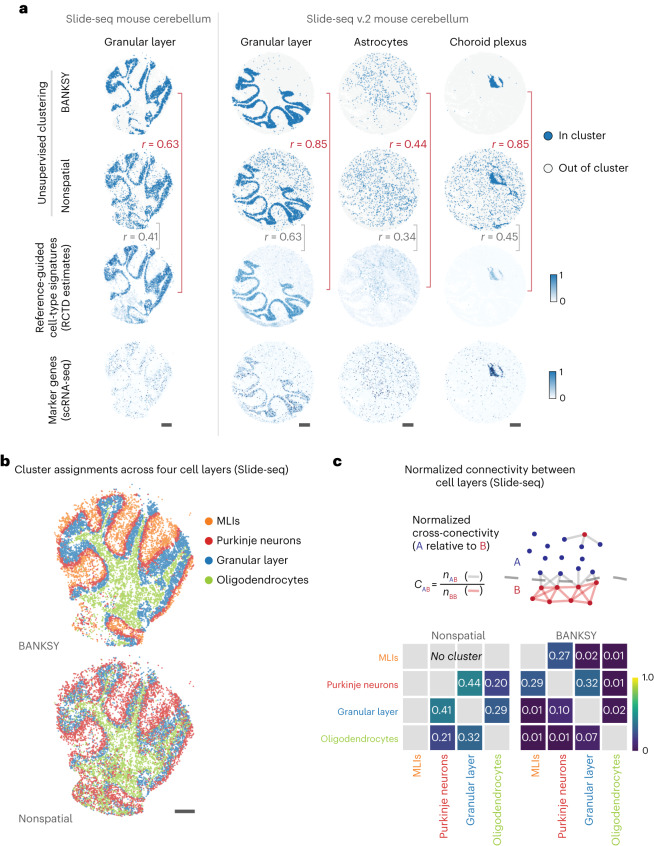
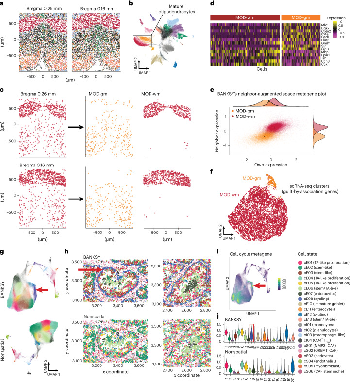
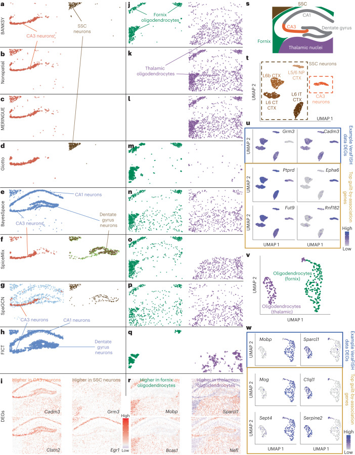
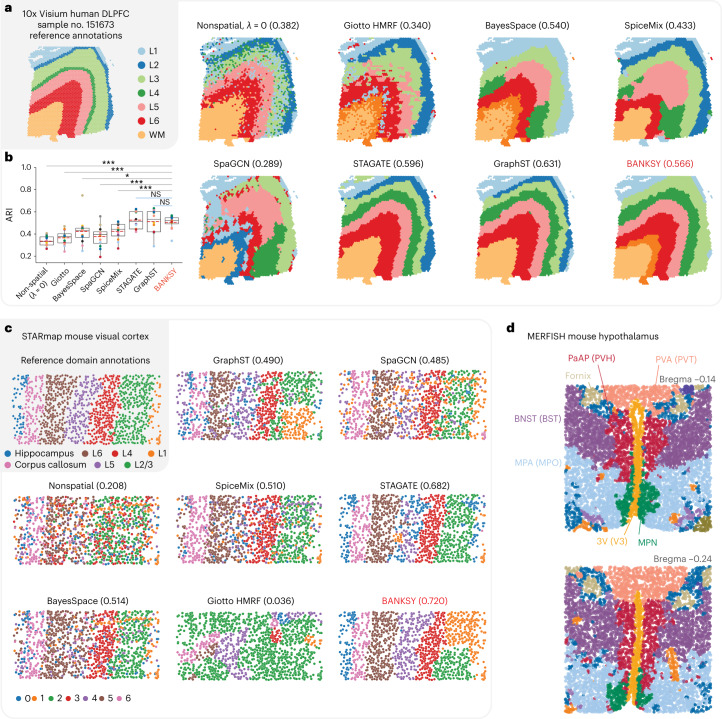
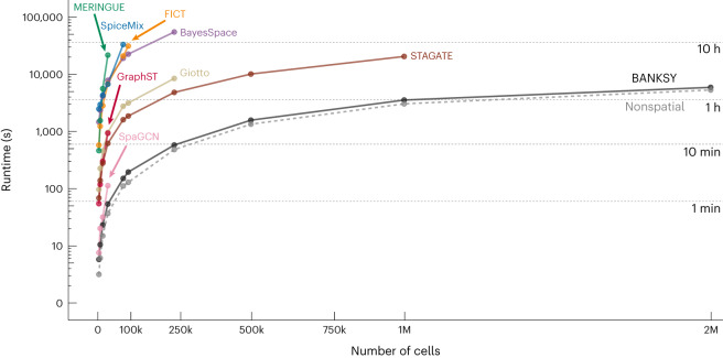
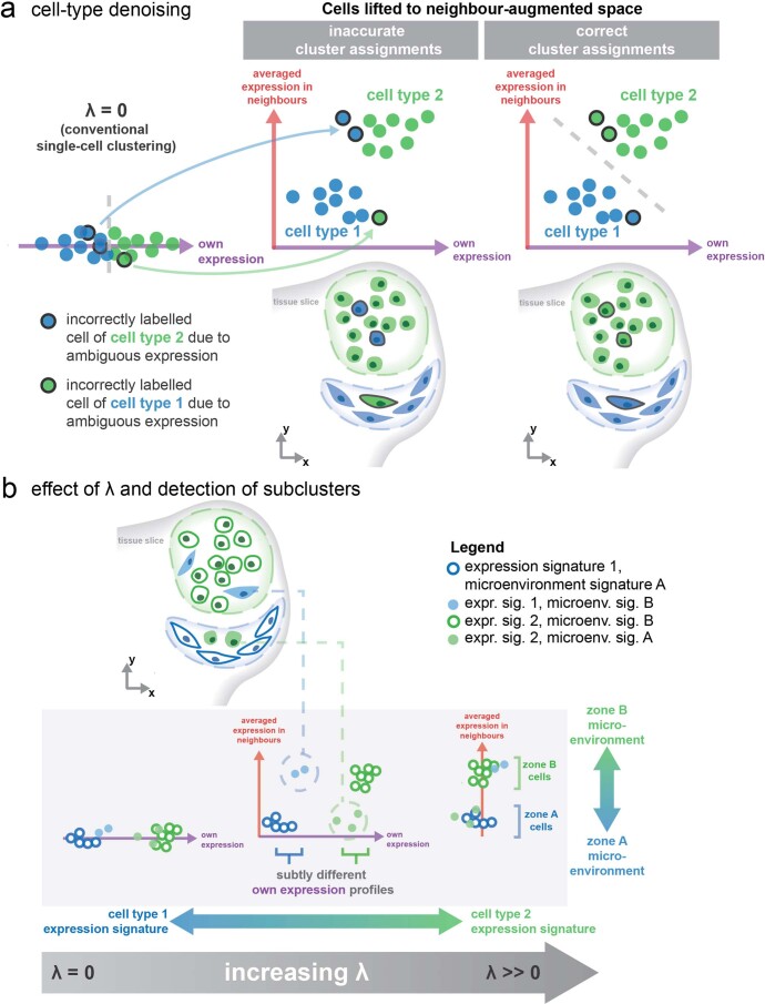
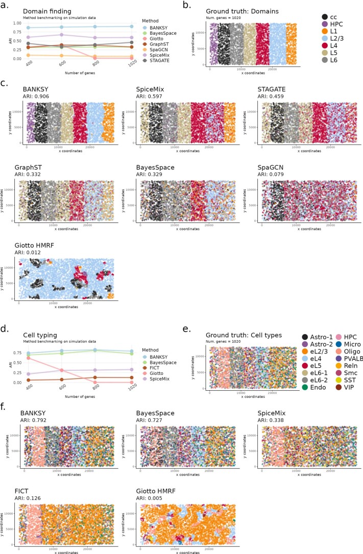
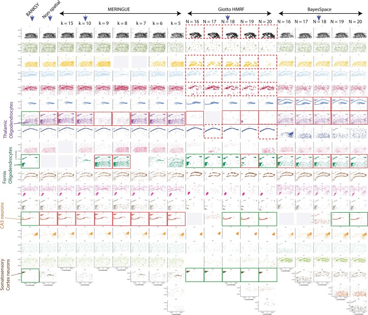
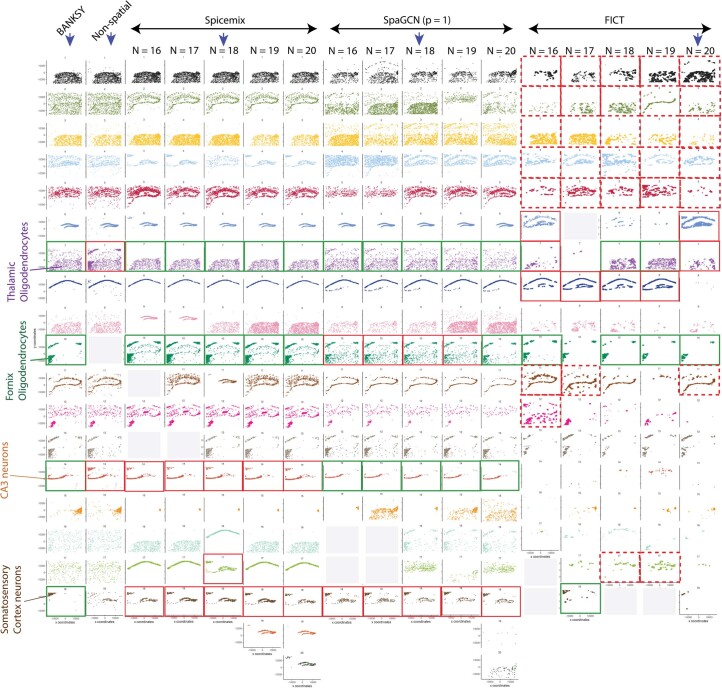
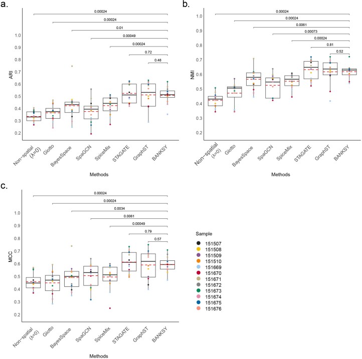
Spatial omics data are clustered to define both cell types and tissue domains. We present Building Aggregates with a Neighborhood Kernel and Spatial Yardstick (BANKSY), an algorithm that unifies these two spatial clustering problems by embedding cells in a product space of their own and the local neighborhood transcriptome, representing cell state and microenvironment, respectively. BANKSY’s spatial feature augmentation strategy improved performance on both tasks when tested on diverse RNA (imaging, sequencing) and protein (imaging) datasets. BANKSY revealed unexpected niche-dependent cell states in the mouse brain and outperformed competing methods on domain segmentation and cell typing benchmarks. BANKSY can also be used for quality control of spatial transcriptomics data and for spatially aware batch effect correction. Importantly, it is substantially faster and more scalable than existing methods, enabling the processing of millions of cell datasets. In summary, BANKSY provides an accurate, biologically motivated, scalable and versatile framework for analyzing spatially resolved omics data.
Overview
The claims and statistics of this paper haven’t been checked yet.
Run the check — free, no account needed. It reads the open-access text and checks the sources it cites.
Integrity signals
Registry
Cites
Statistics
Figures
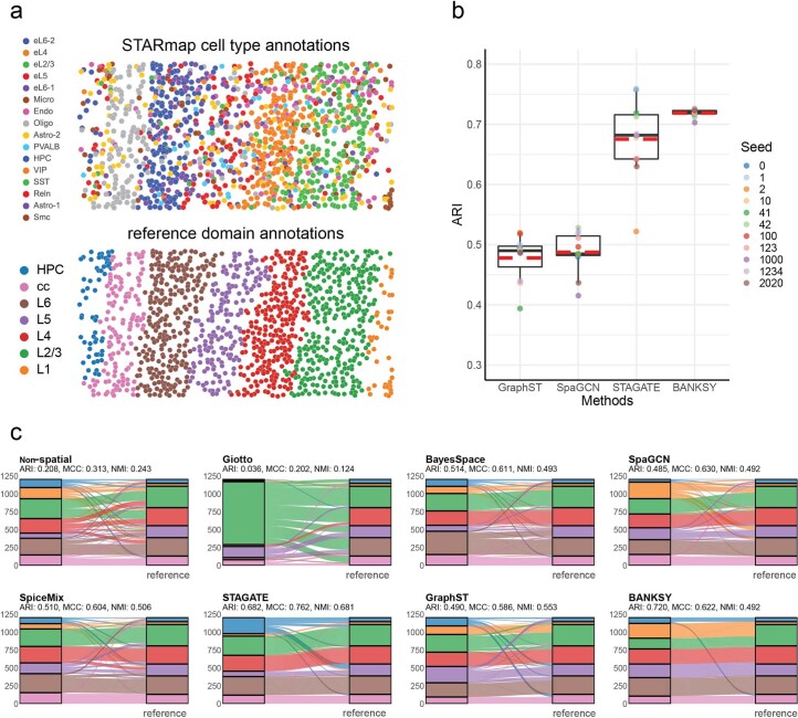
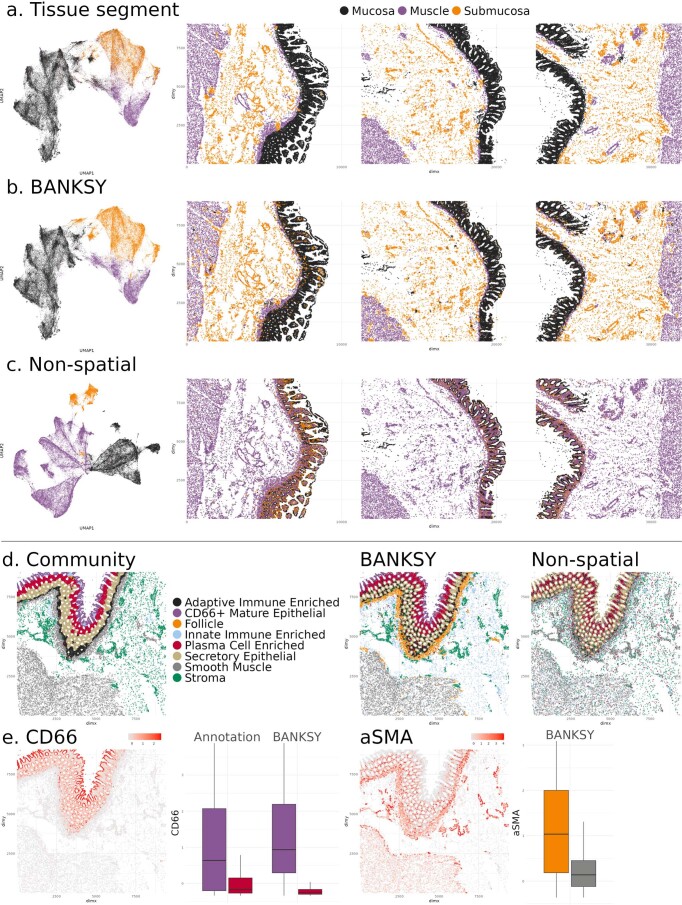
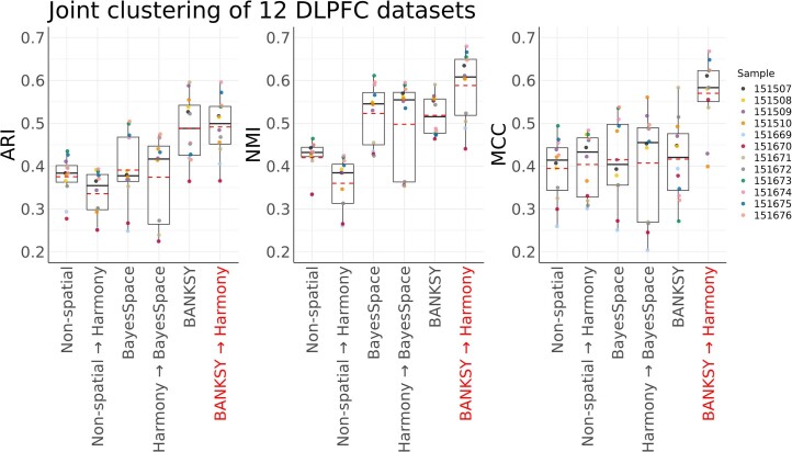
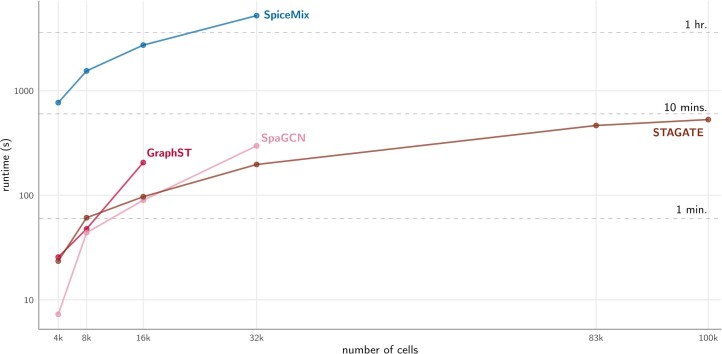
Verification
Claims the paper makes
The claims and statistics of this paper haven’t been checked yet.
Run the check — free, no account needed. It reads the open-access text and checks the sources it cites.
Claims other papers make about this one
Each is checked against this paper’s own text: does it say what the citing paper says it does?
The cited source says what the claim attributes to it.
In the citing paper
“Using BANKSY50-derived spatial domains and Harmony39 correction, we identified 18 reproducible niches shared across 20 samples (Figures S5A–S5C; STAR Methods).”
From Cellular senescence in human liver under normal aging and cancer (2026) · cites this paper as [50]
Self-check
Percentages, sums and reported differences re-computed from the paper’s own numbers.
The self-check needs the paper’s full text.
Citations
Cites
SciPy 1.0: fundamental algorithms for scientific computing in Python
Virtanen, P., Gommers, R., Oliphant, T. E., Haberland, M., Reddy, T., Cournapeau, D., Burovski, E., Peterson, P., Weckesser, W., Bright, J., et al. (2020) · Nature Methods · doi:10.1038/s41592-019-0686-2 · Cited by 38,430
Efficient Estimation of Word Representations in Vector Space
Mikolov, T., Chen, K., Corrado, G., & Dean, J. (2013) · arXiv · doi:10.48550/arxiv.1301.3781 · Cited by 26,568
Self-citationFast unfolding of communities in large networks
Blondel, V. D., Guillaume, J.-L., Lambiotte, R., & Lefebvre, E. (2008) · Journal of Statistical Mechanics: Theory and Experiment · doi:10.1088/1742-5468/2008/10/p10008 · Cited by 21,424
Integrated analysis of multimodal single-cell data
Hao, Y., Hao, S., Andersen-Nissen, E., Mauck, W. M., Zheng, S., Butler, A., Lee, M. J., Wilk, A. J., Darby, C., Zagar, M., et al. (2020) · doi:10.1101/2020.10.12.335331 · Cited by 15,508
Comprehensive Integration of Single-Cell Data
Stuart, T., Butler, A., Hoffman, P., Hafemeister, C., Papalexi, E., Mauck, W. M., Hao, Y., Stoeckius, M., Smibert, P., & Satija, R. (2019) · Cell · doi:10.1016/j.cell.2019.05.031 · Cited by 15,453
The Hungarian method for the assignment problem
Kuhn, H. W. (1955) · Naval Research Logistics Quarterly · doi:10.1002/nav.3800020109 · Cited by 13,120
Fast, sensitive and accurate integration of single-cell data with Harmony
Korsunsky, I., Millard, N., Fan, J., Slowikowski, K., Zhang, F., Wei, K., Baglaenko, Y., Brenner, M., Loh, P.-R., & Raychaudhuri, S. (2019) · Nature Methods · doi:10.1038/s41592-019-0619-0 · Cited by 10,488
SciPy 1.0: fundamental algorithms for scientific computing in Python
Virtanen, P., Gommers, R., Oliphant, T. E., Haberland, M., Reddy, T., Cournapeau, D., Burovski, E., Peterson, P., Weckesser, W., Bright, J., et al. (2019) · Apollo - University of Cambridge Repository · doi:10.17863/cam.64134 · Cited by 10,460
SCANPY: large-scale single-cell gene expression data analysis
Wolf, F. A., Angerer, P., & Theis, F. J. (2018) · Genome Biology · doi:10.1186/s13059-017-1382-0 · Cited by 8,881
Macrophage activation and polarization: nomenclature and experimental guidelines
Murray, P. J., Allen, J. E., Biswas, S. K., Fisher, E. A., Gilroy, D. W., Goerdt, S., Gordon, S., Hamilton, J. A., Ivashkiv, L. B., Lawrence, T., et al. (2014) · Immunity · doi:10.1016/j.immuni.2014.06.008 · Cited by 6,337
Research works printed but not linked: 3
- Huuki-Myers, L. et al. Integrated single cell and unsupervised spatial transcriptomic analysis defines molecular anatomy of the human dorsolateral prefrontal cortex. Preprint at bioRxiv https://doi.org/10.1101/2023.02.15.528722 (2023). · DOI
- Gabor, D. Theory of communication. J. Inst. Electrical Engineering 93, 429–441 (1946).
- Csardi, G. & Nepusz, T. The igraph software package for complex network research. Int. J. Complex Syst. 1695, 1–9 (2006).
Cited by
Sorted by citations
Profiling cell identity and tissue architecture with single-cell and spatial transcriptomics
Gulati, G. S., D'Silva, J. P., Liu, Y., Wang, L., & Newman, A. M. (2024) · Nature Reviews Molecular Cell Biology · doi:10.1038/s41580-024-00768-2 · Cited by 347
Ren, Y., Huang, Z., Zhou, L., Xiao, P., Song, J., He, P., Xie, C., Zhou, R., Li, M., Dong, X., et al. (2023) · Nature Communications · doi:10.1038/s41467-023-36707-6 · Cited by 252
Optimizing Xenium In Situ data utility by quality assessment and best-practice analysis workflows
Salas, S. M., Kuemmerle, L. B., Langseth, C. M., Tiesmeyer, S., Avenel, C., Hu, T., Rehman, H., Grillo, M., Czarnewski, P., Helgadóttir, S., et al. (2025) · Nature Methods · doi:10.1038/s41592-025-02617-2 · Cited by 192
Computational approaches and challenges in spatial transcriptomics
Fang, S., Chen, B., Zhang, Y., Sun, H., Liu, L., Liu, S., Li, Y., & Xu, X. (2023) · Genomics, Proteomics & Bioinformatics · doi:10.1016/j.gpb.2022.10.001 · Cited by 159
Liu, Y., Dai, Y., & Wang, L. (2025) · Cancer Cell · doi:10.1016/j.ccell.2025.12.009 · Cited by 153
Benchmarking clustering, alignment, and integration methods for spatial transcriptomics
Hu, Y., Li, Y., Xie, M., Rao, M., Shen, W., Luo, C., Qin, H., Baek, J., & Zhou, X. (2024) · Genome biology · doi:10.1186/s13059-024-03361-0 · Cited by 106
Quantitative characterization of cell niches in spatially resolved omics data
Birk, S., Bonafonte-Pardàs, I., Feriz, A. M., Boxall, A. R., Agirre, E., Memi, F., Maguza, A., Yadav, A., Armingol, E., Fan, R., et al. (2025) · Nature Genetics · doi:10.1038/s41588-025-02120-6 · Cited by 117
A single-cell, spatial transcriptomic atlas of the Arabidopsis life cycle
Lee, T. A., Illouz‐Eliaz, N., Nobori, T., Xu, J., Jow, B., Nery, J. R., & Ecker, J. R. (2025) · Nature Plants · doi:10.1038/s41477-025-02072-z · Cited by 81
Guo, C., Wang, C., Wang, Z., Liu, F., Liu, L., Yang, Y., Li, X., Guo, B., Lu, R.-Y., Liao, B., et al. (2024) · Nature Communications · doi:10.1038/s41467-024-54685-1 · Cited by 77
Self-citationLee, Y. K., Lee, M., Shin, Y., Kim, K., & Kim, T. J. (2025) · International Journal of Molecular Sciences · doi:10.3390/ijms26093949 · Cited by 80
Fang, S., Xu, M., Cao, L., Liu, X., Bezulj, M., Tan, L., Yuan, Z., Li, Y., Xia, T., Guo, L., et al. (2025) · Nature Communications · doi:10.1038/s41467-025-58079-9 · Cited by 75
Self-citationIdentifying spatial domain by adapting transcriptomics with histology through contrastive learning
Zeng, Y., Yin, R., Luo, M., Chen, J., Pan, Z., Lu, Y., Yu, W., & Yang, Y. (2023) · Briefings in Bioinformatics · doi:10.1093/bib/bbad048 · Cited by 73
Insitutype: likelihood-based cell typing for single cell spatial transcriptomicsPreprint
Danaher, P. J., Zhao, E. J., Yang, Z. G., Ross, D., Gregory, M., Reitz, Z., Kim, T. K., Baxter, S. K., Jackson, S. W., He, S., et al. (2022) · bioRxiv (Cold Spring Harbor Laboratory) · doi:10.1101/2022.10.19.512902 · Cited by 65
Qian, J., Shao, X., Bao, H., Yin, F., Guo, W., Li, C., Li, A., Hua, H., & Fan, X. (2025) · Nature Communications · doi:10.1038/s41467-025-57029-9 · Cited by 62
Spatiotemporal gene expression and cellular dynamics of the developing human heart
Lázár, E., Mauron, R., Andrusivová, Ž., Foyer, J., He, M., Larsson, L., Shakari, N., Salas, S. M., Avenel, C., Sarıyar, S., et al. (2025) · Nature Genetics · doi:10.1038/s41588-025-02352-6 · Cited by 44
SpotSweeper: spatially aware quality control for spatial transcriptomics
Totty, M. S., Hicks, S. C., & Guo, B. (2025) · Nature Methods · doi:10.1038/s41592-025-02713-3 · Cited by 41
Feng, R., Spieth, L., Liu, L., Berghoff, S. A., Franz, J., Liu, Q., Wang, Z., Tiwari, V., Vitale, S., Frerich, S., et al. (2025) · Immunity · doi:10.1016/j.immuni.2025.10.003 · Cited by 40
Plummer, J., Dezem, F. S., Cook, D. P., Park, J., Zhang, L., Liu, Y., Marção, M., DuBose, H., Arjumand, W., Wise, K., et al. (2025) · Nature Biotechnology · doi:10.1038/s41587-025-02811-9 · Cited by 34
Multi-slice spatial transcriptome domain analysis with SpaDo
Duan, B., Chen, S., Cheng, X., & Liu, Q. (2024) · Genome biology · doi:10.1186/s13059-024-03213-x · Cited by 33
Spatial architecture of development and disease
Lázár, E., & Lundeberg, J. (2025) · Nature Reviews Genetics · doi:10.1038/s41576-025-00892-5 · Cited by 38
Neurodevelopmental hijacking of oligodendrocyte lineage programs drives glioblastoma infiltration
Wu, Y., Wu, B. Z., Ellenbogen, Y., Kant, J. A., Yu, P., Li, X., Caloren, L. C., Sotov, V., Tran, C. N., Restrepo, M., et al. (2025) · Developmental Cell · doi:10.1016/j.devcel.2025.04.022 · Cited by 35
Non-invasive profiling of the tumour microenvironment with spatial ecotypes
Zhang, W., Brown, E. L., Usmani, A., Earland, N. J., Kang, M., Olelewe, C., Viswanathan, A., Chauhan, P. S., Steen, C. B., Jeon, H. S., et al. (2026) · Nature · doi:10.1038/s41586-026-10452-4 · Cited by 35
Wang, T., Zhu, H., Zhou, Y., Ding, W., Ding, W., Han, L., & Zhang, X. (2024) · Communications Biology · doi:10.1038/s42003-024-07037-0 · Cited by 29
Prawira, A., Xu, H., Mei, Y., Leow, W. Q., Nasir, N. J. M., Reolo, M. J. Y., Otsuka, M., Rahbari, M., Chen, Z., Weerasooriya, M., et al. (2025) · Gut · doi:10.1136/gutjnl-2025-335084 · Cited by 29
Self-citationYiu, T., Chen, B., Wang, H., Feng, G., Fu, Q.-Q., & Hu, H. (2025) · Journal of Translational Medicine · doi:10.1186/s12967-025-07091-0 · Cited by 37
Deep Learning in Single-cell Analysis
Molho, D., Ding, J., Tang, W., Li, Z., Wen, H., Wang, Y., Venegas, J., Jin, W., Liu, R., Su, R., et al. (2024) · ACM Transactions on Intelligent Systems and Technology · doi:10.1145/3641284 · Cited by 38
Transcriptomic and spatial organization of telencephalic GABAergic neurons
van Velthoven, C. T. J., Gao, Y., Kunst, M., Lee, C., McMillen, D., Chakka, A. B., Casper, T., Clark, M., Chakrabarty, R., Daniel, S. F., et al. (2025) · Nature · doi:10.1038/s41586-025-09296-1 · Cited by 32
High-parameter spatial multi-omics through histology-anchored integration
Liu, Y., Wang, C., Wang, Z., Chen, L., Li, Z., Song, J., Zou, Q., Gao, R., Qian, B., Feng, X., et al. (2025) · Nature Methods · doi:10.1038/s41592-025-02926-6 · Cited by 30
Wang, Y., Liu, Z., & Ma, X. (2024) · Genome biology · doi:10.1186/s13059-024-03272-0 · Cited by 30
Quantitative characterization of tissue states using multiomics and ecological spatial analysis
Ding, D. Y., Tang, Z., Zhu, B., Ren, H., Shalek, A. K., Tibshirani, R. J., & Nolan, G. P. (2025) · Nature Genetics · doi:10.1038/s41588-025-02119-z · Cited by 30
Tang, J., Fan, W., Ruan, Y., Liu, X., Qiu, F., Feng, J., Huang, G., Yan, M., Wang, H., Mu, Q., et al. (2025) · Cancer Cell · doi:10.1016/j.ccell.2025.08.006 · Cited by 25
Donovan, M. L., Jhaveri, N., Ma, N., Cheikh, B. B., DeRosa, J., Mihani, R., Berrell, N., Suen, J. Y., Monkman, J. H., Fraser, J. F., et al. (2024) · STAR Protocols · doi:10.1016/j.xpro.2024.103226 · Cited by 22
Li, Y., Zhang, J., Gao, X., & Zhang, Q. C. (2024) · Cell Systems · doi:10.1016/j.cels.2024.05.001 · Cited by 21
Spatial biology – unravelling complexity within the glioblastoma microenvironment
Robinson, S. D., Filippopoulou, C., Besta, S., Samuels, M. E., Betrán, A. L., Ajamieh, M. A., Vella, V., Jones, W., & Giamas, G. (2025) · Trends in Molecular Medicine · doi:10.1016/j.molmed.2025.01.014 · Cited by 23
Sun, Y., Kong, L., Huang, J., Deng, H., Bian, X., Li, X., Cui, F., Dou, L., Cao, C., Zou, Q., et al. (2024) · Briefings in Functional Genomics · doi:10.1093/bfgp/elae023 · Cited by 23
SpatialLeiden: spatially aware Leiden clustering
Müller‐Bötticher, N., Sahay, S., Eils, R., & Ishaque, N. (2025) · Genome biology · doi:10.1186/s13059-025-03489-7 · Cited by 21
Cancer therapy resistance from a spatial‐omics perspective
Zhang, Y., Yang, C., Chen, X., Wu, L. L., Yuan, Z., Zhang, F., & Qian, B. (2025) · Clinical and Translational Medicine · doi:10.1002/ctm2.70396 · Cited by 19
Spatial perturb-seq: single-cell functional genomics within intact tissue architecture
Shen, K., Seow, W. Y., Keng, C. T., Lim, M. G. L., Lim, D. S., Guo, K., Meliani, A., Hajis, M. I. B., Wang, B., Prabhakar, S., et al. (2026) · Nature Communications · doi:10.1038/s41467-026-69677-6 · Cited by 25
Self-citationSIMVI disentangles intrinsic and spatial-induced cellular states in spatial omics data
Dong, M., Su, D. G., Kluger, H. M., Fan, R., & Kluger, Y. (2025) · Nature Communications · doi:10.1038/s41467-025-58089-7 · Cited by 25
Zuo, C., Xia, J., Xu, Y., Xu, Y., Gao, P., Zhang, J., Wang, Y., & Chen, L. (2025) · Nature Communications · doi:10.1038/s41467-025-60575-x · Cited by 21
Systematic benchmarking of computational methods to identify spatially variable genes
Li, Z., M.Patel, Z., Song, D., Yasa, S. N., Cannoodt, R., Yan, G., Li, J. J., & Pinello, L. (2025) · Genome biology · doi:10.1186/s13059-025-03731-2 · Cited by 47
Ghosh, M., Pilanc, P., Baluszek, S., Jacek, K., Poleszak, K., Szadkowska, P., Lenkiewicz, A. M., Gielniewski, B., Ellert‐Miklaszewska, A., Castro, M. G., et al. (2025) · PLoS Biology · doi:10.1371/journal.pbio.3002893 · Cited by 20
Iwane, K., Nakanishi, Y., Muta, Y., Chen, J., Yasumura, K., Omatsu, M., Aoyama, N., Ikeda, M., Masui, Y., Cai, L., et al. (2025) · Nature Communications · doi:10.1038/s41467-025-66485-2 · Cited by 20
Lapuente-Santana, Ó., Kant, J., & Eduati, F. (2024) · npj Precision Oncology · doi:10.1038/s41698-024-00749-w · Cited by 17
Comparing Xenium 5K and Visium HD data from identical tissue slide at a pathological perspective
Long, M., Hu, T., Wang, W., Gao, J., Wang, N., & Nilsson, M. F. (2025) · Journal of Experimental & Clinical Cancer Research · doi:10.1186/s13046-025-03479-4 · Cited by 16
StaVia: spatially and temporally aware cartography with higher-order random walks for cell atlases
Stassen, S. V., Kobashi, M., Lam, E. Y. M., Huang, Y., Ho, J. W. K., & Tsia, K. K. (2024) · Genome biology · doi:10.1186/s13059-024-03347-y · Cited by 17
Cellular neighborhoods in cancer
Ma, L., Xiong, B., Liu, M., & Tan, K. (2026) · Nature Cancer · doi:10.1038/s43018-025-01107-w · Cited by 16
Learning tissue representation by identification of persistent local patterns in spatial omics data
Tanevski, J., Vulliard, L., Ibarra-Arellano, M. A., Schapiro, D., Hartmann, F. J., & Sáez-Rodríguez, J. (2025) · Nature Communications · doi:10.1038/s41467-025-59448-0 · Cited by 13
Zhu, B., Gao, S., Chen, S., Wang, Y., Yeung, J., Bai, Y., Huang, A. Y., Yeo, Y. Y., Liao, G., Mao, S., et al. (2025) · Nature Immunology · doi:10.1038/s41590-025-02163-1 · Cited by 16
Bridging the dimensional gap from planar spatial transcriptomics to 3D cell atlases
Lin, S., Wang, Z., Cui, Y., Zou, Q., Han, C., Yan, R., Yang, Z., Zhang, W., Gao, R., Song, J., et al. (2025) · Nature Methods · doi:10.1038/s41592-025-02969-9 · Cited by 19
PCA-based spatial domain identification with state-of-the-art performance
Schaub, D. P., Yousefi, B., Kaiser, N., Khatri, R., Puelles, V. G., Krebs, C. F., Panzer, U., & Bonn, S. (2024) · Bioinformatics · doi:10.1093/bioinformatics/btaf005 · Cited by 16
Bridging single cells to organs: Mesoscale modules as fundamental units of tissue function
Chen, Y., Germain, R. N., Hunter, G. L., Kulkarni, R. P., Lander, A. D., Lowenstein, P. R., Purvis, J. E., & Wong, H. S. (2025) · Cell · doi:10.1016/j.cell.2025.10.012 · Cited by 18
Points2Regions : Fast, interactive clustering of imaging‐based spatial transcriptomics data
Andersson, A., Beháňová, A., Avenel, C., Windhager, J., Malmberg, F., & Wählby, C. (2024) · Cytometry Part A · doi:10.1002/cyto.a.24884 · Cited by 14
Liu, X., Qu, C., Liu, C., Zhu, N., Huang, H., Teng, F., Huang, C., Luo, B., Liu, X., Xie, M., et al. (2024) · GigaScience · doi:10.1093/gigascience/giae078 · Cited by 12
Padlock Probe–Based Targeted In Situ Sequencing: Overview of Methods and Applications
Magoulopoulou, A., Salas, S. M., Tiklová, K., Samuelsson, E. R., Hilscher, M. M., & Nilsson, M. (2023) · Annual Review of Genomics and Human Genetics · doi:10.1146/annurev-genom-102722-092013 · Cited by 14
Cellular and Spatial Drivers of Unresolved Injury and Functional Decline in the Human KidneyPreprint
Lake, B. B., Ferreira, R. M., Hansen, J., Menon, R., Basta, J. M., Thiessen‐Philbrook, H., Reinert, S., Fallegger, R., Lagwankar, A. K., Chen, X., et al. (2025) · bioRxiv (Cold Spring Harbor Laboratory) · doi:10.1101/2025.09.26.678707 · Cited by 18
Cellular senescence in human liver under normal aging and cancer
Karpova, A., Li, X., Peng, C.-W., Gallant, K. L., Rapp, D. R., Alligood, D. M., Houston, A. J., Park, A., Targino da Costa, A. L. N., Chou, W.-H., et al. (2026) · Cell Genomics · doi:10.1016/j.xgen.2025.101133 · Cited by 13
Challenges and Opportunities in Analyzing Cancer-Associated Microbiomes
Chia, M., Pop, M., Salzberg, S. L., & Nagarajan, N. (2025) · Cancer Research · doi:10.1158/0008-5472.can-24-3629 · Cited by 11
Zhang, M., Zhang, W., & Ma, X. (2024) · Briefings in Bioinformatics · doi:10.1093/bib/bbae437 · Cited by 11
Sun, Y. H., Zhang, M., Zhao, Y., Yan, Y., Wang, L., Liu, X., Xia, S., Wang, B., Zhang, X., & Wang, Y. (2025) · npj Precision Oncology · doi:10.1038/s41698-025-01005-5 · Cited by 12
Zeng, Y., Yin, R., Luo, M., Chen, J., Pan, Z., Lu, Y., Yu, W., & Yang, Y. (2022) · bioRxiv (Cold Spring Harbor Laboratory) · doi:10.1101/2022.09.30.510297 · Cited by 11
hoodscanR: profiling single-cell neighborhoods in spatial transcriptomics data
Liu, N., Martin, J., Bhuva, D. D., Chen, J., Li, M., Lee, S., Kharbanda, M., Cheng, J., Mohamed, A., Kulasinghe, A., et al. (2026) · Bioinformatics · doi:10.1093/bioinformatics/btag609 · Cited by 10
Rayan, N. A., Aow, J., Lim, M. G. L., Arcego, D. M., Ryan, R., Nourbakhsh, N., de Lima, R. M. S., Craig, K., Zhang, T., Goh, Y. T., et al. (2024) · Molecular Psychiatry · doi:10.1038/s41380-024-02619-0 · Cited by 10
Self-citationMulvey, B., Wang, Y., Divecha, H. R., Bach, S. V., Montgomery, K. D., Cinquemani, S., Chandra, A., Du, Y., Miller, R. A., Kleinman, J. E., et al. (2026) · Cell Reports · doi:10.1016/j.celrep.2025.116877 · Cited by 10
Next‐generation spatial transcriptomics: unleashing the power to gear up translational oncology
Wang, N., Hong, W., Wu, Y., Chen, Z., Bai, M., Wang, W., & Zhu, J. Y. (2024) · MedComm · doi:10.1002/mco2.765 · Cited by 10
Wang, W., Zheng, S., Shin, S. C., Chávez, J., & Yuan, G. (2025) · Genome biology · doi:10.1186/s13059-025-03588-5 · Cited by 9
Li, Y., Filla, T., Györfi, A., Liang, M., Devakumar, V., Micu, A., Chai, H., Bergmann, C., Pecher, A., Henes, J., et al. (2025) · Annals of the Rheumatic Diseases · doi:10.1016/j.ard.2025.06.002 · Cited by 13
Xiao, X., Zhang, L. Y., Zhao, H., & Wang, Z. (2025) · Nature Machine Intelligence · doi:10.1038/s42256-025-01161-0 · Cited by 12
Towards a universal spatial molecular atlas of the mouse brainPreprint
He, Y., Sheng, H., Shi, H., Wang, W. X., Tang, Z., Jia Liu, B., & Wang, X. (2024) · bioRxiv (Cold Spring Harbor Laboratory) · doi:10.1101/2024.05.27.594872 · Cited by 9
Cellular neighborhood analysis in spatial omics reveals new tissue domains and cell subtypes
Ruitenberg, M. J., & Nguyen, Q. H. (2024) · Nature Genetics · doi:10.1038/s41588-023-01646-x · Cited by 7
Han, C., Lin, S., Wang, Z., Cui, Y., Zou, Q., & Yuan, Z. (2025) · Nature Machine Intelligence · doi:10.1038/s42256-025-01097-5 · Cited by 8
Chen, R., Yao, Y., Qian, J., Peng, X. X., Shao, X., & Fan, X. (2025) · iMeta · doi:10.1002/imt2.70084 · Cited by 9
Ma, Y., Cai, Y., Yang, J., Zhao, Z., Chen, Z., Lv, D., Yu, J., Yang, C., Liu, K., Zeng, M., et al. (2025) · Cell Reports · doi:10.1016/j.celrep.2025.116704 · Cited by 8
Spatial transcriptomics: integrating platforms and computational approaches for clinical insights
Song, W., Wang, D., Li, J., & Zhang, R. (2025) · Biotechnology Advances · doi:10.1016/j.biotechadv.2025.108791 · Cited by 8
Mapping biology in space: from spatial transcriptomics platforms to analytical tools and databases
Guo, Z., Wu, R., Li, W., Yang, K., Ying, X., Alinejad-Rokny, H., & Ye, Y. (2026) · 中国科学通报:英文版 · doi:10.1016/j.scib.2026.01.034 · Cited by 12
Miao, Z., Tian, T., Chen, W., Wang, Q., Ma, L., Zhang, D., Xie, M., Yu, Z., Guo, X., Bai, G., et al. (2024) · The Innovation Life · doi:10.59717/j.xinn-life.2024.100097 · Cited by 9
High-parametric protein maps reveal the spatial organization in early-developing human lung
Sarıyar, S., Sountoulidis, A., Hansen, J. N., Salas, S. M., Mardamshina, M., Casals, A. M., Navarro, F. B., Andrusivová, Ž., Li, X., Czarnewski, P., et al. (2024) · Nature Communications · doi:10.1038/s41467-024-53752-x · Cited by 10
Characterization of spatial homogeneous regions in tissues with concordex
Jackson, K. C., Booeshaghi, A. S., Gálvez-Merchán, Á., Moses, L., Chari, T., Kim, A., Hosten-Mittermaier, O., & Pachter, L. S. (2026) · BMC Methods · doi:10.1186/s44330-026-00086-3 · Cited by 8
Single-cell spatial (scs) omics: Recent developments in data analysis
Camacho, J., Armstrong, M., García, L., Díaz, C., & Gómez‐Llorente, C. (2024) · TrAC Trends in Analytical Chemistry · doi:10.1016/j.trac.2024.118109 · Cited by 9
Crosstalk of glutamine metabolism between cancer-associated fibroblasts and cancer cells
Chen, T., Xu, Y., Yang, F., Pan, Y., Ji, N., Li, J., Zeng, X. Y., Chen, Q., Jiang, L., & Shen, Y. (2025) · Cellular Signalling · doi:10.1016/j.cellsig.2025.111874 · Cited by 9
GASTON-Mix: a unified model of spatial gradients and domains using spatial mixture-of-experts
Chitra, U., Dan, S., Krienen, F. M., & Raphael, B. J. (2025) · Bioinformatics · doi:10.1093/bioinformatics/btaf254 · Cited by 7
Xia, C., Cao, Z., & Gao, G. (2025) · Nature Communications · doi:10.1038/s41467-025-63140-8 · Cited by 10
Harnessing the potential of spatial statistics for spatial omics data with pasta
Emons, M., Gunz, S., Crowell, H. L., Mallona, I., Kuehl, M. B., Furrer, R., & Robinson, M. D. (2025) · Nucleic Acids Research · doi:10.1093/nar/gkaf870 · Cited by 11
Dong, K., Gao, Y., Zou, Q., Cui, Y., Han, C., Lin, S., Wang, Z., Tang, C., Cheng, X., Meng, F., et al. (2025) · Genome biology · doi:10.1186/s13059-025-03796-z · Cited by 8
Tang, J., Chen, Z., Qian, K., Huang, S., He, Y., Yin, S., He, X., Ye, B., ZHUANG, Y., Meng, H., et al. (2026) · Nature Cell Biology · doi:10.1038/s41556-025-01838-z · Cited by 8
BANKSY: scalable cell typing and domain segmentation for spatial omics
Singhal, V., & Chou, N. (2024) · Nature Reviews Genetics · doi:10.1038/s41576-024-00743-9 · Cited by 7
Self-citationClustering the cortical laminae: in vivo parcellation
Shamir, I., Assaf, Y., & Ron Shamir, A. (2024) · Brain Structure and Function · doi:10.1007/s00429-023-02748-2 · Cited by 6
Thuilliez, C., Moquin‐Beaudry, G., Khneisser, P., Costa, M. E. M. D., Karkar, S., Boudhouche, H., Drubay, D., Audinot, B., Geoerger, B., Scoazec, J., et al. (2024) · Bioinformatics Advances · doi:10.1093/bioadv/vbae081 · Cited by 9
Wang, J., Gong, R., Heidari, S., Rogers, M., Tani, T., Abe, H., Ichinohe, N., Woodward, A., & Delmas, P. J. (2024) · Neuroinformatics · doi:10.1007/s12021-024-09688-0 · Cited by 6
Yu, Y., Wang, S., Li, J., Yu, M., McCrocklin, K., Kang, J., Ma, A., Ma, Q., Xu, D. H., & Wang, J. (2025) · Nature Communications · doi:10.1038/s41467-025-63141-7 · Cited by 5
Charting the cardiac landscape: Advances in spatial transcriptomics for heart biology
Farah, E. N., Diaz, J. T., Bloomekatz, J. E., & Chi, N. C. (2025) · Seminars in Cell and Developmental Biology · doi:10.1016/j.semcdb.2025.103648 · Cited by 9
Prioritizing perturbation-responsive gene patterns using interpretable deep learning
Cui, Y., & Yuan, Z. (2025) · Nature Communications · doi:10.1038/s41467-025-61476-9 · Cited by 6
The transcriptomic and spatial organization of telencephalic GABAergic neuronal typesPreprint
van Velthoven, C. T. J., Gao, Y., Kunst, M., Lee, C., McMillen, D., Chakka, A. B., Casper, T., Clark, M., Chakrabarty, R., Daniel, S. F., et al. (2024) · bioRxiv (Cold Spring Harbor Laboratory) · doi:10.1101/2024.06.18.599583 · Cited by 6
Mapping and reprogramming human tissue microenvironments with MintFlowPreprint
Akbarnejad, A., Steele, L., Jafree, D. J., Birk, S., Sallese, M. R., Rademaker, K., Boxall, A. R., Rumney, B. M., Tudor, C. L., Patel, M. J., et al. (2025) · bioRxiv (Cold Spring Harbor Laboratory) · doi:10.1101/2025.06.24.661094 · Cited by 10
Gingerich, I. K., Goods, B. A., & Frost, H. R. (2025) · PLoS Computational Biology · doi:10.1371/journal.pcbi.1013759 · Cited by 5
Towards deciphering the bone marrow microenvironment with spatial multi-omics
Yip, R. K. H., Hawkins, E. D., Bowden, R. J., & Rogers, K. L. (2025) · Seminars in Cell and Developmental Biology · doi:10.1016/j.semcdb.2025.01.001 · Cited by 5
Steamboat: Attention-Based Multiscale Delineation of Cellular Interactions in Tissues
Liang, S., Tang, J., Wang, G., & Ma, J. (2025) · Lecture notes in computer science · doi:10.1007/978-3-031-90252-9_55 · Cited by 5
Peng, X., Smithy, J. W., Yosofvand, M., Kostrzewa, C. E., Bleile, M., Ehrich, F. D., Lee, J., Postow, M. A., Callahan, M. K., Panageas, K. S., et al. (2025) · Nature Communications · doi:10.1038/s41467-025-61821-y · Cited by 5
Self-citationSpatial transcriptomics in bone research: navigating hype and hurdles
Sokolowskei, D., Shah, A., Trostle, A. J., Juan, C., Sammarco, M. C., & Tower, R. J. (2025) · Pathology · doi:10.1016/j.pathol.2025.10.003 · Cited by 6
Mitochondrial clone tracing within spatially intact human tissuesPreprint
Bracht, S. A., Rong, J., Gier, R. A., DeMarshall, M., Golden, H., Dhakal, D., McDevitt, J. C., Mo, F., Furth, E. E., Strauss, A. L., et al. (2025) · bioRxiv (Cold Spring Harbor Laboratory) · doi:10.1101/2025.07.11.664452 · Cited by 6
Metadata
- Authors
- Singhal, V., Chou, N., Lee, J., Yue, Y., Liu, J., Chock, W. K., Lin, L., Chang, Y.-C., Teo, E. M. L., Aow, J., Lee, H. K., Chen, K. H., & Prabhakar, S.
- Affiliations
- Agency for Science, Technology and Research (SG)
- National University of Singapore (SG)
- Corresponding author
- Chen, Kok Hao
- Prabhakar, Shyam
- ORCID
- Venue
- Nature Genetics
- Published
- 2024-02-27
- Volume
- 56
- Issue
- 3
- Pages
- 431-441
- ISSN
- 1061-4036 (ISSN-L)
- 1546-1718
- PMC
- PMC10937399
- OpenAlex
- W4392198237
- Cited by
- 282
- References
- 77
- Citations by year
- 2026: 124 · 2025: 103 · 2024: 47
- FWCI
- 37.44
- Type
- journal article
- Language
- en
- Open access
- hybrid
- doi.org
- Locations
- Nature Genetics (publishedVersion, cc-by)
- bioRxiv (Cold Spring Harbor Laboratory) (acceptedVersion, cc-by-nc-nd)
- PubMed Central (publishedVersion, cc-by)
- PubMed (publishedVersion)
- DR-NTU (Nanyang Technological University) (submittedVersion, cc-by)
- Funders
- MOH | National Medical Research Council (OFIRG21jun-0090, OF-YIRG18nov-0014, OFIRG-000618-00)
- Agency for Science, Technology and Research (#H18/01/a0/020, 202D800010, H18/01/a0/020, I1801E0029)
- National Research Foundation Singapore (NRF-CRP25-2020-0001)
- Topics
- Life Sciences › Biochemistry, Genetics and Molecular Biology › Molecular Biology › Single-cell and spatial transcriptomics
- Life Sciences › Biochemistry, Genetics and Molecular Biology › Biophysics › Cell Image Analysis Techniques
- Life Sciences › Biochemistry, Genetics and Molecular Biology › Molecular Biology › Gene expression and cancer classification
- Keywords
- spatial omics, cell typing, spatial clustering, mouse brain, batch effect correction, scalable data analysis
- MeSH terms
- Data Analysis, Algorithms, Animals, RNA, Benchmarking, Gene Expression Profiling, Mice, Transcriptome
About these checks
Registry, retraction, Cites and statistics results are computed, not judged, with one exception: a printed reference that no index lists is read once by a model before it counts as a citation error. Claims are extracted by a model and kept only when their quote appears verbatim in the text; each cited source is then checked, with a model’s help, for whether it says what the claim relies on. Those verdicts can be wrong — read the claim’s quote and the cited source before relying on one.
Statistics and arithmetic are recomputed, not judged.
Confidence bands are shown only for fields calibrated on our annotated test set. None is calibrated yet, so no band is shown.
What counts as a claim ›How we measure confidence ›How we check this ›
Are you an author, or do you see a problem with this page? Request a review
