

The DOI matches its Crossref record; 65 of the 65 references it cites were found in a registry or an index.
Oral administration of Proteus mirabilis damages dopaminergic neurons and motor functions in mice
Open accessAbstract
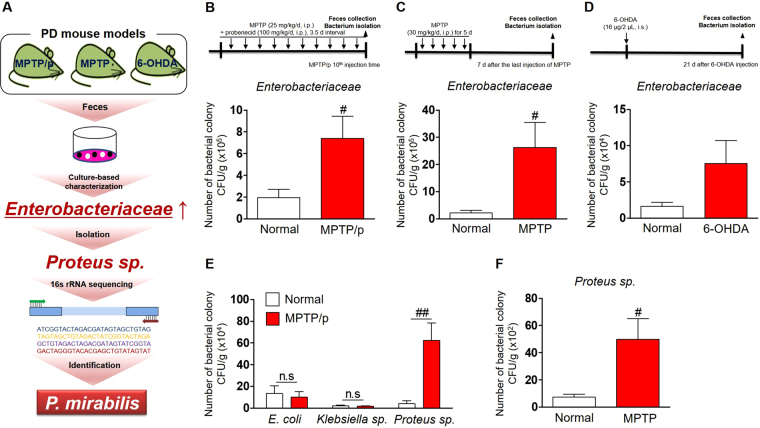
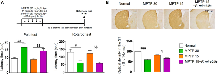
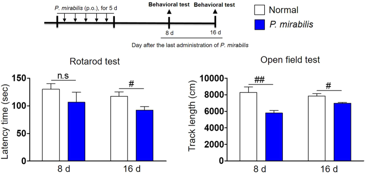
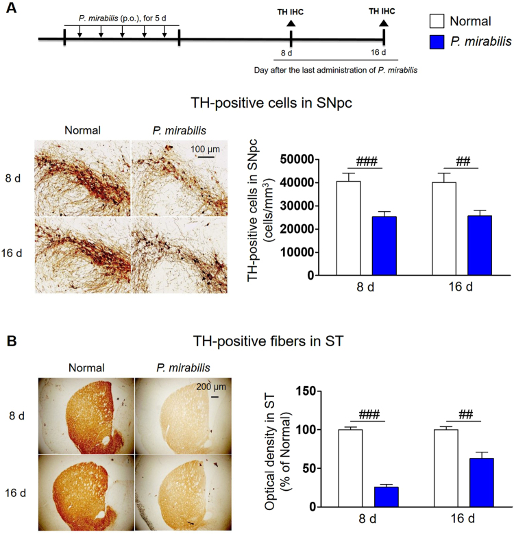
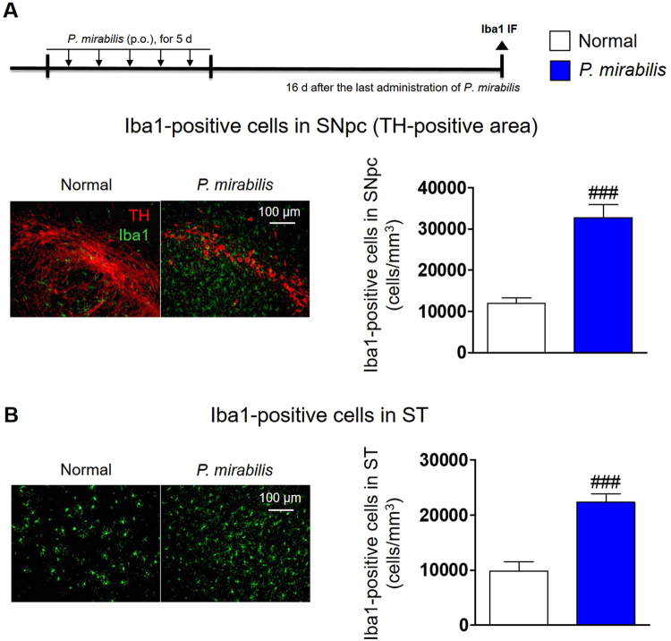
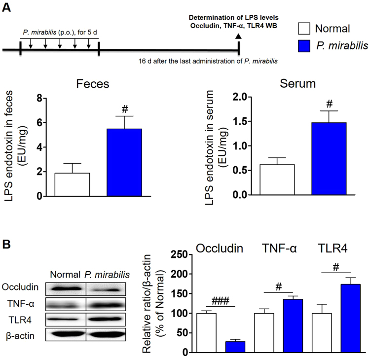
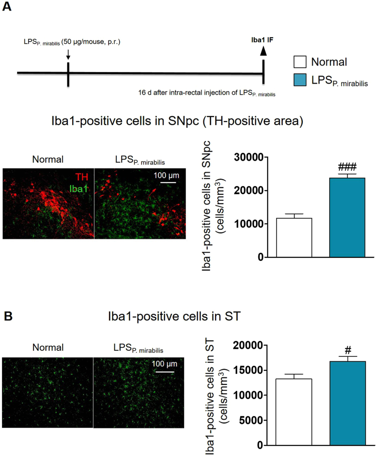
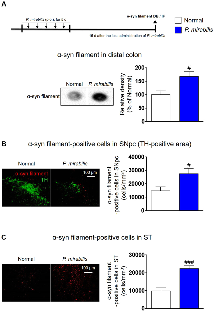
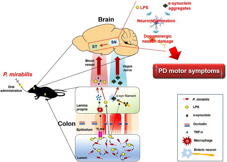
Recently, studies on the relationship between gut dysbiosis and Parkinson’s disease (PD) have increased, but whether a specific gut bacterium may cause PD remains unexplored. Here, we report, for the first time, that a specific gut bacterium directly induces PD symptoms and dopaminergic neuronal damage in the mouse brain. We found that the number of Enterobacteriaceae, particularly Proteus mirabilis, markedly and commonly increased in PD mouse models. Administration of P. mirabilis isolated from PD mice significantly induced motor deficits, selectively caused dopaminergic neuronal damage and inflammation in substantia nigra and striatum, and stimulated α-synuclein aggregation in the brain as well as in the colon. We found that lipopolysaccharides, a virulence factor of P. mirabilis, may be associated in these pathological changes via gut leakage and inflammatory actions. Our results suggest a role of P. mirabilis on PD pathogenesis in the brain.
Overview
The claims and statistics of this paper haven’t been checked yet.
Run the check — free, no account needed. It reads the open-access text and checks the sources it cites.
Integrity signals
Registry
Cites
Statistics
Figures
Verification
Claims the paper makes
The claims and statistics of this paper haven’t been checked yet.
Run the check — free, no account needed. It reads the open-access text and checks the sources it cites.
Claims other papers make about this one
Each is checked against this paper’s own text: does it say what the citing paper says it does?
In the citing paper
“Proteus mirabilis can cause motor dyskinesia, produce neuroinflammation and cause dopaminergic neuronal damage by producing LPS (Choi et al. 2018).”
From Therapeutic effect of GLP-1 engineered strain on mice model of Alzheimer’s disease and Parkinson’s disease (2020) · cites this paper as Choi et al. 2018
In the citing paper
“that may trigger neuroinflammation and lead to pathological changes related to PD by its metabolites (such as lipopolysaccharide) [39]”
From Neuroprotective Effect of Ceftriaxone on MPTP‐Induced Parkinson’s Disease Mouse Model by Regulating Inflammation and Intestinal Microbiota (2021) · cites this paper as [39]
In the citing paper
“Mice exposed to 1-methyl-4-phenyl-1,2,3,6-tetrahydropyridine (MPTP) or 6-hydroxydopamine presented increased amount of Proteus mirabilis in their feces.”
From The Role of Microbiota-Gut-Brain Axis in Neuropsychiatric and Neurological Disorders (2021) · cites this paper as [408]
In the citing paper
“After repeated oral administration of these bacteria isolated from the above-mentioned models to WT mice, the treated animals displayed Parkinson's disease-like symptoms, including impaired motor function, elevated α-synuclein aggregation in the brain and colon as well as dopaminergic neuronal loss [408].”
From The Role of Microbiota-Gut-Brain Axis in Neuropsychiatric and Neurological Disorders (2021) · cites this paper as [408]
In the citing paper
“In the end, in a differently induced model of PD, the repeated oral administration of Proteus mirabilis or its derived LPS to 1-methyl-4-phenyl-1,2,3,6-tetrahydropyridine-treated or young mice was sufficient to induce a PD-like phenotype with motor deficit, loss of dopaminergic neurons in the nigra, brain and gut inflammation with disruption of the intestinal epithelial barrier, and formation of α”
From The gut-brain axis: how microbiota and host inflammasome influence brain physiology and pathology (2020) · cites this paper as [185]
Self-check
Percentages, sums and reported differences re-computed from the paper’s own numbers.
The self-check needs the paper’s full text.
Citations
Cites
The Mouse Brain in Stereotaxic Coordinates
Paxinos, G. (2001) · Cited by 12,859
Staging of brain pathology related to sporadic Parkinson’s disease
Braak, H., Tredici, K. D., Rüb, U., de Vos, R. A. I., Jansen Steur, E. N. H., & Braak, E. (2003) · Neurobiology of Aging · doi:10.1016/s0197-4580(02)00065-9 · Cited by 10,777
From inflammation to sickness and depression: when the immune system subjugates the brain
Dantzer, R., O'Connor, J. C., Freund, G. G., Johnson, R. W., & Kelley, K. W. (2008) · Nature Reviews Neuroscience · doi:10.1038/nrn2297 · Cited by 7,395
Dauer, W., & Przedborski, S. (2003) · Neuron · doi:10.1016/s0896-6273(03)00568-3 · Cited by 5,469
Raetz, C. R. H., & Whitfield, C. (2002) · Annual Review of Biochemistry · doi:10.1146/annurev.biochem.71.110601.135414 · Cited by 4,404
Intestinal mucosal barrier function in health and disease
Turner, J. R. (2009) · Nature reviews. Immunology · doi:10.1038/nri2653 · Cited by 3,952
Gut microbiota regulate motor deficits and neuroinflammation in a model of Parkinson's disease
Sampson, T. R., Debelius, J. W., Thron, T., Janssen, S., Shastri, G. G., Ilhan, Z. E., Challis, C., Schretter, C. E., Rocha, S., Gradinaru, V., et al. (2016) · Cell · doi:10.1016/j.cell.2016.11.018 · Cited by 3,622
Hsiao, E. Y., McBride, S. W., Hsien, S., Sharon, G., Hyde, E. R., McCue, T., Codelli, J. A., Chow, J., Reisman, S. E., Petrosino, J. F., et al. (2013) · Cell · doi:10.1016/j.cell.2013.11.024 · Cited by 3,283
Gut biogeography of the bacterial microbiota
Donaldson, G. P., Lee, S. M., & Mazmanian, S. K. (2016) · Nature Reviews Microbiology · doi:10.1038/nrmicro3552 · Cited by 2,535
Systemic LPS causes chronic neuroinflammation and progressive neurodegeneration
Qin, L., Wu, X., Block, M. L., Liu, Y., Breese, G. R., Hong, J., Knapp, D. J., & Crews, F. T. (2007) · Glia · doi:10.1002/glia.20467 · Cited by 2,144
Research works printed but not linked: 1
- Paxinos, G. & Franklin, K. Paxinos and Franklin’s the Mouse Brain in Stereotaxic Coordinates 4th Edition. 4th edn, (Academic Press, 2012).
Cited by
Sorted by citations
Items 101–170 of 170
Microbe-host interactions: structure and functions of Gram-negative bacterial membrane vesicles
Xiao, M., Li, G., & Yang, H. (2023) · Frontiers in Microbiology · doi:10.3389/fmicb.2023.1225513 · Cited by 13
Szelągowski, A., & Kozakiewicz, M. (2023) · Oxidative Medicine and Cellular Longevity · doi:10.1155/2023/7759053 · Cited by 14
Yang, J., Song, X., Yan, S., Li, Q., & Yang, W. (2026) · Frontiers in Immunology · doi:10.3389/fimmu.2025.1739329 · Cited by 13
Overview of the Gut Microbiome
Blackmer‐Raynolds, L., & Sampson, T. R. (2023) · Seminars in Neurology · doi:10.1055/s-0043-1771463 · Cited by 11
The Intricate Interplay: Microbial Metabolites and the Gut‐Liver‐Brain Axis in Parkinson's Disease
Kishanchand, D. K., A., A. K. K., Chandrababu, K., Philips, C. A., Sivan, U., & Chakrapani, P. S. B. (2025) · Journal of Neuroscience Research · doi:10.1002/jnr.70016 · Cited by 13
Alpha synuclein overexpression can drive microbiome dysbiosis in mice
Sampson, T. R., Wallen, Z. D., Won, W.-J., Standaert, D. G., Payami, H., & Harms, A. S. (2025) · Scientific Reports · doi:10.1038/s41598-024-82139-7 · Cited by 11
Lakosa, A., Rahimian, A., Tomasi, F., Marti, F., Reynolds, L. M., Tochon, L., David, V., Danckaert, A., Canonne, C., Tahraoui, S., et al. (2023) · Neuropsychopharmacology · doi:10.1038/s41386-023-01563-x · Cited by 10
Cross-talk between T-cells and gut-microbiota in neurodegenerative disorders
Pacheco, R. (2019) · Neural Regeneration Research · doi:10.4103/1673-5374.262582 · Cited by 10
Differences in the eyelid and buccal microbiome between open‐angle glaucoma and uveitic glaucoma
Lee, J. W., Lim, S., Shin, J. H., Lee, Y., & Seo, J. H. (2021) · Acta Ophthalmologica · doi:10.1111/aos.14967 · Cited by 12
Self-citationChen, J., Zou, Y., Zheng, T., Huang, S., Guo, L., Lin, J., & Zheng, Q. (2022) · Journal of Agricultural and Food Chemistry · doi:10.1021/acs.jafc.2c05785 · Cited by 9
Potential roles of the gut microbiota in the manifestations of drug use disorders
Wang, Z., Hou, C., Chen, L., Zhang, M., & Luo, W. (2022) · Frontiers in Psychiatry · doi:10.3389/fpsyt.2022.1046804 · Cited by 9
Grahl, M. V. C., Andrade, B. D. S., Perin, A. P. A., Neves, G. A., Duarte, L. D. S., Uberti, A. F., Hohl, K. S., Follmer, C., & Carlini, C. R. (2023) · Microorganisms · doi:10.3390/microorganisms11082042 · Cited by 12
Lynch, C. M. K., Nagpal, J. K., Luczynski, P., Neufeld, K. M., Dinan, T. G., Clarke, G. A., & Cryan, J. F. (2024) · Elsevier eBooks · doi:10.1016/b978-0-323-99971-7.00012-6 · Cited by 9
An, D., & Xu, Y. (2024) · Heliyon · doi:10.1016/j.heliyon.2024.e30175 · Cited by 9
Dopamine and Gastrointestinal Motility
Zheng, L., Liu, S., Zhou, L., Zhang, X., Xiao, Y., & Zhu, J. (2021) · Springer eBooks · doi:10.1007/978-981-33-6586-5_5 · Cited by 9
Kim, H.-Y., Bae, C., Kim, J., Lee, Y., Jeon, H., Kim, H., & Kim, S. (2022) · Antioxidants · doi:10.3390/antiox11010141 · Cited by 10
Huh, E., Choi, J. G., Lee, M. Y., Kim, J. H., Choi, Y., Ju, I. G., Eo, H., Park, M. G., Kim, D.-H., Park, H., et al. (2023) · Frontiers in Molecular Neuroscience · doi:10.3389/fnmol.2023.1201073 · Cited by 8
Self-citationInfectious Microorganisms Seen as Etiologic Agents in Parkinson’s Disease
Stroe, A. Z., Jurja, S., MEHEDINTI, M. C., Stoica, A.-M., Chita, D. S., Stuparu, A. F., & Axelerad, A. D. (2023) · Life · doi:10.3390/life13030805 · Cited by 8
Oh my gut! Is the microbial origin of neurodegenerative diseases real?
Walker, A. C., & Czyż, D. M. (2023) · Infection and Immunity · doi:10.1128/iai.00437-22 · Cited by 11
Rewell, S. S. J., Shad, A., Chen, L., Macowan, M. G., Chu, E., Gandasasmita, N., Casillas-Espinosa, P. M., Li, J., O'Brien, T. J., & Semple, B. D. (2025) · Experimental Neurology · doi:10.1016/j.expneurol.2025.115150 · Cited by 7
Tian, C., Yang, M., Chen, K., Luo, D., Yue, N.-N., Zhao, H., Zhang, Y., Lu, J., Liang, Y.-J., Song, Y., et al. (2025) · Journal of Inflammation Research · doi:10.2147/jir.s514838 · Cited by 7
Wang, S., Chen, W., Shang, Y., Ren, L., Zhang, X., Guo, Y., & Zhang, C. (2022) · Forensic Sciences Research · doi:10.1080/20961790.2022.2046368 · Cited by 7
Rahman, A. N. A., Elshopakey, G. E., Alsaqufi, A. S., Mansour, A. T., Alkhamis, Y. A., Hassanien, H. A., Abbas, A., El-Murr, A. E., Ibrahim, R. E., & Mansour, M. F. (2025) · Aquaculture Reports · doi:10.1016/j.aqrep.2025.103126 · Cited by 6
Moiseyenko, A., Antonello, G., Schonhoff, A. M., Boktor, J. C., Long, K., Dirks, B., Oguienko, A. D., Winnett, A. V., Simpson, P., Daeizadeh, D., et al. (2026) · npj Parkinson s Disease · doi:10.1038/s41531-026-01287-x · Cited by 8
Neto, J. R. M., Lima, V. D. A., & Follmer, C. (2023) · FEBS Journal · doi:10.1111/febs.17027 · Cited by 6
Neto, D. P. A., & Sant’Ana, A. S. (2023) · Fungal Biology Reviews · doi:10.1016/j.fbr.2023.100327 · Cited by 6
Gonçalves, P. D. C., Cerqueira, E. C., Lima, V. D. A., & Follmer, C. (2023) · Expert Review of Neurotherapeutics · doi:10.1080/14737175.2023.2196014 · Cited by 6
Probiotic: A Gut Microbiota-Based Therapeutic Approaches for the Treatment of Parkinson’s Disease
Rout, M., Prusty, S. K., & Kar, D. M. (2024) · Current Clinical Pharmacology · doi:10.2174/0127724328332572241219102122 · Cited by 5
Gröger, L., Rishik, S., Ludwig, N., Beganovic, A., Koch, M., Rheinheimer, S., Hart, M., König, P., Trampert, T., Paul, P., et al. (2026) · Gut Microbes · doi:10.1080/19490976.2026.2630482 · Cited by 5
Parkinson’s disease beyond the brain: implications for treatments
Naeini, S. M., Lopez, M. D., Fraser, P. E., & Tandon, A. (2025) · Frontiers in Aging Neuroscience · doi:10.3389/fnagi.2025.1600782 · Cited by 4
The Gut-Brain Axis (GBA): Implications for Brain Longevity
Gautam, P., Vishwakarma, R. K., & Pathak, A. (2025) · Nutritional neurosciences · doi:10.1007/978-981-95-2790-8_9 · Cited by 4
Microbial Trojan Horses: Virulence Factors as Key Players in Neurodegenerative Diseases
Grahl, M. V. C., Hohl, K. S., Smaniotto, T. Â., & Carlini, C. R. (2025) · Molecules · doi:10.3390/molecules30030687 · Cited by 5
Choi, Y., Shin, Y.-J., Huh, E., Lee, H., Park, S., Kim, J. H., Lee, S., Park, Y., Park, M. G., Kim, D., et al. (2025) · Brain Behavior and Immunity · doi:10.1016/j.bbi.2025.106093 · Cited by 3
Self-citationChi, F., Chen, K., Yin, Y., Hakovirta, J. R., & Saris, P. E. J. (2026) · Sustainable Microbiology · doi:10.1093/sumbio/qvag002 · Cited by 5
Liu, W., Zhou, W., Zhao, P., Wu, T., Gu, H., Li, Y., Zhong, C., Bai, H., Zhao, N., & Huang, X. (2024) · Molecular Neurobiology · doi:10.1007/s12035-024-04076-w · Cited by 3
Nonenzymatic properties of ureases
Grahl, M. V. C., Uberti, A. F., & Carlini, C. R. (2024) · Elsevier eBooks · doi:10.1016/b978-0-323-91800-8.00013-8 · Cited by 3
Hernández-García, J., Muro-Reche, P., & Orenes‐Piñero, E. (2025) · Molecular and Cellular Biochemistry · doi:10.1007/s11010-025-05271-6 · Cited by 3
Detection and Transmission of Proteus mirabilis in Immunodeficient Mice
Tierce, R., Winn, A. A., Albers, T. M., Poueymirou, W. T., Levee, E. M., Woods, S. E., & Reddyjarugu, B. (2022) · Journal of the American Association for Laboratory Animal Science · doi:10.30802/aalas-jaalas-21-000104 · Cited by 3
Lee, H., Kim, J. H., Choi, Y., Lee, M., Park, H., & Oh, M. S. (2025) · The Journal of Nutritional Biochemistry · doi:10.1016/j.jnutbio.2025.110116 · Cited by 2
Self-citationNarrative Review: Gut Microbiota and Its Impact on α‐syn Function in Parkinson's Disease
Salinas-Velarde, I. D., Donaciano-Domínguez, J., Oros‐Pantoja, R., Garrido, J. F. A., González‐Cervantes, R. M., Munguía‐Cervantes, J. E., Gómez‐López, M., Bustos‐Martínez, J., & Piña, A. E. S. (2025) · MicrobiologyOpen · doi:10.1002/mbo3.70173 · Cited by 3
Merchak, A. R., Herrick, M. K., Houser, M. C., Keating, C. E., Chang, J., & Tansey, M. G. (2026) · Frontiers in Cellular Neuroscience · doi:10.3389/fncel.2026.1794784 · Cited by 2
Immunology and microbiome: Implications for motor systems
Mahjoub, Y., & Martino, D. (2023) · Handbook of clinical neurology · doi:10.1016/b978-0-323-98818-6.00001-7 · Cited by 2
The gut microbiome in animal models of Parkinson’s disease
Morais, L. H., Fonseca, M. D. C., & Sampson, T. R. (2024) · Elsevier eBooks · doi:10.1016/b978-0-443-13550-7.00010-0 · Cited by 2
Mora-Ortiz, M., García-Fernández, H., Pérez‐Martínez, P., & Camargo, A. (2024) · Open Research Europe · doi:10.12688/openreseurope.18568.1 · Cited by 2
Comprehensive review of in vitro gut-brain axis models in Parkinson’s disease research
Cauba, J. N., Woo, J., Wiggins, R. W., & Mito, S. (2025) · Brain Organoid and Systems Neuroscience Journal · doi:10.1016/j.bosn.2025.03.002 · Cited by 2
Intestinal dysbiosis leads to the reduction in neurochemical production in Parkinson’s disease (PD)
Kumar, R., & Kumar, R. (2025) · International review of neurobiology · doi:10.1016/bs.irn.2025.03.004 · Cited by 3
Carlini, C. R., Olivera‐Severo, D., & Ligabue‐Braun, R. (2024) · Elsevier eBooks · doi:10.1016/b978-0-323-91800-8.00014-x · Cited by 1
The Role of Microbes for Triggering Neurological Diseases
Wang, K. (2023) · Highlights in Science Engineering and Technology · doi:10.54097/hset.v36i.5714 · Cited by 1
Ureases: neurotoxicity of Canavalia ensiformis ureases in the rodent and insect nervous systems
de Almeida, C. G. M., Carrazoni, T., & Carlini, C. R. (2024) · Elsevier eBooks · doi:10.1016/b978-0-443-23763-8.00072-5 · Cited by 1
Dehnow, A. H., & Krishna, M. S. (2020) · Advances in Zoology and Botany · doi:10.13189/azb.2020.080202 · Cited by 1
Intestine microbiota and neurodegenerative diseases: Can microbiota affect the brain?
Meirelles, E. F., & Grahl, M. V. C. (2020) · Open Journal of Biological Sciences · doi:10.17352/ojbs.000019
Interplay between gut bacteria and Parkinson’s disease medication
van Kessel, S. P. (2021) · University of Groningen research database (University of Groningen / Centre for Information Technology) · doi:10.33612/diss.182831964
Could JC virus be involved in the onset of multiple system atrophy? A hypothesis
Linard, M., Foubert‐Samier, A., Pacaud, J., & Helmer, C. (2023) · Parkinsonism & Related Disorders · doi:10.1016/j.parkreldis.2023.105358
HUMAN MICROBIOME: VILLAIN OR HERO?
Rodrigues, A. R. D. L., de Azevedo, N. S., & do Amaral, V. (2023) · International Journal of Health Science · doi:10.22533/at.ed.1593702304095
Conectando o intestino e o cérebro
Neto, D. P. A. (2021) · doi:10.47749/t/unicamp.2021.1236325
Proteus mirabilis, diseases, and tryptamine
Paley, E. L. (2024) · Elsevier eBooks · doi:10.1016/b978-0-443-28830-2.00011-7
Manjarres, Z., Ugalde, V., Prado, C. E., Castro-Córdova, P., Chovar, O., Valdés, I., Espinoza, A., Valenzuela, S., Ricca, M., Martínez-Hernández, J. E., et al. (2024) · Research Square · doi:10.21203/rs.3.rs-4707767/v1
Mukherjee, S., Grouza, V., Tchung, A., Even, A., Yaqubi, M., Tuznik, M., Cannon, T., Recinto, S. J., Gavino, C., Bourque, M.-J., et al. (2024) · bioRxiv (Cold Spring Harbor Laboratory) · doi:10.1101/2024.12.24.630165
Choi, Y.-C., Choi, J. G., Huh, E., Eo, H., Shin, Y. J., Park, M. G., Kim, D., & Oh, M. S. (2025) · Beneficial Microbes · doi:10.1163/18762891-bja00066
Self-citationMethylglyoxal Aggregates α-Synuclein by Inhibiting SIRT1-Hif-1α in Intestinal Enteroendocrine Cells
Huh, E., Kim, J. M., Kim, S. H., Choi, Y., Park, M. G., & Oh, M. S. (2025) · International Journal of Toxicology · doi:10.1177/10915818251378724
Self-citationFOOD, MICROBES, AND THE MIND: INTERVENTIONS AND TECH-FOR-HEALTH IMPLICATIONS
Pabis, W., Biesaga, P., Kotte, K., Jadanowski, O., Łebek, K., Sobota, W., Piskorz, P., Litworska-Sójka, D., Komsta, B., & Lipiec, J. (2025) · International Journal of Innovative Technologies in Social Science · doi:10.31435/ijitss.3(47).2025.3841
Das, A., Vivek, V., & Aich, P. (2025) · Discover Immunity. · doi:10.1007/s44368-025-00016-5
Bai, X.-B., Xu, S., Zhou, L.-J., Xiaoqian, M., Li, Y.-L., Chen, Y.-L., Jiang, Y.-H., Lin, W., Chen, B.-Y., Du, L., et al. (2023) · Figshare · doi:10.6084/m9.figshare.24586069
Park, S. H., Park, S. M., Suh, J. W., Kim, J. Y., Sohn, J. W., & Yoon, Y. K. (2026) · Annals of Clinical Microbiology and Antimicrobials · doi:10.1186/s12941-026-00850-x
Arthur, K., Amoah, R., Danquah, J., & Mante, P. K. (2026) · BMC Microbiology · doi:10.1186/s12866-026-04901-3
Neto, D. P. A., Virgílio-da-Silva, J. V., Correia, J. S., Láscaris, M. P. S., Silva, J. V. M., Barbosa, C. A., Machado, B. D. P., Sforça, M. L., da Silva, G. H. R., Mariutti, L. R. B., et al. (2026) · Food Bioscience · doi:10.1016/j.fbio.2026.109094
Gallegos-Altamirano, E., Jorge, C.-A., Erandi, P. F., Guevara‐Salinas, A., Emiliano, G.-V., Valeria, M.-R., Diana, Á.-L., Rodríguez‐Violante, M., Ichikawa-Escamilla, E., Yamil, R.-M. A., et al. (2026) · Brain Behavior and Immunity · doi:10.1016/j.bbi.2026.106840
Mukherjee, S., Grouza, V., Tchung, A., Even, A., Yaqubi, M., Tuznik, M., Cannon, T., Recinto, S. J., Gavino, C., Bourque, M.-J., et al. (2026) · PLoS Pathogens · doi:10.1371/journal.ppat.1014315 · Cited by 1
Proteobacteria: A Toolbox for Clinicians
Fusco, W. G., Margarita, E., Capuano, E., Severino, A., Porcari, S., Varca, S., Galasso, T., Lener, E., Gasbarrini, A., & Ianiro, G. (2026) · Microorganisms · doi:10.3390/microorganisms14102238
Wang, Y.-F., Hu, J.-Y., Li, B.-W., Xu, S., & He, X.-M. (2026) · ACS Applied Materials and Interfaces · doi:10.1021/acsami.6c13475
Metadata
- Authors
- Choi, J. G., Kim, N., Ju, I. G., Eo, H., Lim, S.-M., Jang, S.-E., Kim, D.-H., & Oh, M. S.
- Affiliations
- Kyung Hee University (KR)
- Corresponding author
- Kim, Dong-Hyun
- Oh, Myung Sook
- ORCID
- Venue
- Scientific Reports
- Published
- 2018-01-19
- Volume
- 8
- Issue
- 1
- Pages
- 1275-1275
- ISSN
- 2045-2322 (ISSN-L)
- PMC
- PMC5775305
- OpenAlex
- W2782946155
- Cited by
- 170
- References
- 65
- Citations by year
- 2026: 12 · 2025: 19 · 2024: 23 · 2023: 32 · 2022: 23 · 2021: 25 · 2020: 19 · 2019: 14 · 2018: 3
- FWCI
- 10.36
- Type
- journal article
- Language
- en
- Open access
- gold
- doi.org
- Locations
- Scientific Reports (publishedVersion, cc-by)
- PubMed Central (publishedVersion, cc-by)
- PubMed (publishedVersion)
- DOAJ (DOAJ: Directory of Open Access Journals) (submittedVersion)
- Funders
- National Research Foundation (2015R1A2A2A01004341)
- National Research Foundation of Korea (NRF-2017R1A5A2014768, 2017R1A5A2014768, NRF-2015R1A2A2A01004341)
- Ministry of Science and ICT, South Korea (NRF-2017R1A5A2014768, 2017R1A5A2014768)
- Topics
- Health Sciences › Medicine › Clinical Neurology › Parkinson's Disease Mechanisms and Treatments
- Life Sciences › Biochemistry, Genetics and Molecular Biology › Molecular Biology › Gut microbiota and health
- Life Sciences › Neuroscience › Neurology › Neuroinflammation and Neurodegeneration Mechanisms
- Keywords
- Proteus mirabilis, Parkinson's disease, dopaminergic neurons, motor impairment, gut dysbiosis, Enterobacteriaceae, lipopolysaccharide, leaky gut, neuroinflammation, substantia nigra, striatum, alpha-synuclein aggregation, gut-brain axis, oral administration
- MeSH terms
- Gastrointestinal Microbiome, Animals, Colon, Corpus Striatum, Lipopolysaccharides, Male, Mice, Inbred C57BL, Movement, Proteus mirabilis, Substantia Nigra, MPTP Poisoning, Mice, Dopaminergic Neurons
About these checks
Registry, retraction, Cites and statistics results are computed, not judged, with one exception: a printed reference that no index lists is read once by a model before it counts as a citation error. Claims are extracted by a model and kept only when their quote appears verbatim in the text; each cited source is then checked, with a model’s help, for whether it says what the claim relies on. Those verdicts can be wrong — read the claim’s quote and the cited source before relying on one.
Statistics and arithmetic are recomputed, not judged.
Confidence bands are shown only for fields calibrated on our annotated test set. None is calibrated yet, so no band is shown.
What counts as a claim ›How we measure confidence ›How we check this ›
Are you an author, or do you see a problem with this page? Request a review
