

The DOI matches its Crossref record; 59 of the 59 references it cites were found in a registry or an index.
Brain Cell Type Specific Gene Expression and Co-expression Network Architectures
Open accessAbstract
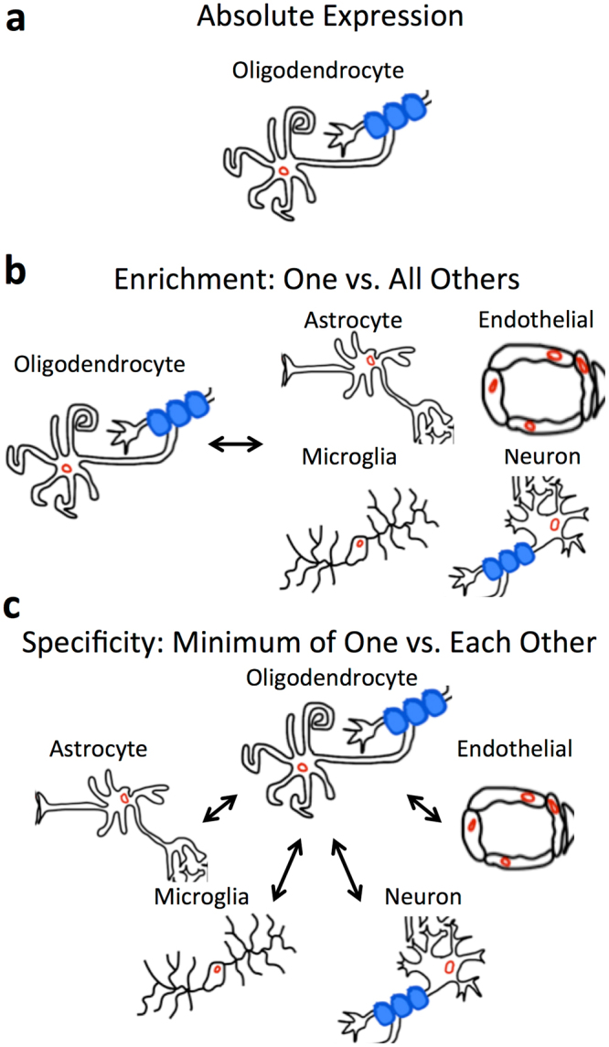
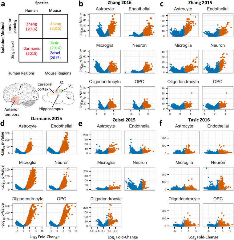
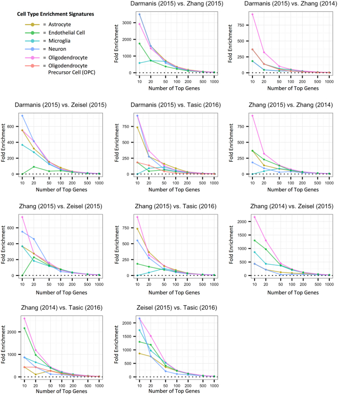
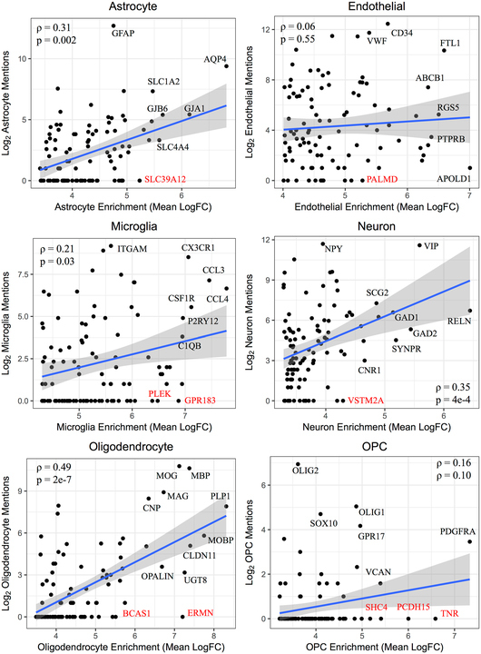
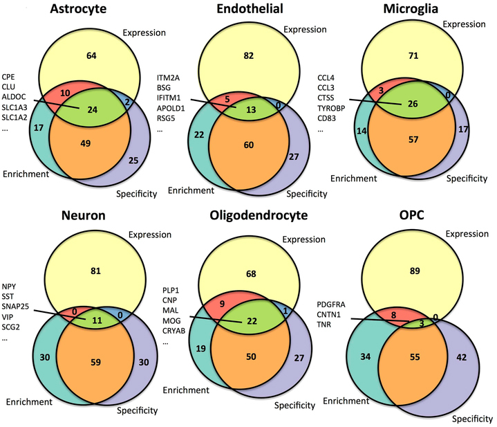
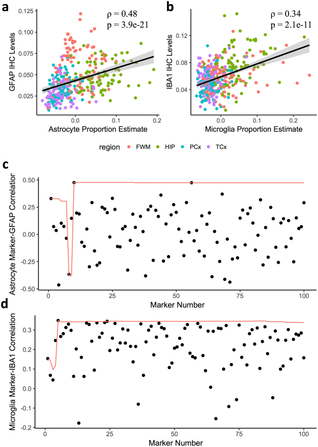
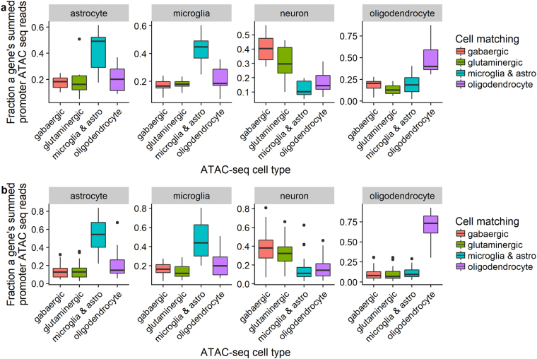
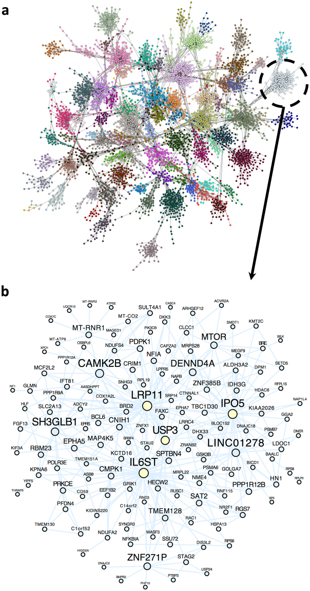
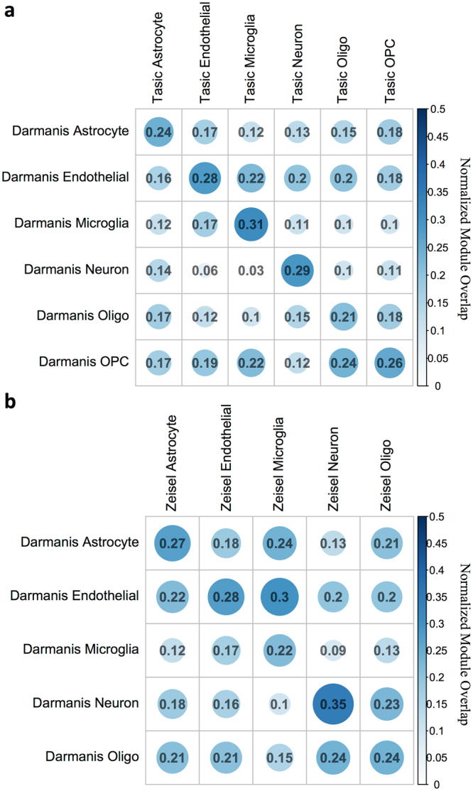
Elucidating brain cell type specific gene expression patterns is critical towards a better understanding of how cell-cell communications may influence brain functions and dysfunctions. We set out to compare and contrast five human and murine cell type-specific transcriptome-wide RNA expression data sets that were generated within the past several years. We defined three measures of brain cell type-relative expression including specificity, enrichment, and absolute expression and identified corresponding consensus brain cell “signatures,” which were well conserved across data sets. We validated that the relative expression of top cell type markers are associated with proxies for cell type proportions in bulk RNA expression data from postmortem human brain samples. We further validated novel marker genes using an orthogonal ATAC-seq dataset. We performed multiscale coexpression network analysis of the single cell data sets and identified robust cell-specific gene modules. To facilitate the use of the cell type-specific genes for cell type proportion estimation and deconvolution from bulk brain gene expression data, we developed an R package, BRETIGEA. In summary, we identified a set of novel brain cell consensus signatures and robust networks from the integration of multiple datasets and therefore transcend limitations related to technical issues characteristic of each individual study.
Overview
The claims and statistics of this paper haven’t been checked yet.
Run the check — free, no account needed. It reads the open-access text and checks the sources it cites.
Integrity signals
Registry
Cites
Statistics
Figures
Verification
Claims the paper makes
The claims and statistics of this paper haven’t been checked yet.
Run the check — free, no account needed. It reads the open-access text and checks the sources it cites.
Claims other papers make about this one
Each is checked against this paper’s own text: does it say what the citing paper says it does?
The source backs part of the claim, not all of it — or backs it for a different population, condition or comparison.
In the citing paper
“The cell clusters were annotated manually using marker genes that were conserved between both conditions as astrocytes, microglia, neurons, endothelial cells, ependymal cells, and mixed glia (Supplementary Data 1) (Arneson et al., 2018;McKenzie et al., 2018).”
From Single-cell resolution analysis of the crosstalk between chemogenically activated astrocytes and microglia (2020) · cites this paper as McKenzie et al., 2018
Experimental — accuracy not yet measuredThe cited source says what the claim attributes to it.
In the citing paper
“Canonical CNS cell type markers were compiled from previous studies66–69.”
From APOE4 to APOE2 allelic switching in mice improves Alzheimer’s disease-related metabolic signatures, neuropathology and cognition (2025) · cites this paper as [66]
The cited source says what the claim attributes to it.
In the citing paper
“To estimate the cell-type proportion of bulk tissue RNA-seq data used in this study, we perform a cell-type deconvolution on each sample using the brain cell–type marker signatures provided by the BRETIGEA R package (26).”
From Molecular subtyping of Alzheimer’s disease using RNA sequencing data reveals novel mechanisms and targets (2021) · cites this paper as [26]
The cited source says what the claim attributes to it.
In the citing paper
“All cell clusters were annotated manually based on marker genes that were conserved between the saccharin group and the CNO group and gene lists present in literature (Arneson et al., 2018;McKenzie et al., 2018).”
From Single-cell resolution analysis of the crosstalk between chemogenically activated astrocytes and microglia (2020) · cites this paper as McKenzie et al., 2018
The cited source says what the claim attributes to it.
In the citing paper
“We used a consensus top ranked cell type-specific marker set from a recent meta-analysis of multiple cell type-specific and single cell RNAseq datasets (McKenzie et al., 2018).”
From Cholesterol and matrisome pathways dysregulated in astrocytes and microglia (2022) · cites this paper as McKenzie et al., 2018
Self-check
Percentages, sums and reported differences re-computed from the paper’s own numbers.
The self-check needs the paper’s full text.
Citations
Cites
Controlling the False Discovery Rate: A Practical and Powerful Approach to Multiple Testing
Benjamini, Y., & Hochberg, Y. (1995) · Journal of the Royal Statistical Society Series B: Statistical Methodology · doi:10.1111/j.2517-6161.1995.tb02031.x · Cited by 109,482
STAR: ultrafast universal RNA-seq aligner
Dobin, A., Davis, C. A., Schlesinger, F., Drenkow, J., Zaleski, C., Jha, S., Batut, P., Chaisson, M., & Gingeras, T. R. (2013) · Bioinformatics · doi:10.1093/bioinformatics/bts635 · Cited by 51,709
Robinson, M. D., McCarthy, D. J., & Smyth, G. K. (2010) · Bioinformatics · doi:10.1093/bioinformatics/btp616 · Cited by 41,364
featureCounts: an efficient general purpose program for assigning sequence reads to genomic features
Liao, Y., Smyth, G. K., & Shi, W. (2014) · Bioinformatics · doi:10.1093/bioinformatics/btt656 · Cited by 27,219
Tissue-based map of the human proteome
Uhlén, M., Fagerberg, L., Hallström, B. M., Lindskog, C., Oksvold, P., Mardinoglu, A., Sivertsson, Å., Kampf, C., Sjöstedt, E., Asplund, A., et al. (2015) · Science · doi:10.1126/science.1260419 · Cited by 15,286
Highly parallel genome-wide expression profiling of individual cells using nanoliter droplets.
Macosko, E. Z., Basu, A., Satija, R., Nemesh, J., Shekhar, K., Goldman, M., Tirosh, I., Bialas, A. R., Kamitaki, N., Martersteck, E. M., et al. (2015) · Cell · doi:10.1016/j.cell.2015.05.002 · Cited by 7,110
Astrocytes: Biology and pathology
Sofroniew, M. V., & Vinters, H. V. (2010) · Acta Neuropathologica · doi:10.1007/s00401-009-0619-8 · Cited by 5,196
Zhang, Y., Chen, K., Sloan, S. A., Bennett, M. L., Scholze, A. R., O'Keeffe, S., Phatnani, H. P., Guarnieri, P., Caneda, C., Ruderisch, N., et al. (2014) · The Journal of Neuroscience · doi:10.1523/jneurosci.1860-14.2014 · Cited by 4,875
Cahoy, J. D., Emery, B., Kaushal, A., Foo, L. C., Zamanian, J. L., Christopherson, K. S., Xing, Y., Lubischer, J. L., Krieg, P. A., Krupenko, S. A., et al. (2008) · The Journal of Neuroscience · doi:10.1523/jneurosci.4178-07.2008 · Cited by 3,129
Cell types in the mouse cortex and hippocampus revealed by single-cell RNA-seq
Zeisel, A., Muñoz-Manchado, A. B., Codeluppi, S., Lönnerberg, P., La Manno, G., Juréus, A., Marques, S., Munguba, H., He, L., Betsholtz, C., et al. (2015) · Science · doi:10.1126/science.aaa1934 · Cited by 3,124
Cited by
Sorted by citations
Items 101–200 of 492
Sokolowski, M., & Wasserman, D. (2020) · European Neuropsychopharmacology · doi:10.1016/j.euroneuro.2020.06.002 · Cited by 29
Kathuria, A., Lopez-Lengowski, K., Watmuff, B., & Karmacharya, R. (2020) · Stem Cells and Development · doi:10.1089/scd.2020.0069 · Cited by 30
Miedema, S. S. M., Mol, M. O., Koopmans, F., Hondius, D., van Nierop, P., Menden, K., Mestdagh, C. F. D. V., van Rooij, J., Ganz, A. B., Paliukhovich, I., et al. (2022) · Acta Neuropathologica Communications · doi:10.1186/s40478-022-01387-8 · Cited by 27
“Cell type enrichment analysis Cell type enrichment analysis can help to stratify data from mixed cell populations, without the need for physical cell sorting [4, 45].”
Lewy pathology formation in patient-derived GBA1 Parkinson’s disease midbrain organoids
Frattini, E., Faustini, G., Lopez, G., Carsana, E. V., Tosi, M., Trezzi, I., Magni, M., Soldà, G., Straniero, L., Facchi, D., et al. (2024) · Brain · doi:10.1093/brain/awae365 · Cited by 31
Therapeutic strategies for autism: targeting three levels of the central dogma of molecular biology
Hong, D., & Iakoucheva, L. M. (2023) · Translational Psychiatry · doi:10.1038/s41398-023-02356-y · Cited by 32
“Furthermore, the brain is a more complex organ with greater complexity in cell types and significantly varying levels of gene expression between each type, making cell-specificity an additional concern in NDD therapeutics [50].”
BackgroundFunctional variants identify sex-specific genes and pathways in Alzheimer’s Disease
Bourquard, T., Lee, K. D., Al‐Ramahi, I., Pham, M., Shapiro, D., Lagisetty, Y., Soleimani, S., Mota, S., Wilhelm, K. O., Samieinasab, M., et al. (2023) · Nature Communications · doi:10.1038/s41467-023-38374-z · Cited by 32
“For the coexpression analysis of single cell RNAseq, cell type speci fi c WGCNA networks were obtained from 154 .”
Almeida, M. C., Eger, S. J., He, C., Audouard, M., Nikitina, A. A., Glasauer, S. M. K., Han, D., Mejía‐Cupajita, B., Acosta‐Uribe, J., Villalba‐Moreno, N. D., et al. (2024) · Neuron · doi:10.1016/j.neuron.2024.02.009 · Cited by 34
The importance of identified neurons in gastropod molluscs to neuroscience
Katz, P. S., & Quinlan, P. D. (2018) · Current Opinion in Neurobiology · doi:10.1016/j.conb.2018.10.009 · Cited by 28
Cholesterol and matrisome pathways dysregulated in human APOE ε4 gliaPreprint
Julia, T. C. W., Liang, S., Lü, Q., Pipalia, N. H., Chao, M. J., Shi, Y., Bertelsen, S. E., Kapoor, M., Marcora, E. M., Sikora, E., et al. (2019) · bioRxiv (Cold Spring Harbor Laboratory) · doi:10.1101/713362 · Cited by 29
Self-citationCDSeqR: fast complete deconvolution for gene expression data from bulk tissues
Kang, K., Huang, C. D., Li, Y., Umbach, D. M., & Li, L. (2021) · BMC Bioinformatics · doi:10.1186/s12859-021-04186-5 · Cited by 26
“e Expression level of PLP1 (Oligodendrocyte marker [41]) in the clusters of CDSeq-estimated cell types (cell type number is 7); f Expression level of PLP1 in human”
BackgroundJohnson, T. S., Xiang, S., Dong, T., Huang, Z., Cheng, M., Wang, T., Yang, K., Ni, D., Huang, K., & Zhang, J. (2021) · Scientific Reports · doi:10.1038/s41598-020-79740-x · Cited by 26
Iacoangeli, A., Fogh, I., Selvackadunco, S., Topp, S. D., Shatunov, A., van Rheenen, W., Khleifat, A. A., Opie-Martin, S., Ratti, A., Calvo, A., et al. (2021) · Brain Communications · doi:10.1093/braincomms/fcab236 · Cited by 25
“BRETIGEA was used to estimate cell composition per sample using BRETIGEA’s cell marker dataset (50 markers).(29) To analysis differences in cell composition by type across samples, we used a linear regression model to compare cell-type estimates between ALS and controls controlling for variables age of death, post-mor…”
MethodNúñez-Ríos, D. L., Martínez‐Magaña, J. J., Nagamatsu, S. T., Andrade-Brito, D., Forero, D. A., Orozco, C. A., & Montalvo‐Ortiz, J. L. (2022) · Biomedicines · doi:10.3390/biomedicines10051107 · Cited by 27
“Brain cell-type specific gene expression patterns of human and murine RNA expression data sets were recently reported showing the top 1000 cell type-specific genes from astrocytes, oligodendrocytes, microglia, neuron and endothelial cells [149].”
BackgroundOue, H., Yamazaki, Y., Qiao, W., Yuanxin, C., Ren, Y., Kurti, A., Shue, F., Parsons, T. M., Perkerson, R. B., Kawatani, K., et al. (2023) · JCI Insight · doi:10.1172/jci.insight.163822 · Cited by 26
“scriptome analysis on astrocytes, endothelial cells, microglia, neurons, oligodendrocytes (25), and vas-”
MethodPergola, G., Parihar, M., Sportelli, L., Bharadwaj, R. A., Borcuk, C., Radulescu, E., Bellantuono, L., Blasi, G., Chen, Q., Kleinman, J. E., et al. (2023) · Science Advances · doi:10.1126/sciadv.ade2812 · Cited by 26
Short-term sleep deprivation in mice induces B cell migration to the brain compartment
Korin, B., Avraham, S., Azulay‐Debby, H., Farfara, D., Hakim, F., & Rolls, A. (2019) · SLEEP · doi:10.1093/sleep/zsz222 · Cited by 28
A cAMP-Related Gene Network in Microglia Is Inversely Regulated by Morphine Tolerance and Withdrawal
Coffey, K. R., Lesiak, A. J., Marx, R. E., Vo, E. K., Garden, G. A., & Neumaier, J. F. (2021) · Biological Psychiatry Global Open Science · doi:10.1016/j.bpsgos.2021.07.011 · Cited by 27
BrainBase: a curated knowledgebase for brain diseases
Liu, L., Zhang, Y., Niu, G., Li, Q., Zhao, L., Zhu, T., Feng, C., Liu, X., Zhang, Y., Xu, T., et al. (2021) · Nucleic Acids Research · doi:10.1093/nar/gkab987 · Cited by 24
Wang, N., Langfelder, P., Stricos, M., Ramanathan, L., Richman, J., Vaca, R., Plascencia, M., Gu, X., Zhang, S., Tamai, K., et al. (2022) · Neuron · doi:10.1016/j.neuron.2022.09.028 · Cited by 25
Lü, Y., Girardi, C. S., Fitz, N. F., McGuire, M. R., Ostach, M. A., Mamun-Or-Rashid, A. N. M., Lefterov, I. M., & Koldamova, R. P. (2023) · Cell Reports · doi:10.1016/j.celrep.2023.112785 · Cited by 27
“A list of marker genes for different cell types, generated based on our previous studies 25 and other publications 75-79 were used as a reference for cell type annotation (Table S1).”
Hecker, M., Boxberger, N., Illner, N., Fitzner, B., Schröder, I., Winkelmann, A., Dudešek, A., Meister, S., Koczan, D., Lorenz, P., et al. (2019) · PLoS Genetics · doi:10.1371/journal.pgen.1007961 · Cited by 26
Mol, M. O., Miedema, S. S. M., Swieten, J. C. V., van Rooij, J., & Dopper, E. G. P. (2021) · International Journal of Molecular Sciences · doi:10.3390/ijms221910298 · Cited by 23
Integrated analysis of robust sex-biased gene signatures in human brain
Wapeesittipan, P., & Joshi, A. (2023) · Biology of Sex Differences · doi:10.1186/s13293-023-00515-w · Cited by 24
“gene sets [28] and calculated significant overlap using different thresholds for both male and female-biased genes (see "Methods").”
BackgroundPetralia, M. C., Ciurleo, R., Saraceno, A., Pennisi, M., Basile, M. S., Fagone, P., Bramanti, P., Nicoletti, F., & Cavalli, E. (2020) · Genes · doi:10.3390/genes11040390 · Cited by 25
“In order to elucidate the involvement of the different brain cell types in SCZ pathology, we retrieved consensus brain cell gene signatures for astrocytes, microglia, neurons and oligodendrocytes, from the work by McKenzie and colleagues [26].”
MethodKim, Y. W., Al‐Ramahi, I., Koire, A. M., Wilson, S. J., Konecki, D. M., Mota, S., Soleimani, S., Botas, J., & Lichtarge, O. (2020) · Alzheimer s & Dementia · doi:10.1002/alz.12240 · Cited by 22
Transcriptomic and Cellular Decoding of Regional Brain Vulnerability to Neurodevelopmental DisordersPreprint
Seidlitz, J., Nadig, A., Liu, S., Bethlehem, R. A. I., Vértes, P. E., Morgan, S. E., Váša, F., Romero-García, R., Lalonde, F. M., CLASEN, L. S., et al. (2019) · bioRxiv (Cold Spring Harbor Laboratory) · doi:10.1101/573279 · Cited by 25
Zhukovsky, P., Tio, E. S., Coughlan, G., Bennett, D. A., Wang, Y., Hohman, T. J., Pizzagalli, D. A., Mulsant, B. H., Voineskos, A. N., & Felsky, D. (2024) · Nature Communications · doi:10.1038/s41467-024-49430-7 · Cited by 25
Whitney, K. R., Song, W., Sharma, A., Dangoor, D. K., Farrell, K. W., Krassner, M. M., Ressler, H. W., Christie, T. D., Kandoi, S., Walker, R. H., et al. (2024) · Acta Neuropathologica · doi:10.1007/s00401-024-02823-w · Cited by 23
Self-citationIslam, S., Kamila, S., & Chattopadhyay, A. (2022) · Journal of Environmental Science and Health Part C · doi:10.1080/26896583.2022.2158675 · Cited by 24
“astrocytes, oligodendrocytes, microglia, endothelial cells, oligodendrocyte precursor cells (NG2 cells), ependymal cells, smooth muscle cells, and pericytes, that contribute to the complexity of the brain.(133) Most of the cells in the brain tissue remain in the mitotic phase throughout life and henceforth are potenti…”
BackgroundTranscription factors NFIA and NFIB induce cellular differentiation in high-grade astrocytoma
Chen, K., Bridges, C. R., Lynton, Z., Lim, J. W. C., Stringer, B. W., Rajagopal, R., Wong, K., Ganesan, D., Ariffin, H., Day, B. W., et al. (2019) · Journal of Neuro-Oncology · doi:10.1007/s11060-019-03352-3 · Cited by 23
Hentrich, T., Wassouf, Z., Ehrhardt, C., Haas, E., Mills, J. D., Aronica, E., Outeiro, T. F., Hübener‐Schmid, J., Rieß, O., Casadei, N., et al. (2020) · Aging · doi:10.18632/aging.103935 · Cited by 23
“To further explore the link to oligodendrocytes, the 38 DEGs were examined for their cell type-specific expression in human against reference data for neurons, astrocytes, microglia, oligodendrocytes, and endothelial cells [19].”
BackgroundPostmortem Interval Leads to Loss of Disease-Specific Signatures in Brain Tissue
Olney, K. C., Gibson, K. A., Cadiz, M. P., Rahimzadeh, N., Swarup, V., & Fryer, J. D. (2025) · eneuro · doi:10.1523/eneuro.0505-24.2025 · Cited by 22
“Cell type annotation was conducted using established brain cell type markers (McKenzie et al., 2018) and the top genes from Seurat ’ s FindMarkers function.”
Esmail, S., & Danter, W. R. (2021) · Frontiers in Aging Neuroscience · doi:10.3389/fnagi.2021.643889 · Cited by 22
“…to evaluate the performance of the AD simulations compared to the aiWBO (Romito-DiGiacomo et al., 2007; Santos et al., 2011; Raja et al., 2016; Arbor, 2017; Gerakis and Hetz, 2019; Madadi et al., 2019; Brighi et al., 2020; Papaspyropoulos et al., 2020; Ribeiro et al., 2020; Silva et al., 2020).”
Antiviral response within different cell types of the CNS
Telikani, Z., Monson, E. A., Hofer, M. J., & Helbig, K. J. (2022) · Frontiers in Immunology · doi:10.3389/fimmu.2022.1044721 · Cited by 23
“Sendai virus BE(2)-C Neurons – TLR3, MDA5 RIG-I IRF3 IFNb, NFKB, PI3K – Human Murine In vitro (41)”
BackgroundInfluentialCell type-specific circular RNA expression in human glial cells
Curry‐Hyde, A., Gray, L. G., Chen, B. J., Ueberham, U., Arendt, T., & Janitz, M. (2020) · Genomics · doi:10.1016/j.ygeno.2020.09.042 · Cited by 21
Pathways linking Alzheimer’s disease risk genes expressed highly in microglia
Hodges, A. K., Piers, T. M., Collier, D., Cousins, O., & Pocock, J. M. (2021) · Neuroimmunology and Neuroinflammation · doi:10.20517/2347-8659.2020.60 · Cited by 21
“org/brain/([162]) and http://research-pub.”
Blood and brain gene expression signatures of chronic intermittent ethanol consumption in mice
Ferguson, L. B., Roberts, A. J., Mayfield, R. D., & Messing, R. O. (2022) · PLoS Computational Biology · doi:10.1371/journal.pcbi.1009800 · Cited by 22
Sarnowski, C., Huan, T., Ma, Y., Joehanes, R., Beiser, A., DeCarli, C. S., Heard‐Costa, N. L., Levy, D. A., Lin, H., Liu, C., et al. (2023) · Clinical Epigenetics · doi:10.1186/s13148-023-01589-4 · Cited by 22
Güvenek, A., Parikshak, N., Zamolodchikov, D., Gelfman, S., Moscati, A., Dobbyn, L., Stahl, E. A., Shuldiner, A. R., & Coppola, G. (2024) · Communications Biology · doi:10.1038/s42003-024-06684-7 · Cited by 23
“PARVG expression has been recently reported as higher in microglia compared to other cells 48 , and upregulated in the brains of AD patients 44 .”
Chen, X., Duan, H.-F., Xiao, L., & Gan, J. (2018) · Frontiers in Genetics · doi:10.3389/fgene.2018.00565 · Cited by 22
Ghisai, S. A., van Hijfte, L., Vallentgoed, W., Tesileanu, C. M. S., de Heer, I. J., Kros, J. M., Sanson, M., Gorlia, T., Wick, W., Vogelbaum, M. A., et al. (2024) · Acta Neuropathologica · doi:10.1007/s00401-024-02811-0 · Cited by 21
“In all datasets, the primary source of variation (PC1) was likely associated with tumour purity as demonstrated by the expression of neuronal genes, with neuron marker genes like SLC12A5 , TMEM130 and SV2B ranking among the top 50 contributors [25].”
Valdes, P., Caldwell, A. B., Liu, Q., Fitzgerald, M. Q., Ramachandran, S., Karch, C. M. M., Dominantly Inherited Alzheimer Network (DIAN), Adams, S., Allegri, R., Araki, A., et al. (2025) · Alzheimer s Research & Therapy · doi:10.1186/s13195-024-01659-6 · Cited by 25
“Markers were selected from neuronal cell identity marker resources and published literature sources for GABAergic [43–49], dopaminergic [50–55], glutamatergic [44, 56–58], synaptic [43, 48], pan-neuronal [59, 60], amyloid-associated, and cortical layer [61, 62] neuron types.”
Single-cell-resolution transcriptome map of human, chimpanzee, bonobo, and macaque brainsPreprint
Khrameeva, E. E., Kurochkin, I., Han, D., Guijarro, P., Kanton, S., Santel, M., Qian, Z., Rong, S., Mazin, P., Bulat, M., et al. (2019) · bioRxiv (Cold Spring Harbor Laboratory) · doi:10.1101/764936 · Cited by 19
Microglial-targeting induced by intranasal linalool during neurological protection postischemia
Barrera-Sandoval, Á. M., Osorio, E., & Cardona‐Gómez, G. P. (2019) · European Journal of Pharmacology · doi:10.1016/j.ejphar.2019.172420 · Cited by 19
Shared brain transcriptomic signature in TDP-43 type A FTLD patients with or without GRN mutations
Pottier, C. P., Mateiu, L. M., Baker, M., DeJesus‐Hernandez, M., Vicente, C. T., Finch, N. A., Tian, S., van Blitterswijk, M., Murray, M. E., Ren, Y., et al. (2021) · Brain · doi:10.1093/brain/awab437 · Cited by 20
Exploring ITM2A as a new potential target for brain delivery
Cegarra, C., Chaves, C., Déon, C., Do, T. M., Dumas, B., Frenzel, A., Kuhn, P., Roudières, V., Guillemot, J.-C., & Lesuisse, D. (2022) · Fluids and Barriers of the CNS · doi:10.1186/s12987-022-00321-3 · Cited by 19
“An analysis of five human and murine cell type-specific transcriptome-wide RNA expression datasets generated within the past several years also identified ITM2A as one of the top expressed genes in endothelial cells [31].”
BackgroundPark, S. W., Seo, M. K., Webster, M. J., Lee, J. G., & Kim, S. (2022) · Translational Psychiatry · doi:10.1038/s41398-022-01944-8 · Cited by 20
“The data was generated using 5 human cell-type-specific transcriptome-wide RNA expression data sets further validated using an orthogonal ATAC-seq dataset [27].”
MethodJones, A. R., Iacoangeli, A., Adey, B. N., Bowles, H., Shatunov, A., Troakes, C., Garson, J. A., McCormick, A., & Al‐Chalabi, A. (2021) · Scientific Reports · doi:10.1038/s41598-021-93742-3 · Cited by 21
Golden, L. R., Siano, D. S., Stephens, I. O., MacLean, S. M., Saito, K., Nolt, G. L., Funnell, J. L., Pallerla, A. V., Lee, S., Smith, C., et al. (2025) · Nature Neuroscience · doi:10.1038/s41593-025-02094-y · Cited by 18
“Canonical CNS cell type markers were compiled from previous studies 66–69 .”
Wittrahm, R., Takalo, M., Marttinen, M., Kuulasmaa, T., Mäkinen, P. M., Kemppainen, S., Martiskainen, H., Rauramaa, T. H., Pike, I., Leinonen, V., et al. (2021) · Cells · doi:10.3390/cells10040860 · Cited by 20
Gallina, A. L., Rykaczewska, U., Wirka, R. C., Caravaca, A. S., Shavva, V. S., Youness, M., Karadimou, G., Lengquist, M., Razuvaev, A., Paulsson‐Berne, G., et al. (2021) · Frontiers in Cardiovascular Medicine · doi:10.3389/fcvm.2021.655869 · Cited by 18
Current Perspectives: Evidence to Date on BTK Inhibitors in the Management of Multiple Sclerosis
Contentti, E. C., & Correale, J. D. (2022) · Drug Design Development and Therapy · doi:10.2147/dddt.s348129 · Cited by 18
Demin, K. A., Krotova, N. A., Ilyin, N. P., Galstyan, D. S., Kolesnikova, T. O., Strekalova, T. V., de Abreu, M. S., Petersen, E. V., Zabegalov, K. N., & Kalueff, A. V. (2022) · Scientific Reports · doi:10.1038/s41598-022-22688-x · Cited by 19
Kaurani, L., Islam, M. R., Heilbronner, U., Krüger, D. M., Zhou, J., Methi, A., Strauss, J., Pradhan, R., Schröder, S., Burkhardt, S., et al. (2024) · The EMBO Journal · doi:10.1038/s44318-024-00067-8 · Cited by 17
“Published single-cell data (McKenzie et al, 2018) were utilized to explore neuron, astrocyte-, and microglia-speci fi c expression of genes.”
Pletenev, I. A., Bazarevich, M., Zagirova, D., Kononkova, A. D., Cherkasov, A. V., Efimova, O., Tiukacheva, E. A., Morozov, K. V., Ulianov, K. A., Komkov, D., et al. (2024) · Nucleic Acids Research · doi:10.1093/nar/gkae271 · Cited by 18
MAPT haplotype-associated transcriptomic changes in progressive supranuclear palsy
Ressler, H. W., Humphrey, J., Vialle, R. A., Babrowicz, B. M., Kandoi, S., Raj, T., Dickson, D. W., Ertekin‐Taner, N., Crary, J. F., & Farrell, K. W. (2024) · Acta Neuropathologica Communications · doi:10.1186/s40478-024-01839-3 · Cited by 17
“To perform gene expression deconvolution analysis, we used BRETIGEA (BRain cEll Type specIfic Gene Expression Analysis) [49].”
MethodTayran, H., Yılmaz, E., Bhattarai, P., Min, Y., Wang, X., Ma, Y., Wang, N., Jeong, I., Nelson, N. N., Kassara, N., et al. (2024) · Cell Genomics · doi:10.1016/j.xgen.2024.100642 · Cited by 18
Rate of brain aging and APOE ε4 are synergistic risk factors for Alzheimer’s disease
Glorioso, C. A., Pfenning, A. R., Lee, S. S., Bennett, D. A., Sibille, E. L., Kellis, M., & Guarente, L. (2019) · Life Science Alliance · doi:10.26508/lsa.201900303 · Cited by 18
“In addition, there is little change in the CM or PE cohorts in transcripts de fi ned as neuronal-speci fi c or glial-speci fi c (McKenzie et al, 2018), suggesting there is not a signi fi cant loss of neurons in PFC, consistent with earlier data (Haug et al, 1984; Morrison & Hof, 1997; Yankner et al,…”
BackgroundMethodResultInfluentialVallentgoed, W., Hoogstrate, Y., van Garderen, K. A., van Hijfte, L., van Dijk, E., Kouwenhoven, M. C. M., Niers, J. M., Draaisma, K., Martin, I., de Leng, W. W. J., et al. (2025) · Nature Cancer · doi:10.1038/s43018-025-01023-z · Cited by 20
“For healthy cell types, the top 500 genes were selected from a reference gene list 73 ; for the tumor cell subclusters, marker genes were used from Ventreicher et al. 74 .”
Chaves, C., Do, T.-M., Cegarra, C., Roudières, V., Tolou, S., Thill, G., Rocher, C., Didier, M., & Lesuisse, D. (2020) · Pharmaceutics · doi:10.3390/pharmaceutics12100967 · Cited by 18
Cervera‐Juanes, R. P., Darakjian, P., Ball, M., Kohama, S. G., & Urbanski, H. F. (2021) · GeroScience · doi:10.1007/s11357-021-00453-8 · Cited by 14
Nagamatsu, S. T., Rompala, G. R., Hurd, Y. L., Núñez-Ríos, D. L., & Montalvo‐Ortiz, J. L. (2023) · Frontiers in Psychiatry · doi:10.3389/fpsyt.2022.1078894 · Cited by 17
“Brain cell-type enrichment analysis was carried out using transcriptomic data from human postmortem brain samples to characterize the cell-type enrichment of OUDassociated differential mCpHs in neurons, microglia, astrocytes, oligodendrocytes, and endothelial cell type-specific markers (30).”
MethodSelf-citationEvolutionary and biomedical implications of sex differences in the primate brain transcriptome
DeCasien, A. R., Chiou, K. L., Testard, C., Mercer, A., Valle, J. E. N.-D., Bauman, S. E., González, O., Stock, M. K., Ruiz-Lambides, A. V., Martínez, M. I., et al. (2024) · Cell Genomics · doi:10.1016/j.xgen.2024.100589 · Cited by 17
Kim, G. S., & Uddin, M. (2020) · Biology of Sex Differences · doi:10.1186/s13293-020-00288-6 · Cited by 17
“conserved consensus brain cell marker sets identified across mouse and human datasets [27] and implements”
BackgroundKumar, T., Sethuraman, R., Mitra, S., Ravindran, B., & Narayanan, M. (2023) · PLoS Computational Biology · doi:10.1371/journal.pcbi.1011022 · Cited by 18
Slota, J. A., Medina, S. J., Frost, K. L., & Booth, S. A. (2022) · Frontiers in Neuroscience · doi:10.3389/fnins.2022.918811 · Cited by 16
Transcriptomics of interstitial lung disease: a systematic review and meta-analysis
He, D. X., Guler, S. A., Shannon, C. P., Ryerson, C. J., & Tebbutt, S. J. (2024) · European Respiratory Journal · doi:10.1183/13993003.01070-2024 · Cited by 17
“0.3) [21] by using the top 20 gene markers for cell types described in GSE122960 [22] on the ComBat-corrected gene expression data matrix.”
InfluentialMultiomic single-cell profiling identifies critical regulators of postnatal brain
Clarence, T., Bendl, J. M., Cao, X. D., Wang, X., Zheng, S., Hoffman, G. E., Kozlenkov, A., Hong, A., Iskhakova, M., Jaiswal, M. K., et al. (2025) · Nature Genetics · doi:10.1038/s41588-025-02083-8 · Cited by 20
“This is in line with previous reports on cell-type composition changes in the human brain 18–20 .”
Self-citationZhao, X., Zhang, F., Kandel, S. R., Brau, F., & He, J. J. (2022) · Cell Reports · doi:10.1016/j.celrep.2022.110765 · Cited by 16
“Specifically, we took the top 6,000 ranked cell-type-enriched mouse genes (McKenzie et al., 2018), mapped them with the RNA-seq transcriptome database from purified brain cells (Zhang et al.”
MethodMwikali, K., Pance, A., Mwangi, S., Goulding, D., Kemp, A. C., Rono, M. K., Ochola‐Oyier, L. I., Bull, P. C., Bejon, P., Rayner, J. C., et al. (2023) · Nature Communications · doi:10.1038/s41467-023-42103-x · Cited by 16
Min, Y., Wang, X., İş, Ö., Patel, T., Gao, J., Reddy, J. S., Quicksall, Z., Nguyen, T., Lin, S., Tutor‐New, F. Q., et al. (2023) · Nature Communications · doi:10.1038/s41467-023-42626-3 · Cited by 20
Astrocytes Drive Divergent Metabolic Gene Expression in Humans and Chimpanzees
Zintel, T. M., Pizzollo, J., Claypool, C. G., & Babbitt, C. C. (2023) · Genome Biology and Evolution · doi:10.1093/gbe/evad239 · Cited by 18
Tian, G., Bartas, K., Hui, M., Chen, L., Vasquez, J. J., Azouz, G., Derdeyn, P., Manville, R. W., Ho, E. L., Fang, A. S., et al. (2024) · Neuron · doi:10.1016/j.neuron.2024.07.018 · Cited by 22
Dynamic Lamin B1-Gene Association During Oligodendrocyte Progenitor Differentiation
Yattah, C., Hernandez, M., Huang, D., Park, H., Liao, W., & Casaccia, P. (2020) · Neurochemical Research · doi:10.1007/s11064-019-02941-y · Cited by 16
Self-citationFew Is Enough: Task-Augmented Active Meta-learning for Brain Cell Classification
Yuan, P., Mobiny, A., Jahanipour, J., Li, X., Cicalese, P. A., Roysam, B., Patel, V. M., Maric, D., & Nguyen, H. V. (2020) · Lecture notes in computer science · doi:10.1007/978-3-030-59710-8_36 · Cited by 15
Mengaziol, J., Dunn, A. D., Salimando, G. J., Wooldridge, L. M., Creus‐Muncunill, J., Eacret, D., Chen, C., Bland, K., Liu‐Chen, L., Ehrlich, M. E., et al. (2022) · PLoS ONE · doi:10.1371/journal.pone.0270317 · Cited by 15
“Enrichment for genes characteristic of brain cell types [21] was calculated for using a hypergeometric test.”
MethodKisaretova, P. E., Tsybko, A. S., Bondar, N. P., & Reshetnikov, V. V. (2023) · Biomedicines · doi:10.3390/biomedicines11020289 · Cited by 15
“Gpr84 and Ptprc are microgliaspecific genes [57] and encode receptors involved in the regulation of neuroinflammation.”
BackgroundMolecular differences in brain regional vulnerability to aging between males and females
Zhou, X., Cao, J., Zhu, L., Farrell, K. W., Wang, M., Guo, L., Yang, J., McKenzie, A. T., Crary, J. F., Cai, D., et al. (2023) · Frontiers in Aging Neuroscience · doi:10.3389/fnagi.2023.1153251 · Cited by 16
Self-citationDifferential usage of DNA modifications in neurons, astrocytes, and microglia
Tooley, K. B., Chucair‐Elliott, A. J., Ocañas, S. R., Machalinski, A. H., Pham, K. D., Hoolehan, W., Kulpa, A. M., Stanford, D. R., & Freeman, W. M. (2023) · Epigenetics & Chromatin · doi:10.1186/s13072-023-00522-6 · Cited by 13
“Cell type specific marker gene lists were generated from the re-analysis of lists published by McKenzie et al. [117] of immunopuri-fied [118] and high throughput single cell data from mice [119, 120].”
MethodAlzheimer’s disease rewires gene coexpression networks coupling different brain regions
Mitra, S., Kailash, B. P., Srivatsan, C. R., Saikumar, N. V., Philip, P., & Narayanan, M. (2024) · npj Systems Biology and Applications · doi:10.1038/s41540-024-00376-y · Cited by 19
“Human marker genes (markers_df_human_brain data frame) for the four major cell-types were obtained from the BRETIGEA (BRain cEll Type specI fi c Gene Expression Analysis) meta-analysis study 62 .”
MethodLoss of TREM2 diminishes CAA despite an overall increase of amyloid load in Tg‐SwDI mice
Zhong, R., Xu, Y., Williams, J. W., & Li, L. (2024) · Alzheimer s & Dementia · doi:10.1002/alz.14222 · Cited by 16
“The marker genes generated from these cell types were compared to reported marker genes 45–48 to confirm the cell type prediction (Figure 6B-C).”
Sharpe, A. L., Trzeciak, M., Eliason, N. L., Blankenship, H. E., Byrd, B. A. M., Douglas, P., Freeman, W. M., & Beckstead, M. J. (2021) · Addiction Biology · doi:10.1111/adb.13120 · Cited by 15
Briel, N., Ruf, V., Pratsch, K., Roeber, S., Widmann, J., Mielke, J., Dorostkar, M. M., Windl, O., Arzberger, T., Herms, J. W., et al. (2022) · Acta Neuropathologica · doi:10.1007/s00401-022-02483-8 · Cited by 19
Just a SNP away: The future of in vivo massively parallel reporter assay
Degner, K. N., Bell, J., Jones, S. D., & Won, H. (2024) · Cell Insight · doi:10.1016/j.cellin.2024.100214 · Cited by 14
Pola, I., Ashton, N. J., Bastiani, M. A. D., Brum, W. S., Rahmouni, N., Tan, K., Machado, L. S., Servaes, S., Stevenson, J., Tissot, C., et al. (2025) · Alzheimer s & Dementia · doi:10.1002/alz.70171 · Cited by 15
“64 This spatial consistency supports the hypothesis that these neuroinflammatory proteins and the underlying AD processes converge in these regions and could therefore processes in the disease process.”
Esmail, S., & Danter, W. R. (2021) · Biomedicines · doi:10.3390/biomedicines9040440 · Cited by 15
Figueiredo, R. Q., del Ser, S. D., Raschka, T., Hofmann‐Apitius, M., Kodamullil, A. T., Mubeen, S., & Domingo‐Fernándéz, D. (2022) · BMC Bioinformatics · doi:10.1186/s12859-022-04765-0 · Cited by 14
“Similarly, McKenzie et al. [24] analyzed co-expression changes in different cell types of the brain, discovering significant cell type-specific expression signatures, while also finding well-known cell type marker genes among the most enriched genes across cell types.”
BackgroundReshetnikov, V. V., Kisaretova, P. E., & Bondar, N. P. (2022) · International Journal of Molecular Sciences · doi:10.3390/ijms232213792 · Cited by 16
“Our dataset was tested for enrichment with genes specific to neurons, astrocytes, microglia, endothelial cells, oligodendrocytes, and oligodendrocyte precursor cells according to recently published data [34].”
MethodExploratory analysis of L1 retrotransposons expression in autism
Spirito, G., Filosi, M., Domenici, E., Mangoni, D., Gustincich, S., & Sanges, R. (2023) · Molecular Autism · doi:10.1186/s13229-023-00554-5 · Cited by 15
“For each sample, negatively correlated genes resulted enriched for neural markers from single cell studies [65] and for SFARI genes.”
BackgroundStructural brain preservation: a potential bridge to future medical technologies
McKenzie, A. T., Zeleznikow-Johnston, A., Sparks, J. S., Nnadi, O., Smart, J., Wiley, K., Cerullo, M. A., de Wolf, A., Minerva, F., Risco, R., et al. (2024) · Frontiers in Medical Technology · doi:10.3389/fmedt.2024.1400615 · Cited by 15
Self-citationAn assessment of the transport mechanism and intraneuronal stability of l-carnosine
Lopachev, A. V., Абаимов, Д. А., Filimonov, I. S., Куличенкова, К. Н., & Федорова, Т. Н. (2021) · Amino Acids · doi:10.1007/s00726-021-03094-5 · Cited by 13
Regenerative Neurology and Regenerative Cardiology: Shared Hurdles and Achievements
Mitrečić, D., Hribljan, V., Jagečić, D., Isaković, J., Lamberto, F., Horánszky, A., Zana, M., Földes, G., Zavan, B., Pivoriūnas, A., et al. (2022) · International Journal of Molecular Sciences · doi:10.3390/ijms23020855 · Cited by 13
“These findings confirm the potential of EVs as a therapeutic tool against various diseases and disorders of the CNS.”
BackgroundInfluentialHussain, T., Sanchez, K. G., Crayton, J., Saha, D., Jeter, C. R., Lu, Y., Abba, M. C., Seo, R., Noebels, J. L., Fonken, L. K., et al. (2023) · Progress in Neurobiology · doi:10.1016/j.pneurobio.2023.102425 · Cited by 11
“The R package BRETIGEA (Brain cell type specific gene expression analysis) was employed to estimate relative brain cell type proportions from the RNA-seq profiles (McKenzie, 2018).”
The stability of the myelinating oligodendrocyte transcriptome is regulated by the nuclear lamina
Pruvost, M., Patzig, J., Yattah, C., Selcen, I., Hernandez, M., Park, H., Moyon, S., Liu, S., Morioka, M. S., Shopland, L. S., et al. (2023) · Cell Reports · doi:10.1016/j.celrep.2023.112848 · Cited by 11
“25 34 At an ultrastructural level, the process of differentiation is characterized by the transition from predominantly euchromatic nuclei in progenitors to electron-dense nuclei in myelinating oligodendrocytes, which are characterized by large masses of peripheral heterochromatin, in the proximity…”
Self-citationCross-species comparative hippocampal transcriptomics in Alzheimer’s disease
Bastiani, M. A. D., Bellaver, B., Carello‐Collar, G., Zimmermann, M., Kunach, P., Lima‐Filho, R. A. S., Forner, S., Martini, A. C., Pascoal, T. A., Lourenco, M. V., et al. (2023) · iScience · doi:10.1016/j.isci.2023.108671 · Cited by 13
İş, Ö., Min, Y., Wang, X., Oatman, S. R., Daniel, A. A., & Ertekin‐Taner, N. (2024) · Glia · doi:10.1002/glia.24652 · Cited by 13
Transcriptomic signatures of oxytosis/ferroptosis are enriched in Alzheimer’s disease
Currais, A. J. M., Sanchez, K. L., Soriano‐Castell, D., Dar, N. J., Evensen, K. G., Soriano, S., & Maher, P. A. (2025) · BMC Biology · doi:10.1186/s12915-025-02235-6 · Cited by 14
“We used three measures of human and mouse brain cell type-relative expression (top 1000 genes), including specificity, enrichment, and absolute expression [53].”
An analytical method for the identification of cell type-specific disease gene modules
Guan, J., Lin, Y., Wang, Y., Gao, J., & Ji, G. (2021) · Journal of Translational Medicine · doi:10.1186/s12967-020-02690-5 · Cited by 12
Jha, M., Roy, S., & Kalita, J. K. (2020) · Computers in Biology and Medicine · doi:10.1016/j.compbiomed.2020.104023 · Cited by 12
Baik, S., Selvaraji, S., Fann, D. Y., Poh, L., Jo, D., Herr, D. R., Zhang, S. R., Kim, H. A., Silva, M. D., Lai, M. K. P., et al. (2021) · Aging · doi:10.18632/aging.203123 · Cited by 12
“McKenzie and colleagues [34] had compared murine cell-type specific transcriptome-wide RNA expression data sets from three different groups [35–37].”
Background
Metadata
- Authors
- McKenzie, A. T., Wang, M., Hauberg, M. E., Fullard, J. F., Kozlenkov, A., Keenan, A., Hurd, Y. L., Dracheva, S., Casaccia, P., Roussos, P., & Zhang, B.
- Affiliations
- Icahn School of Medicine at Mount Sinai (US)
- Aarhus University (DK)
- James J. Peters VA Medical Center (US)
- City University of New York (US)
- Corresponding author
- Zhang, Bin
- ORCID
- Venue
- Scientific Reports
- Published
- 2018-06-11
- Volume
- 8
- Issue
- 1
- Pages
- 8868-8868
- ISSN
- 2045-2322 (ISSN-L)
- PMC
- PMC5995803
- OpenAlex
- W2806989085
- Cited by
- 481
- References
- 59
- Citations by year
- 2026: 41 · 2025: 59 · 2024: 84 · 2023: 70 · 2022: 60 · 2021: 78 · 2020: 56 · 2019: 28 · 2018: 5
- FWCI
- 15.03
- Type
- journal article
- Language
- en
- Update notices
- correction (2021-09-24)
- Open access
- gold
- doi.org
- Locations
- Scientific Reports (publishedVersion, cc-by)
- Europe PMC (PubMed Central) (publishedVersion, cc-by)
- PubMed Central (publishedVersion, cc-by)
- PubMed (publishedVersion)
- DOAJ (DOAJ: Directory of Open Access Journals) (submittedVersion, cc-by-sa)
- Scopus (Elsevier) (submittedVersion)
- Funders
- U.S. Department of Veterans Affairs (I01 BX002876, I01BX002395, I01 BX003625)
- Lundbeckfonden (R155-2014-1724, R248‐2017‐2003)
- National Institutes of Health
- National Institute on Aging (R01AG057907, F30AG052261, R01AG046170, U01AG046170, RF1AG054014, RF1AG057440, U01AG052411, R01AG050986)
- National Institute of Mental Health (R01MH109677)
- National Institute on Drug Abuse (P01DA008227)
- National Institute of Neurological Disorders and Stroke (R37NS042925, R01NS052738)
- Topics
- Life Sciences › Biochemistry, Genetics and Molecular Biology › Molecular Biology › Single-cell and spatial transcriptomics
- Life Sciences › Biochemistry, Genetics and Molecular Biology › Molecular Biology › Gene expression and cancer classification
- Life Sciences › Biochemistry, Genetics and Molecular Biology › Molecular Biology › Bioinformatics and Genomic Networks
- Keywords
- cell type markers, co-expression network, single-cell transcriptomics, cell type deconvolution, cell type signatures, intercellular communication, postmortem brain
- MeSH terms
- Animals, Female, Genetic Markers, Humans, Male, Mice, Inbred C57BL, Neuroglia, Neurons, Temporal Lobe, Gene Expression Profiling, Databases, Nucleic Acid, Endothelial Cells, Mice, Gene Regulatory Networks, Single-Cell Analysis, Transcriptome, Datasets as Topic
About these checks
Registry, retraction, Cites and statistics results are computed, not judged, with one exception: a printed reference that no index lists is read once by a model before it counts as a citation error. Claims are extracted by a model and kept only when their quote appears verbatim in the text; each cited source is then checked, with a model’s help, for whether it says what the claim relies on. Those verdicts can be wrong — read the claim’s quote and the cited source before relying on one.
Statistics and arithmetic are recomputed, not judged.
Confidence bands are shown only for fields calibrated on our annotated test set. None is calibrated yet, so no band is shown.
What counts as a claim ›How we measure confidence ›How we check this ›
Are you an author, or do you see a problem with this page? Request a review
