

The DOI matches its Crossref record; 59 of the 59 references it cites were found in a registry or an index.
Brain Cell Type Specific Gene Expression and Co-expression Network Architectures
Open accessAbstract
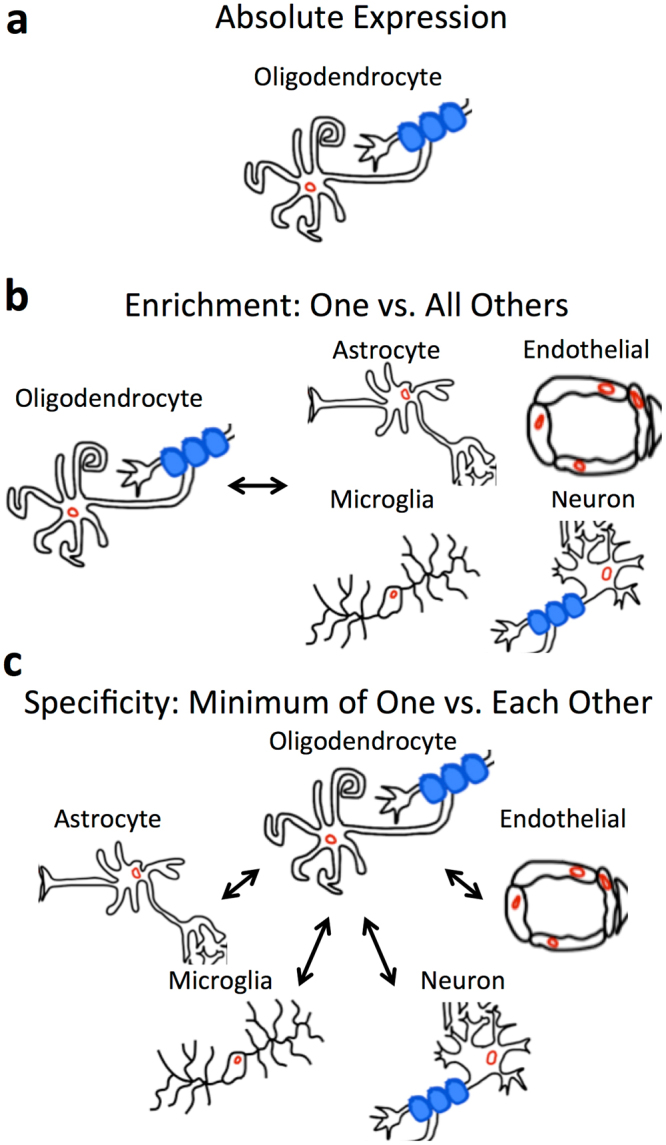
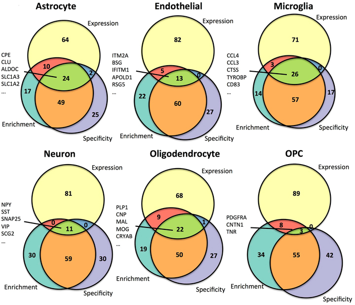
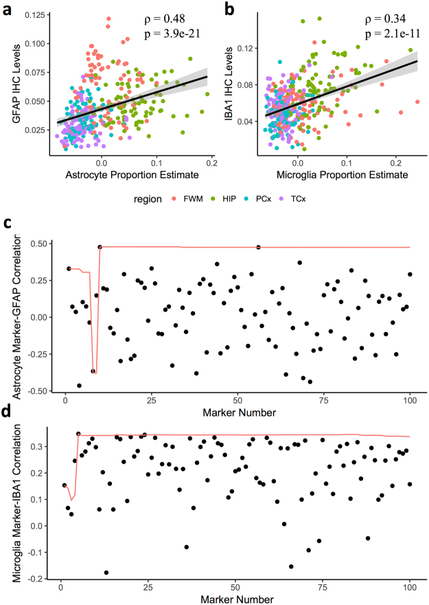
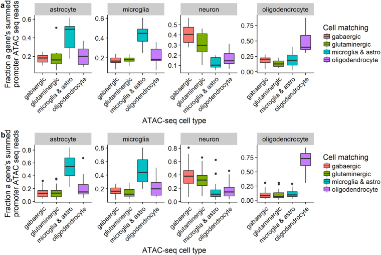
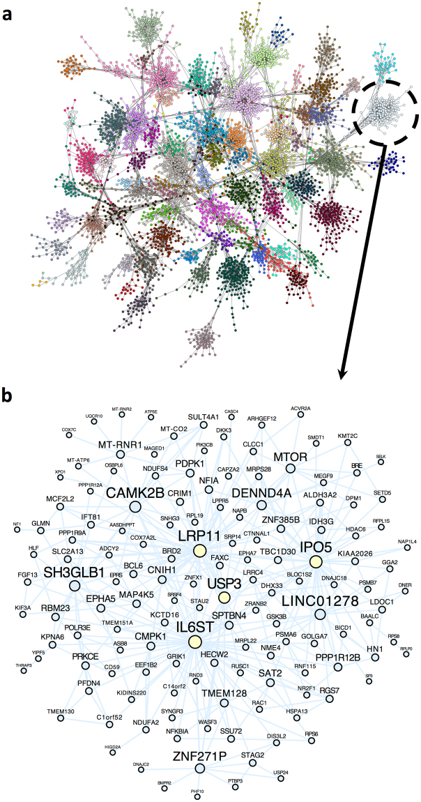
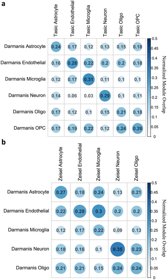
Elucidating brain cell type specific gene expression patterns is critical towards a better understanding of how cell-cell communications may influence brain functions and dysfunctions. We set out to compare and contrast five human and murine cell type-specific transcriptome-wide RNA expression data sets that were generated within the past several years. We defined three measures of brain cell type-relative expression including specificity, enrichment, and absolute expression and identified corresponding consensus brain cell “signatures,” which were well conserved across data sets. We validated that the relative expression of top cell type markers are associated with proxies for cell type proportions in bulk RNA expression data from postmortem human brain samples. We further validated novel marker genes using an orthogonal ATAC-seq dataset. We performed multiscale coexpression network analysis of the single cell data sets and identified robust cell-specific gene modules. To facilitate the use of the cell type-specific genes for cell type proportion estimation and deconvolution from bulk brain gene expression data, we developed an R package, BRETIGEA. In summary, we identified a set of novel brain cell consensus signatures and robust networks from the integration of multiple datasets and therefore transcend limitations related to technical issues characteristic of each individual study.
Overview
The claims and statistics of this paper haven’t been checked yet.
Run the check — free, no account needed. It reads the open-access text and checks the sources it cites.
Integrity signals
Registry
Cites
Statistics
Figures
Verification
Claims the paper makes
The claims and statistics of this paper haven’t been checked yet.
Run the check — free, no account needed. It reads the open-access text and checks the sources it cites.
Claims other papers make about this one
Each is checked against this paper’s own text: does it say what the citing paper says it does?
The source backs part of the claim, not all of it — or backs it for a different population, condition or comparison.
In the citing paper
“The cell clusters were annotated manually using marker genes that were conserved between both conditions as astrocytes, microglia, neurons, endothelial cells, ependymal cells, and mixed glia (Supplementary Data 1) (Arneson et al., 2018;McKenzie et al., 2018).”
From Single-cell resolution analysis of the crosstalk between chemogenically activated astrocytes and microglia (2020) · cites this paper as McKenzie et al., 2018
Experimental — accuracy not yet measuredThe cited source says what the claim attributes to it.
In the citing paper
“Canonical CNS cell type markers were compiled from previous studies66–69.”
From APOE4 to APOE2 allelic switching in mice improves Alzheimer’s disease-related metabolic signatures, neuropathology and cognition (2025) · cites this paper as [66]
The cited source says what the claim attributes to it.
In the citing paper
“To estimate the cell-type proportion of bulk tissue RNA-seq data used in this study, we perform a cell-type deconvolution on each sample using the brain cell–type marker signatures provided by the BRETIGEA R package (26).”
From Molecular subtyping of Alzheimer’s disease using RNA sequencing data reveals novel mechanisms and targets (2021) · cites this paper as [26]
The cited source says what the claim attributes to it.
In the citing paper
“All cell clusters were annotated manually based on marker genes that were conserved between the saccharin group and the CNO group and gene lists present in literature (Arneson et al., 2018;McKenzie et al., 2018).”
From Single-cell resolution analysis of the crosstalk between chemogenically activated astrocytes and microglia (2020) · cites this paper as McKenzie et al., 2018
The cited source says what the claim attributes to it.
In the citing paper
“We used a consensus top ranked cell type-specific marker set from a recent meta-analysis of multiple cell type-specific and single cell RNAseq datasets (McKenzie et al., 2018).”
From Cholesterol and matrisome pathways dysregulated in astrocytes and microglia (2022) · cites this paper as McKenzie et al., 2018
Self-check
Percentages, sums and reported differences re-computed from the paper’s own numbers.
The self-check needs the paper’s full text.
Citations
Cites
Controlling the False Discovery Rate: A Practical and Powerful Approach to Multiple Testing
Benjamini, Y., & Hochberg, Y. (1995) · Journal of the Royal Statistical Society Series B: Statistical Methodology · doi:10.1111/j.2517-6161.1995.tb02031.x · Cited by 109,482
STAR: ultrafast universal RNA-seq aligner
Dobin, A., Davis, C. A., Schlesinger, F., Drenkow, J., Zaleski, C., Jha, S., Batut, P., Chaisson, M., & Gingeras, T. R. (2013) · Bioinformatics · doi:10.1093/bioinformatics/bts635 · Cited by 51,709
Robinson, M. D., McCarthy, D. J., & Smyth, G. K. (2010) · Bioinformatics · doi:10.1093/bioinformatics/btp616 · Cited by 41,364
featureCounts: an efficient general purpose program for assigning sequence reads to genomic features
Liao, Y., Smyth, G. K., & Shi, W. (2014) · Bioinformatics · doi:10.1093/bioinformatics/btt656 · Cited by 27,219
Tissue-based map of the human proteome
Uhlén, M., Fagerberg, L., Hallström, B. M., Lindskog, C., Oksvold, P., Mardinoglu, A., Sivertsson, Å., Kampf, C., Sjöstedt, E., Asplund, A., et al. (2015) · Science · doi:10.1126/science.1260419 · Cited by 15,286
Highly parallel genome-wide expression profiling of individual cells using nanoliter droplets.
Macosko, E. Z., Basu, A., Satija, R., Nemesh, J., Shekhar, K., Goldman, M., Tirosh, I., Bialas, A. R., Kamitaki, N., Martersteck, E. M., et al. (2015) · Cell · doi:10.1016/j.cell.2015.05.002 · Cited by 7,110
Astrocytes: Biology and pathology
Sofroniew, M. V., & Vinters, H. V. (2010) · Acta Neuropathologica · doi:10.1007/s00401-009-0619-8 · Cited by 5,196
Zhang, Y., Chen, K., Sloan, S. A., Bennett, M. L., Scholze, A. R., O'Keeffe, S., Phatnani, H. P., Guarnieri, P., Caneda, C., Ruderisch, N., et al. (2014) · The Journal of Neuroscience · doi:10.1523/jneurosci.1860-14.2014 · Cited by 4,875
Cahoy, J. D., Emery, B., Kaushal, A., Foo, L. C., Zamanian, J. L., Christopherson, K. S., Xing, Y., Lubischer, J. L., Krieg, P. A., Krupenko, S. A., et al. (2008) · The Journal of Neuroscience · doi:10.1523/jneurosci.4178-07.2008 · Cited by 3,129
Cell types in the mouse cortex and hippocampus revealed by single-cell RNA-seq
Zeisel, A., Muñoz-Manchado, A. B., Codeluppi, S., Lönnerberg, P., La Manno, G., Juréus, A., Marques, S., Munguba, H., He, L., Betsholtz, C., et al. (2015) · Science · doi:10.1126/science.aaa1934 · Cited by 3,124
Cited by
Sorted by citations
Grubman, A., Chew, G., Ouyang, J. F., Sun, G., Choo, X. Y., McLean, C., Simmons, R. K., Buckberry, S., Vargas-Landin, D. B., Poppe, D., et al. (2019) · Nature Neuroscience · doi:10.1038/s41593-019-0539-4 · Cited by 956
Spatial Transcriptomics and In Situ Sequencing to Study Alzheimer’s Disease
Chen, W.-T., Lu, A., Craessaerts, K., Pavie, B., Sala Frigerio, C., Corthout, N., Qian, X., Laláková, J., Kühnemund, M., Voytyuk, I., et al. (2020) · Cell · doi:10.1016/j.cell.2020.06.038 · Cited by 949
Johnson, E. C. B., Dammer, E. B., Duong, D. M., Ping, L., Zhou, M., Yin, L., Higginbotham, L. A., Guajardo, A., White, B., Troncoso, J. C., et al. (2020) · Nature Medicine · doi:10.1038/s41591-020-0815-6 · Cited by 1,001
Self-citationCholesterol and matrisome pathways dysregulated in astrocytes and microglia
TCW, J., Qian, L., Pipalia, N. H., Chao, M. J., Liang, S. A., Shi, Y., Jain, B. R., Bertelsen, S. E., Kapoor, M., Marcora, E., et al. (2022) · Cell · doi:10.1016/j.cell.2022.05.017 · Cited by 411
Self-citationZhao, J., Fu, Y., Yamazaki, Y., Ren, Y., Davis, M. D., Liu, C.-C., Lu, W., Wang, X., Chen, K., Cherukuri, Y., et al. (2020) · Nature Communications · doi:10.1038/s41467-020-19264-0 · Cited by 374
Transcriptional signature in microglia associated with Aβ plaque phagocytosis
Grubman, A., Choo, X. Y., Chew, G., Ouyang, J. F., Sun, G., Croft, N. P., Rossello, F. J., Simmons, R., Buckberry, S., Landin, D. V., et al. (2021) · Nature Communications · doi:10.1038/s41467-021-23111-1 · Cited by 357
Transcriptomic and cellular decoding of regional brain vulnerability to neurogenetic disorders
Seidlitz, J., Nadig, A., Liu, S., Bethlehem, R. A. I., Vértes, P. E., Morgan, S. E., Váša, F., Romero-García, R., Lalonde, F. M., CLASEN, L. S., et al. (2020) · Nature Communications · doi:10.1038/s41467-020-17051-5 · Cited by 349
Neff, R. A., Wang, M., Vatansever, S., Guo, L., Ming, C., Wang, Q., Wang, E., Horgusluoglu-Moloch, E., Song, W.-M., Li, A., et al. (2021) · Science Advances · doi:10.1126/sciadv.abb5398 · Cited by 306
“Previous work by our laboratory has shown that brain cell–type surrogate proportion values (SPVs), including the proportion of neurons in a sample, can be inferred from bulk RNA-seq data when combined with measurements of brain cell type–specific gene expression patterns (26) and may serve as a marker of neuronal loss…”
BackgroundMethodInfluentialSelf-citationTranscriptome analysis reveals tumor microenvironment changes in glioblastoma
Hoogstrate, Y., Draaisma, K., Ghisai, S. A., van Hijfte, L., Barin, N., de Heer, I., Coppieters, W., van den Bosch, T. P. P., Bolleboom, A., Gao, Z., et al. (2023) · Cancer Cell · doi:10.1016/j.ccell.2023.02.019 · Cited by 240
“To assess whether upregulation of C4 and C3 originates from the heterogeneously tumor-intrinsic NPC- and OPC-like meta-features, we compared their expression with independent single-cell data, annotated using celltype-specific marker genes.(17) For this we sequenced and analyzed glioblastoma snRNA-seq samples from the…”
MethodSingle-nucleus RNA-seq identifies Huntington disease astrocyte states
Al-Dalahmah, O., Sosunov, A. A., Shaik, A., Ofori, K., Liu, Y., Vonsattel, J. P., Adorjan, I., Menon, V., & Goldman, J. E. (2020) · Acta Neuropathologica Communications · doi:10.1186/s40478-020-0880-6 · Cited by 264
“used to classify nuclei are based on the literature [36, 63], modified, and are provided (Additional file 2: Table S1).”
MethodMapping gene transcription and neurocognition across human neocortex
Hansen, J. Y., Markello, R. D., Vogel, J. W., Seidlitz, J., Bzdok, D., & Mišić, B. (2021) · Nature Human Behaviour · doi:10.1038/s41562-021-01082-z · Cited by 208
Lee, S., Devanney, N. A., Golden, L. R., Smith, C. T., Schwartz, J. L., Walsh, A. E., Clarke, H. A., Goulding, D. S., Allenger, E. J., Morillo-Segovia, G., et al. (2023) · Cell Reports · doi:10.1016/j.celrep.2023.112196 · Cited by 203
Ullman, J. C., Arguello, A., Getz, J. A., Bhalla, A., Mahon, C. S., Wang, J., Giese, T., Bedard, C., Kim, D. J., Blumenfeld, J. R., et al. (2020) · Science Translational Medicine · doi:10.1126/scitranslmed.aay1163 · Cited by 205
A single-nuclei RNA sequencing study of Mendelian and sporadic AD in the human brain
Del-Aguila, J. L., Li, Z., Dube, U., Mihindukulasuriya, K. A., Budde, J. P., Fernandez, M. V., Ibanez, L., Bradley, J., Wang, F., Bergmann, K., et al. (2019) · Alzheimer's Research & Therapy · doi:10.1186/s13195-019-0524-x · Cited by 183
Wang, M., Li, A., Sekiya, M., Beckmann, N. D., Quan, X., Schrode, N., Fernando, M. B., Yu, A., Zhu, L., Cao, J., et al. (2021) · Neuron · doi:10.1016/j.neuron.2020.11.002 · Cited by 185
“type used the top 500 ranked consensus cell type-specific genes derived from a metaanalysis of 5 cell type-specific or single cell RNA-seq datasets (McKenzie et al., 2018).”
InfluentialSelf-citationTrapecar, M., Wogram, E., Svoboda, D., Communal, C., Omer, A., Lungjangwa, T., Sphabmixay, P., Velazquez, J., Schneider, K., Wright, C. W., et al. (2021) · Science Advances · doi:10.1126/sciadv.abd1707 · Cited by 176
Maitra, M., Mitsuhashi, H., Rahimian, R., Chawla, A., Yang, J., Fiori, L. M., Davoli, M. A., Perlman, K., Aouabed, Z., Mash, D. C., et al. (2023) · Nature Communications · doi:10.1038/s41467-023-38530-5 · Cited by 168
Single-cell-resolution transcriptome map of human, chimpanzee, bonobo, and macaque brains
Khrameeva, E., Kurochkin, I., Han, D., Guijarro, P., Kanton, S., Santel, M., Qian, Z., Rong, S., Mazin, P., Sabirov, M., et al. (2020) · Genome Research · doi:10.1101/gr.256958.119 · Cited by 162
“Functional changes acquired on the human lineage are likely to be mediated by alterations in gene expression and cell-type composition in particular brain structures (O ’ Bleness et al. 2012; Sousa et al. 2017b; McKenzie et al. 2018).”
BackgroundInfluentialReceptor-mediated transcytosis for brain delivery of therapeutics: receptor classes and criteria
Haqqani, A. S., Bélanger, K., & Stanimirovic, D. B. (2024) · Frontiers in Drug Delivery · doi:10.3389/fddev.2024.1360302 · Cited by 153
A Parkinson's disease CircRNAs Resource reveals a link between circSLC8A1 and oxidative stress
Hanan, M., Simchovitz, A., Yayon, N., Vaknine, S., Cohen‐Fultheim, R., Karmon, M., Madrer, N., Rohrlich, T. M., Maman, M., Bennett, E. R., et al. (2020) · EMBO Molecular Medicine · doi:10.15252/emmm.201911942 · Cited by 130
Buchholz, S., & Zempel, H. (2024) · Alzheimer s & Dementia · doi:10.1002/alz.13784 · Cited by 154
Astrocyte transcriptomic changes along the spatiotemporal progression of Alzheimer’s disease
Serrano-Pozo, A., Li, H., Li, Z., Muñoz-Castro, C., Jaisa-aad, M., Healey, M. A., Welikovitch, L. A., Jayakumar, R., Bryant, A. G., Noori, A., et al. (2024) · Nature Neuroscience · doi:10.1038/s41593-024-01791-4 · Cited by 140
Seney, M. L., Kim, S.-M., Glausier, J. R., Hildebrand, M. A., Xue, X., Zong, W., Wang, J., Shelton, M. A., Phan, B. N., Srinivasan, C., et al. (2021) · Biological Psychiatry · doi:10.1016/j.biopsych.2021.06.007 · Cited by 135
“To estimate cell-type fractions from bulk RNA-seq, Digital Sorting Algorithm (31) deconvolved astrocytes, endothelial cells, microglia, neurons, and oligodendrocytes from BRETIGEA (32).”
Hanan, M., Simchovitz, A., Yayon, N., Vaknine, S., Cohen‐Fultheim, R., Karmon, M., Madrer, N., Rohrlich, T. M., Maman, M., Bennett, E. R., et al. (2020) · EMBO Molecular Medicine · doi:10.15252/emmm.202013551 · Cited by 128
“We next utilized single-cell RNA sequencing data from human brains to determine the cell types expressing these DE genes (McKenzie et al , 2018).”
SARS-CoV-2 promotes microglial synapse elimination in human brain organoids
Samudyata, S., Oliveira, A. O., Malwade, S., de Sousa, N. R., Goparaju, S. K., Gracias, J., Orhan, F., Steponaviciute, L., Schalling, M., Sheridan, S. D., et al. (2022) · Molecular Psychiatry · doi:10.1038/s41380-022-01786-2 · Cited by 125
Single cell transcriptome profiling of the human alcohol-dependent brain
Brenner, E., Tiwari, G. R., Kapoor, M., Liu, Y., Brock, A., & Mayfield, R. D. (2020) · Human Molecular Genetics · doi:10.1093/hmg/ddaa038 · Cited by 118
Decoding the Transcriptional Response to Ischemic Stroke in Young and Aged Mouse Brain
Androvic, P., Kirdajova, D., Tureckova, J., Zucha, D., Rohlova, E., Abaffy, P., Kriska, J., Valny, M., Anderova, M., Kubista, M., et al. (2020) · Cell Reports · doi:10.1016/j.celrep.2020.107777 · Cited by 118
Tau as a Biomarker of Neurodegeneration
Holper, S., Watson, R., & Yassi, N. (2022) · International Journal of Molecular Sciences · doi:10.3390/ijms23137307 · Cited by 121
“Under physiological conditions, tau exists in multiple neuronal compartments [1], is present at basal levels in the brain’s interstitial fluid [2], is expressed by several glial cell types [3], and has various non-microtubule-associated functions [4].”
BackgroundElyanow, R., Dumitrascu, B., Engelhardt, B. E., & Raphael, B. J. (2020) · Genome Research · doi:10.1101/gr.251603.119 · Cited by 106
Deconvolving the contributions of cell-type heterogeneity on cortical gene expression
Patrick, E., Taga, M. F., Ergün, A., Ng, B., Casazza, W., Cimpean, M., Yung, C. J., Schneider, J. A., Bennett, D. A., Gaiteri, C., et al. (2020) · PLoS Computational Biology · doi:10.1371/journal.pcbi.1008120 · Cited by 101
Glioblastoma epigenome profiling identifies SOX10 as a master regulator of molecular tumour subtype
Wu, Y., Fletcher, M., Gu, Z., Wang, Q., Costa, B., Bertoni, A., Man, K.-H., Schlotter, M., Felsberg, J., Mangei, J., et al. (2020) · Nature Communications · doi:10.1038/s41467-020-20225-w · Cited by 101
Wang, M., Song, W., Chen, M., Wang, Q., Zhou, X., Xu, P., Krek, A., Yoon, Y., Ho, L., Orr, M. E., et al. (2022) · Molecular Neurodegeneration · doi:10.1186/s13024-022-00517-z · Cited by 104
“This is exemplified by VCAN, an oligodendrocyte progenitor cell marker [143], and APOE, whose polymorphism is a major genetic risk determinant of AD [144] and a marker for astrocyte and activated microglia [8] (Fig.”
BackgroundSelf-citationAssociation of a common genetic variant with Parkinson’s disease is mediated by microglia
Langston, R. G., Beilina, A., Reed, X., Kaganovich, A., Singleton, A. B., Blauwendraat, C., Gibbs, J. R., & Cookson, M. R. (2022) · Science Translational Medicine · doi:10.1126/scitranslmed.abp8869 · Cited by 93
Reid, M. J., Beltran-Lobo, P., Johnson, L., Perez‐Nievas, B. G., & Noble, W. J. (2020) · Frontiers in Neurology · doi:10.3389/fneur.2020.572850 · Cited by 98
GJA1 (connexin43) is a key regulator of Alzheimer’s disease pathogenesis
Kajiwara, Y., Wang, E., Wang, M., Sin, W. C., Brennand, K., Schadt, E. E., Naus, C. C. G., Buxbaum, J. D., & Zhang, B. (2018) · Acta Neuropathologica Communications · doi:10.1186/s40478-018-0642-x · Cited by 99
Self-citationSpatial transcriptomic clocks reveal cell proximity effects in brain ageing
Sun, E., Zhou, O. Y., Hauptschein, M., Rappoport, N., Xu, L., Negredo, P. N., Liu, L. A., Rando, T. A., Zou, J., & Brunet, A. (2024) · Nature · doi:10.1038/s41586-024-08334-8 · Cited by 106
“The cell-type and subtype marker genes included all markers in a suggested panel by Vizgen; markers determined from literature review focused on cells of the brain vasculature 5,82,83 , NSCs and neuro-blasts 3,5,84,85 , and immune cells 3,5,86,87 .”
Midbrain organoids with an SNCA gene triplication model key features of synucleinopathy
Mohamed, N., Sirois, J., Ramamurthy, J., Mathur, M., Lépine, P., Deneault, É., Maussion, G., Nicouleau, M., Chen, C. X. ‐., Abdian, N., et al. (2021) · Brain Communications · doi:10.1093/braincomms/fcab223 · Cited by 91
“Clusters were annotated using a combination of automated annotation (scCATCH,(37) ClustifyR(38) and SingleR(39)) and Seurat’s FindMarkers function, the top20 markers were selected for each cluster and cross referenced to expression databases manually.(40,41) Clusters were annotated from 14 clusters; some groups of neu…”
MethodHartl, C., Ramaswami, G., Pembroke, W. G., Muller, S., Pintacuda, G., Saha, A., Parsana, P., Battle, A. J., Lage, K., & Geschwind, D. H. (2021) · Nature Neuroscience · doi:10.1038/s41593-021-00887-5 · Cited by 86
Reemst, K., Kracht, L., Kotah, J. M., Rahimian, R., van Irsen, A. A. S., Sotomayor, G. C., Verboon, L. N., Brouwer, N., Simard, S., Turecki, G., et al. (2022) · Translational Psychiatry · doi:10.1038/s41398-022-02265-6 · Cited by 80
“The exact implications of this differential trajectory for ELS microglia remain to be determined, but it might underlie how ELS modulates the response to later-life challenges, such as other forms of stress and infection reported by others and us [27, 31, 32, 82, 87].”
BackgroundIturria‐Medina, Y., Khan, A. F., Adewale, Q., Shirazi, A. H., & Initiative, T. A. D. N. (2019) · Brain · doi:10.1093/brain/awz400 · Cited by 79
Gleichman, A. J., Kawaguchi, R., Sofroniew, M. V., & Carmichael, S. T. (2023) · Nature Communications · doi:10.1038/s41467-023-42746-w · Cited by 82
Cell-type-specific epigenetic effects of early life stress on the brain
Rahman, M. F., & McGowan, P. O. (2022) · Translational Psychiatry · doi:10.1038/s41398-022-02076-9 · Cited by 75
“In the brain, the primary categorization of cell types includes neurons, glia (microglia, astrocytes, oligodendrocytes), pericytes and brain epithelial cells [89, 90], however cells in these broad categories can be subdivided based on cellular functions.”
BackgroundNeurophysiological signatures of cortical micro-architecture
Shafiei, G., Fulcher, B., Voytek, B., Satterthwaite, T. D., Baillet, S., & Mišić, B. (2023) · Nature Communications · doi:10.1038/s41467-023-41689-6 · Cited by 81
Delbridge, A. R. D., Huh, D., Brickelmaier, M., Burns, J. C., Roberts, C., Challa, R., Raymond, N., Cullen, P., Carlile, T. M., Ennis, K. A., et al. (2020) · Frontiers in Cellular Neuroscience · doi:10.3389/fncel.2020.592005 · Cited by 72
“Supporting these observations the expression of published cell-type marker genesets (McKenzie et al., 2018) revealed reduced expression of neuronal marker genes over time and, conversely, an increase in expression of microglia marker genes (Figure 1C).”
BackgroundMethodInfluentialX-chromosome regulation and sex differences in brain anatomy
Raznahan, A., & Distèche, C. M. (2020) · Neuroscience & Biobehavioral Reviews · doi:10.1016/j.neubiorev.2020.10.024 · Cited by 69
“specific cell types across species(Hodge et al., 2019; McKenzie et al., 2018).”
Deciphering cellular transcriptional alterations in Alzheimer’s disease brains
Wang, X., Allen, M., Li, S., Quicksall, Z., Patel, T., Carnwath, T., Reddy, J. S., Carrasquillo, M. M., Lincoln, S., Nguyen, T., et al. (2020) · Molecular Neurodegeneration · doi:10.1186/s13024-020-00392-6 · Cited by 65
Brain Organoids as Tools for Modeling Human Neurodevelopmental Disorders
Adams, J. W., Cugola, F. R., & Muotri, A. R. (2019) · Physiology · doi:10.1152/physiol.00005.2019 · Cited by 68
Gupta, C., Chandrashekar, P. B., Jin, T., He, C., Khullar, S., Chang, Q., & Wang, D. (2022) · Journal of Neurodevelopmental Disorders · doi:10.1186/s11689-022-09438-w · Cited by 66
“Gene coexpression networks have been applied to study the human brain at bulk and single-cell levels [153, 154] and have various applications in neurological and neuropsychiatric conditions (reviewed in [155, 156]).”
BackgroundPatel, T., Carnwath, T., Wang, X., Allen, M., Lincoln, S., Lewis‐Tuffin, L. J., Quicksall, Z., Lin, S., Tutor‐New, F. Q., Ho, C. C. G., et al. (2022) · Aging Cell · doi:10.1111/acel.13606 · Cited by 61
Zhao, J., Lu, W., Ren, Y., Fu, Y., Martens, Y. A., Shue, F., Davis, M. D., Wang, X., Chen, K., Li, F., et al. (2021) · Acta Neuropathologica · doi:10.1007/s00401-021-02361-9 · Cited by 58
“DEGs cell‑type distribution analysis of RNA‑seq data Cell proportion was estimated using the CIBERSORT program [45] and marker genes described in BRETIGEA [42].”
BackgroundMethodInfluentialInfants’ cortex undergoes microstructural growth coupled with myelination during development
Natu, V. S., Rosenke, M., Wu, H., Querdasi, F. R., Kular, H., Lopez-Alvarez, N., Grotheer, M., Berman, S., Mezer, A., & Kalanit Grill-spector, A. (2021) · Communications Biology · doi:10.1038/s42003-021-02706-w · Cited by 58
Glial mitochondrial function and dysfunction in health and neurodegeneration
McAvoy, K., & Kawamata, H. (2019) · Molecular and Cellular Neuroscience · doi:10.1016/j.mcn.2019.103417 · Cited by 59
Belonwu, S., Li, Y., Bunis, D. G., Rao, A. A., Solsberg, C. W., Tang, A., Fragiadakis, G. K., Dubal, D. B., Oskotsky, T., & Sirota, M. (2021) · Molecular Neurobiology · doi:10.1007/s12035-021-02591-8 · Cited by 55
Extralysosomal cathepsin B in central nervous system: Mechanisms and therapeutic implications
Ni, J., Lan, F., Xu, Y. C., Nakanishi, H., & Li, X. (2022) · Brain Pathology · doi:10.1111/bpa.13071 · Cited by 56
The long noncoding RNA FEDORA is a cell type– and sex-specific regulator of depression
Issler, O., van der Zee, Y. Y., Ramakrishnan, A., Xia, S.-H., Zinsmaier, A. K., Tan, C., Li, W., Browne, C. J., Walker, D. M., Salery, M., et al. (2022) · Science Advances · doi:10.1126/sciadv.abn9494 · Cited by 51
Browne, C. J., Futamura, R., Minier-Toribio, A. M., Hicks, E. M., Ramakrishnan, A., Martínez-Rivera, F. J., Estill, M. S., Godino, A., Parise, E. M., Torres‐Berrío, A., et al. (2023) · Science Advances · doi:10.1126/sciadv.adg8558 · Cited by 51
Self-citationHoll, D., Hau, W. F., Julien, A., Banitalebi, S., Kalkitsas, J., Savant, S., Llorens-Bobadilla, E., Hérault, Y., Pavlovic, G., Amiry-Moghaddam, M. R., et al. (2024) · Nature Neuroscience · doi:10.1038/s41593-024-01678-4 · Cited by 52
“To classify the sorted populations, we used marker profiles for pericytes, fibroblasts, vSMCs, astrocytes, oligodendrocytes and oligodendrocyte progenitor cells 19 and compared marker gene expression between clusters.”
Milind, N., Preuß, C., Haber, A., Ananda, G., Mukherjee, S., Cai, J., Shapley, S. M., Tyler, A. L., Logsdon, B. A., Crane, P. K., et al. (2020) · PLoS Genetics · doi:10.1371/journal.pgen.1008775 · Cited by 51
Consequences and opportunities arising due to sparser single-cell RNA-seq datasets
Bouland, G. A., Mahfouz, A. E., & Reinders, M. J. T. (2023) · Genome biology · doi:10.1186/s13059-023-02933-w · Cited by 52
“the count based PCs, in which cells are colored according to the binary representation of the marker genes AQP4 (E) and TNR (H) which are known markers for astrocytes and OPCs respectively [21].”
BackgroundMethodAgboola, O. S., Hu, X., Shan, Z., Wu, Y., & Lei, L. (2021) · Stem Cell Research & Therapy · doi:10.1186/s13287-021-02369-8 · Cited by 53
Patel, D., Zhang, X., Farrell, J. J., Chung, J., Stein, T. D., Lunetta, K. L., & Farrer, L. A. (2021) · Translational Psychiatry · doi:10.1038/s41398-021-01373-z · Cited by 51
McFarland, K. N., & Chakrabarty, P. (2022) · Neurotherapeutics · doi:10.1007/s13311-021-01179-3 · Cited by 48
“Furthermore, multiple deconvolution methods utilize results from scRNA-seq or snRNA-seq studies to identify cell-specific changes within bulk gene expression datasets [142, 143].”
MethodDay-night and seasonal variation of human gene expression across tissues
Wucher, V., Sodaei, R., Amador, R., Irimia, M., & Guigó, R. (2023) · PLoS Biology · doi:10.1371/journal.pbio.3001986 · Cited by 51
“…changes in human brain’s cytoarchitecture, we used gene expression profiles of cell type–specific markers for a variety of brain cell types [59]: astrocytes, neurons, oligodendrocytes, microglia, and endothelial cells ( A ) Distribution of the number of tissues (x-axis, log 10 scale) in…”
İş, Ö., Wang, X., Reddy, J. S., Min, Y., Yılmaz, E., Bhattarai, P., Patel, T., Bergman, J., Quicksall, Z., Heckman, M. G., et al. (2024) · Nature Communications · doi:10.1038/s41467-024-48926-6 · Cited by 55
Layer-By-Layer: The Case for 3D Bioprinting Neurons to Create Patient-Specific Epilepsy Models
Antill‐O'Brien, N., Bourke, J., & O’Connell, C. D. (2019) · Materials · doi:10.3390/ma12193218 · Cited by 49
“There are thought to be 50–250 neuronal subtypes [164].”
BackgroundConinx, E., Chew, Y. C., Yang, X., Guo, W., Coolkens, A., Baatout, S., Moons, L., Verslegers, M., & Quintens, R. (2020) · Aging · doi:10.18632/aging.104056 · Cited by 46
Cell type-specific potential pathogenic genes and functional pathways in Alzheimer’s Disease
Wang, X., & Li, L. (2021) · BMC Neurology · doi:10.1186/s12883-021-02407-1 · Cited by 47
Type I interferon shapes brain distribution and tropism of tick-borne flavivirus
Chotiwan, N., Rosendal, E., Willekens, S. M. A., Schexnaydre, E. E., Nilsson, E., Lindqvist, R., Hahn, M., Mihai, I. S., Morini, F., Zhang, J., et al. (2023) · Nature Communications · doi:10.1038/s41467-023-37698-0 · Cited by 43
Dang, M. T., Gonzalez, M. V., Gaonkar, K. S., Rathi, K. S., Young, P. A., Arif, S., Zhai, L., Alam, Z., Devalaraja, S., To, T. K. J., et al. (2021) · Cell Reports · doi:10.1016/j.celrep.2021.108917 · Cited by 45
“We ran function findCells() using SVD method to calculate SPVs and all 1000 marker genes for brain cell types provided in BRETIGEA package along with 317 genes for monocyte.”
MethodInfluentialBarkBase: Epigenomic Annotation of Canine Genomes
Megquier, K., Genereux, D. P., Hekman, J. P., Swofford, R., Turner-Maier, J., Johnson, J., Alonso, J., Li, X., Morrill, K. M., Anguish, L. J., et al. (2019) · Genes · doi:10.3390/genes10060433 · Cited by 42
“This is consistent with the tight clustering of cerebellum samples as a distinct clade on our tissue dendrogram (Figure 5A) and with previous reports that cerebellum is a complex tissue composed of a large number of cell types with transcriptomic profiles distinct from those in other organs [96].”
ResultHuang, Y., Wang, M., Ni, H., Zhang, J., Li, A., Hu, B., Junqueira Alves, C., Wahane, S., Rios de Anda, M., Ho, L., et al. (2024) · Nature Neuroscience · doi:10.1038/s41593-024-01664-w · Cited by 42
“78) and the Molecular Signatures Database (MSigDB) 79,80 with Fisher exact test.”
Self-citationCell-type-specific expression of tRNAs in the brain regulates cellular homeostasis
Kapur, M., Molumby, M. J., Guzman, C., Heinz, S., & Ackerman, S. L. (2024) · Neuron · doi:10.1016/j.neuron.2024.01.028 · Cited by 43
Kirmiz, M., Gillies, T. E., Dickson, E. J., & Trimmer, J. S. (2019) · Journal of Biological Chemistry · doi:10.1074/jbc.ra119.007635 · Cited by 40
Exploring lithium’s transcriptional mechanisms of action in bipolar disorder: a multi-step study
Akkouh, I. A., Skrede, S., Holmgren, A., Ersland, K. M., Hansson, L. J., Bahrami, S., Andreassen, O. A., Steen, V. M., Djurovic, S., & Hughes, T. (2019) · Neuropsychopharmacology · doi:10.1038/s41386-019-0556-8 · Cited by 39
“Each of the marker genes was assigned a cell-type specific expression value based on three transcriptome-wide RNA expression murine data sets [18].”
Astrocyte Molecular Clock Function in the Nucleus Accumbens Is Important for Reward-Related Behavior
Becker‐Krail, D. D., Ketchesin, K. D., Burns, J. N., Zong, W., Hildebrand, M. A., DePoy, L. M., Vadnie, C. A., Tseng, G. C., Logan, R. W., Huang, Y. H., et al. (2022) · Biological Psychiatry · doi:10.1016/j.biopsych.2022.02.007 · Cited by 44
Forrest, S. L., Lee, S., Nassir, N., Martínez-Valbuena, I., Sackmann, V., Li, J., Ahmed, A., Tartaglia, M. C., Ittner, L. M., Lang, A. E., et al. (2023) · Acta Neuropathologica · doi:10.1007/s00401-023-02604-x · Cited by 44
van Hijfte, L., Geurts, M., Vallentgoed, W., Eilers, P. H. C., Smitt, P. A. E. S., Debets, R., & French, P. J. (2022) · iScience · doi:10.1016/j.isci.2022.105760 · Cited by 41
“13 For T cells, a separate list of 48 marker genes was used.”
BackgroundMethodMultiscale proteomic modeling reveals protein networks driving Alzheimer’s disease pathogenesis
Wang, E., Yu, K., Cao, J., Wang, M., Katsel, P., Song, W., Wang, Z., Li, Y., Wang, X., Wang, Q., et al. (2025) · Cell · doi:10.1016/j.cell.2025.08.038 · Cited by 45
Self-citationLi, H., Funk, C. C., McFarland, K. N., Dammer, E. B., Allen, M., Carrasquillo, M. M., Levites, Y., Chakrabarty, P., Burgess, J. D., Wang, X., et al. (2021) · Alzheimer s & Dementia · doi:10.1002/alz.12254 · Cited by 42
Mendez, E. F., Wei, H., Hu, R., Stertz, L., Fries, G. R., Wu, X., Najera, K., Monterey, M. D., Lincoln, C. M. M., Kim, J.-W., et al. (2021) · Molecular Psychiatry · doi:10.1038/s41380-021-01259-y · Cited by 40
“The R package BRETIGEA (20) was used to compute relative cell type proportion surrogate variables for the same cell types, using the top 50 cell-type specific genes for each cell type (Supplementary Table 2).”
InfluentialSadeghi, I., Gispert, J. D., Palumbo, E., Muñoz-Aguirre, M., Wucher, V., D’Argenio, V., Santpere, G., Navarro, A., Guigó, R., & Vilor‐Tejedor, N. (2022) · Computational and Structural Biotechnology Journal · doi:10.1016/j.csbj.2022.08.037 · Cited by 42
Chucair‐Elliott, A. J., Ocañas, S. R., Stanford, D. R., Ansere, V. A., Buettner, K. B., Porter, H. L., Eliason, N. L., Reid, J. J., Sharpe, A. L., Stout, M. B., et al. (2020) · Communications Biology · doi:10.1038/s42003-020-01418-x · Cited by 39
Aditi, A., Downing, S. M., Schreiner, P. A., Kwak, Y. D., Li, Y., Shaw, T. I., Russell, H. R., & McKinnon, P. J. (2021) · Neuron · doi:10.1016/j.neuron.2021.09.040 · Cited by 42
Multi-omics signatures of alcohol use disorder in the dorsal and ventral striatum
Zillich, L., Poisel, E., Frank, J., Foo, J. C., Friske, M. M., Streit, F., Sirignano, L., Heilmann‐Heimbach, S., Heimbach, A., Hoffmann, P., et al. (2022) · Translational Psychiatry · doi:10.1038/s41398-022-01959-1 · Cited by 39
DNA damage response in neurodevelopment and neuromaintenance
Qing, X., Zhang, G., & Wang, Z. (2022) · FEBS Journal · doi:10.1111/febs.16535 · Cited by 42
Heo, G., Xu, Y., Wang, E., Ali, M., Oh, H. S., Losada, P. M., Anastasi, F., Escalante, A. G., Puerta, R., Song, S., et al. (2025) · Nature Aging · doi:10.1038/s43587-025-00872-8 · Cited by 51
“The specificity of cell types in the modules was evaluated with the hypergeo-metric test using the human cell-type gene signatures 63 as a reference, following the method described in previous papers 64,65 .”
Self-citationNassir, N., Bankapur, A., Samara, B., Ali, A. Y., Ahmed, A., Inuwa, I. M., Zarrei, M., Shabestari, S. A. S., Albanna, A., Howe, J., et al. (2021) · Human Genomics · doi:10.1186/s40246-021-00368-7 · Cited by 38
Rayan, N. A., Kumar, V., Aow, J., Rastegar, N., Lim, M. G. L., O’Toole, N., Aliwarga, E., Arcego, D. M., Yeo, G. H. T., Wong, J. Y., et al. (2022) · Molecular Psychiatry · doi:10.1038/s41380-022-01725-1 · Cited by 37
“…two hippocampal sub-regions highlights 1) the substantial differences between dorDG and venDG in their molecular responses; 2) increased energy metabolism as a prominent mechanism of fl uoxetine action; and 3) glial subtypes, inhibitory neurons and mossy cells as cellular effectors of fl uoxetine.”
Tumor heterogeneity and tumor-microglia interactions in primary and recurrent IDH1-mutant gliomas
Blanco-Carmona, E., Narayanan, A., Hernandez, I., Nieto, J. C., Elosua-Bayés, M., Sun, X., Schmidt, C., Pamir, M. N., Özduman, K., Herold‐Mende, C. C., et al. (2023) · Cell Reports Medicine · doi:10.1016/j.xcrm.2023.101249 · Cited by 43
Haghani, A., Cacciottolo, M., Doty, K. R., D’Agostino, C., Thorwald, M. A., Safi, N., Levine, M. E., Sioutas, C., Town, T., Forman, H. J., et al. (2020) · eLife · doi:10.7554/elife.54822 · Cited by 40
Lam, I., Ndayisaba, A., Lewis, A. J., Fu, Y., Sagredo, G. T., Kuzkina, A., Zaccagnini, L., Celikag, M., Sandoe, J., Sanz, R. L., et al. (2024) · Neuron · doi:10.1016/j.neuron.2024.06.002 · Cited by 37
Zhou, Z., Bai, J., Zhong, S., Zhang, R., Kang, K., Zhang, X., Xu, Y., Zhao, C., & Zhao, M. (2021) · Oxidative Medicine and Cellular Longevity · doi:10.1155/2021/5555634 · Cited by 36
“To assess the proportion of cell types in the bulk tissue RNA-seq data in our study, a cell-type deconvolution on each sample was conducted by the brain cell-type marker signatures provided by the BRETIGEA R package [29].”
Promoter DNA hypermethylation – Implications for Alzheimer’s disease
Liu, Y., Wang, M., Marcora, E. M., Zhang, B., & Goate, A. (2019) · Neuroscience Letters · doi:10.1016/j.neulet.2019.134403 · Cited by 37
“Since promoter CpG hypermethylation specifically correlates with transcriptional repression, by integrating published brain cell type-specific gene expression human data (Supplementary Table S2), which has only been published for cognitively normal patients [65,66], we aimed to assess the functions of genes in relevan…”
BackgroundSelf-citationCastelli, L. M., Cutillo, L., Souza, C. D. S., Sánchez-Martínez, Á., Granata, I., Lin, Y.-H., Myszczynska, M. A., Heath, P. R., Livesey, M. R., Ning, K., et al. (2021) · Molecular Neurodegeneration · doi:10.1186/s13024-021-00475-y · Cited by 34
“Based on cell type-specific transcriptome databases from human-derived brain cells [45] and mouse brains [46], we identified the top 20-expressed transcripts in our datasets that are present within (i) the top 1000-specific human-derived neurons and 2034enriched mouse brain neurons; (ii) the top 354-specific human-der…”
BackgroundChiou, K. L., DeCasien, A. R., Rees, K. P., Testard, C., Spurrell, C. H., Gogate, A. A., Pliner, H. A., Tremblay, S., Mercer, A., Whalen, C. J., et al. (2022) · Nature Neuroscience · doi:10.1038/s41593-022-01197-0 · Cited by 36
Rompala, G. R., Nagamatsu, S. T., Martínez‐Magaña, J. J., Núñez-Ríos, D. L., Wang, J., Girgenti, M. J., Krystal, J. H., Gelernter, J. E., Alvarez, V. E., Benedek, D. M., et al. (2023) · Nature Communications · doi:10.1038/s41467-023-40285-y · Cited by 33
“Cell type-speci fi c gene markers used for enrichment analysis were derived from McKenzie et al. 55 .”
Self-citationGenes associated with cognitive performance in the Morris water maze: an RNA-seq study
Reshetnikov, V. V., Kisaretova, P. E., Ershov, N., Shulyupova, A. S., Oshchepkov, D. Y., Klimova, N. V., Ivanchikhina, A. V., Merkulova, T. I., & Bondar, N. P. (2020) · Scientific Reports · doi:10.1038/s41598-020-78997-6 · Cited by 32
Identification of cell-type-specific marker genes from co-expression patterns in tissue samples
Qiu, Y., Wang, J., Lei, J., & Roeder, K. (2021) · Bioinformatics · doi:10.1093/bioinformatics/btab257 · Cited by 33
“…sets, we estimate the cell type fractions for each observation, using three marker gene selection methods: the proposed MarkerPen, the supervised method based on single-cell or single-nucleus reference data, and a fixed set of marker genes given by the BRETIGEA R package (McKenzie et al., 2018).”
MethodReich, M., Simon, M. J., Polke, B., Paris, I., Werner, G., Schrader, C. M., Spieth, L., Davis, S. S., Robinson, S., de Melo, G. L., et al. (2024) · Science Translational Medicine · doi:10.1126/scitranslmed.adj7308 · Cited by 32
“S11, C and D) ( 52 ).”
Pathak, G. A., Singh, K., Wendt, F. R., Miller‐Fleming, T. W., Overstreet, C. M., Koller, D., Tylee, D. S., Angelis, F. D., Cabrera‐Mendoza, B., Levey, D. F., et al. (2022) · Molecular Psychiatry · doi:10.1038/s41380-022-01488-9 · Cited by 28
Metadata
- Authors
- McKenzie, A. T., Wang, M., Hauberg, M. E., Fullard, J. F., Kozlenkov, A., Keenan, A., Hurd, Y. L., Dracheva, S., Casaccia, P., Roussos, P., & Zhang, B.
- Affiliations
- Icahn School of Medicine at Mount Sinai (US)
- Aarhus University (DK)
- James J. Peters VA Medical Center (US)
- City University of New York (US)
- Corresponding author
- Zhang, Bin
- ORCID
- Venue
- Scientific Reports
- Published
- 2018-06-11
- Volume
- 8
- Issue
- 1
- Pages
- 8868-8868
- ISSN
- 2045-2322 (ISSN-L)
- PMC
- PMC5995803
- OpenAlex
- W2806989085
- Cited by
- 481
- References
- 59
- Citations by year
- 2026: 41 · 2025: 59 · 2024: 84 · 2023: 70 · 2022: 60 · 2021: 78 · 2020: 56 · 2019: 28 · 2018: 5
- FWCI
- 15.03
- Type
- journal article
- Language
- en
- Update notices
- correction (2021-09-24)
- Open access
- gold
- doi.org
- Locations
- Scientific Reports (publishedVersion, cc-by)
- Europe PMC (PubMed Central) (publishedVersion, cc-by)
- PubMed Central (publishedVersion, cc-by)
- PubMed (publishedVersion)
- DOAJ (DOAJ: Directory of Open Access Journals) (submittedVersion, cc-by-sa)
- Scopus (Elsevier) (submittedVersion)
- Funders
- U.S. Department of Veterans Affairs (I01 BX002876, I01BX002395, I01 BX003625)
- Lundbeckfonden (R155-2014-1724, R248‐2017‐2003)
- National Institutes of Health
- National Institute on Aging (R01AG057907, F30AG052261, R01AG046170, U01AG046170, RF1AG054014, RF1AG057440, U01AG052411, R01AG050986)
- National Institute of Mental Health (R01MH109677)
- National Institute on Drug Abuse (P01DA008227)
- National Institute of Neurological Disorders and Stroke (R37NS042925, R01NS052738)
- Topics
- Life Sciences › Biochemistry, Genetics and Molecular Biology › Molecular Biology › Single-cell and spatial transcriptomics
- Life Sciences › Biochemistry, Genetics and Molecular Biology › Molecular Biology › Gene expression and cancer classification
- Life Sciences › Biochemistry, Genetics and Molecular Biology › Molecular Biology › Bioinformatics and Genomic Networks
- Keywords
- cell type markers, co-expression network, single-cell transcriptomics, cell type deconvolution, cell type signatures, intercellular communication, postmortem brain
- MeSH terms
- Animals, Female, Genetic Markers, Humans, Male, Mice, Inbred C57BL, Neuroglia, Neurons, Temporal Lobe, Gene Expression Profiling, Databases, Nucleic Acid, Endothelial Cells, Mice, Gene Regulatory Networks, Single-Cell Analysis, Transcriptome, Datasets as Topic
About these checks
Registry, retraction, Cites and statistics results are computed, not judged, with one exception: a printed reference that no index lists is read once by a model before it counts as a citation error. Claims are extracted by a model and kept only when their quote appears verbatim in the text; each cited source is then checked, with a model’s help, for whether it says what the claim relies on. Those verdicts can be wrong — read the claim’s quote and the cited source before relying on one.
Statistics and arithmetic are recomputed, not judged.
Confidence bands are shown only for fields calibrated on our annotated test set. None is calibrated yet, so no band is shown.
What counts as a claim ›How we measure confidence ›How we check this ›
Are you an author, or do you see a problem with this page? Request a review
