

The DOI matches its Crossref record; 59 of the 59 references it cites were found in a registry or an index.
Brain Cell Type Specific Gene Expression and Co-expression Network Architectures
Open accessAbstract
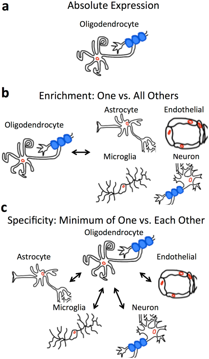
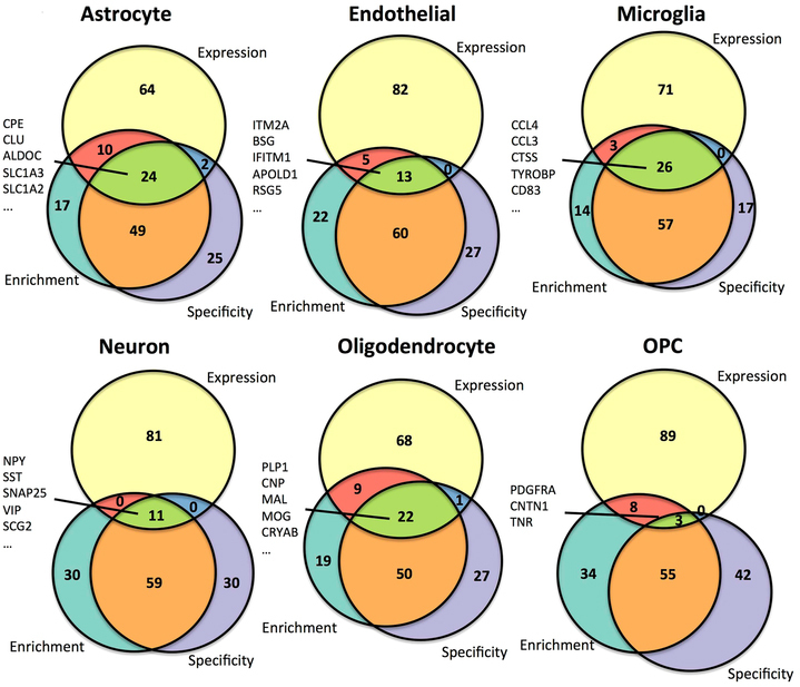
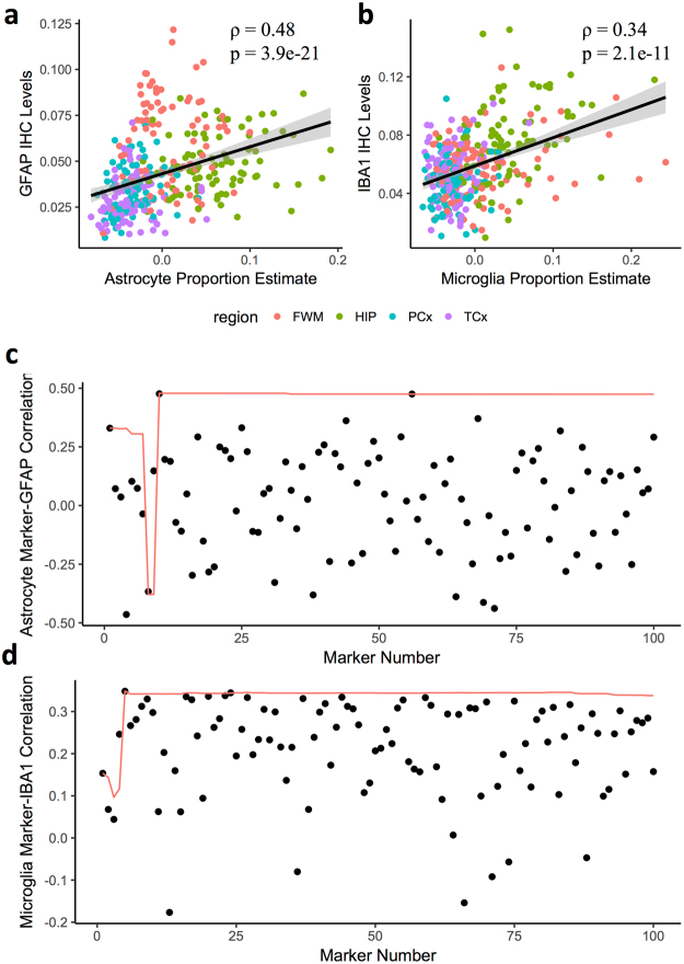
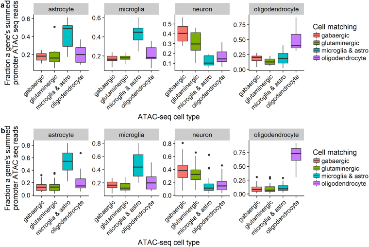
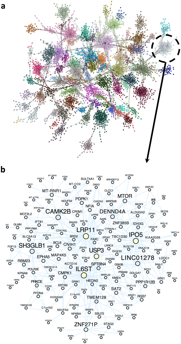
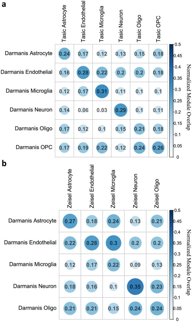
Elucidating brain cell type specific gene expression patterns is critical towards a better understanding of how cell-cell communications may influence brain functions and dysfunctions. We set out to compare and contrast five human and murine cell type-specific transcriptome-wide RNA expression data sets that were generated within the past several years. We defined three measures of brain cell type-relative expression including specificity, enrichment, and absolute expression and identified corresponding consensus brain cell “signatures,” which were well conserved across data sets. We validated that the relative expression of top cell type markers are associated with proxies for cell type proportions in bulk RNA expression data from postmortem human brain samples. We further validated novel marker genes using an orthogonal ATAC-seq dataset. We performed multiscale coexpression network analysis of the single cell data sets and identified robust cell-specific gene modules. To facilitate the use of the cell type-specific genes for cell type proportion estimation and deconvolution from bulk brain gene expression data, we developed an R package, BRETIGEA. In summary, we identified a set of novel brain cell consensus signatures and robust networks from the integration of multiple datasets and therefore transcend limitations related to technical issues characteristic of each individual study.
Overview
The claims and statistics of this paper haven’t been checked yet.
Run the check — free, no account needed. It reads the open-access text and checks the sources it cites.
Integrity signals
Registry
Cites
Statistics
Figures
Verification
Claims the paper makes
The claims and statistics of this paper haven’t been checked yet.
Run the check — free, no account needed. It reads the open-access text and checks the sources it cites.
Claims other papers make about this one
Each is checked against this paper’s own text: does it say what the citing paper says it does?
The source backs part of the claim, not all of it — or backs it for a different population, condition or comparison.
In the citing paper
“The cell clusters were annotated manually using marker genes that were conserved between both conditions as astrocytes, microglia, neurons, endothelial cells, ependymal cells, and mixed glia (Supplementary Data 1) (Arneson et al., 2018;McKenzie et al., 2018).”
From Single-cell resolution analysis of the crosstalk between chemogenically activated astrocytes and microglia (2020) · cites this paper as McKenzie et al., 2018
Experimental — accuracy not yet measuredThe cited source says what the claim attributes to it.
In the citing paper
“Canonical CNS cell type markers were compiled from previous studies66–69.”
From APOE4 to APOE2 allelic switching in mice improves Alzheimer’s disease-related metabolic signatures, neuropathology and cognition (2025) · cites this paper as [66]
The cited source says what the claim attributes to it.
In the citing paper
“To estimate the cell-type proportion of bulk tissue RNA-seq data used in this study, we perform a cell-type deconvolution on each sample using the brain cell–type marker signatures provided by the BRETIGEA R package (26).”
From Molecular subtyping of Alzheimer’s disease using RNA sequencing data reveals novel mechanisms and targets (2021) · cites this paper as [26]
The cited source says what the claim attributes to it.
In the citing paper
“All cell clusters were annotated manually based on marker genes that were conserved between the saccharin group and the CNO group and gene lists present in literature (Arneson et al., 2018;McKenzie et al., 2018).”
From Single-cell resolution analysis of the crosstalk between chemogenically activated astrocytes and microglia (2020) · cites this paper as McKenzie et al., 2018
The cited source says what the claim attributes to it.
In the citing paper
“We used a consensus top ranked cell type-specific marker set from a recent meta-analysis of multiple cell type-specific and single cell RNAseq datasets (McKenzie et al., 2018).”
From Cholesterol and matrisome pathways dysregulated in astrocytes and microglia (2022) · cites this paper as McKenzie et al., 2018
Self-check
Percentages, sums and reported differences re-computed from the paper’s own numbers.
The self-check needs the paper’s full text.
Citations
Cites
Controlling the False Discovery Rate: A Practical and Powerful Approach to Multiple Testing
Benjamini, Y., & Hochberg, Y. (1995) · Journal of the Royal Statistical Society Series B: Statistical Methodology · doi:10.1111/j.2517-6161.1995.tb02031.x · Cited by 109,482
STAR: ultrafast universal RNA-seq aligner
Dobin, A., Davis, C. A., Schlesinger, F., Drenkow, J., Zaleski, C., Jha, S., Batut, P., Chaisson, M., & Gingeras, T. R. (2013) · Bioinformatics · doi:10.1093/bioinformatics/bts635 · Cited by 51,709
Robinson, M. D., McCarthy, D. J., & Smyth, G. K. (2010) · Bioinformatics · doi:10.1093/bioinformatics/btp616 · Cited by 41,364
featureCounts: an efficient general purpose program for assigning sequence reads to genomic features
Liao, Y., Smyth, G. K., & Shi, W. (2014) · Bioinformatics · doi:10.1093/bioinformatics/btt656 · Cited by 27,219
Tissue-based map of the human proteome
Uhlén, M., Fagerberg, L., Hallström, B. M., Lindskog, C., Oksvold, P., Mardinoglu, A., Sivertsson, Å., Kampf, C., Sjöstedt, E., Asplund, A., et al. (2015) · Science · doi:10.1126/science.1260419 · Cited by 15,286
Highly parallel genome-wide expression profiling of individual cells using nanoliter droplets.
Macosko, E. Z., Basu, A., Satija, R., Nemesh, J., Shekhar, K., Goldman, M., Tirosh, I., Bialas, A. R., Kamitaki, N., Martersteck, E. M., et al. (2015) · Cell · doi:10.1016/j.cell.2015.05.002 · Cited by 7,110
Astrocytes: Biology and pathology
Sofroniew, M. V., & Vinters, H. V. (2010) · Acta Neuropathologica · doi:10.1007/s00401-009-0619-8 · Cited by 5,196
Zhang, Y., Chen, K., Sloan, S. A., Bennett, M. L., Scholze, A. R., O'Keeffe, S., Phatnani, H. P., Guarnieri, P., Caneda, C., Ruderisch, N., et al. (2014) · The Journal of Neuroscience · doi:10.1523/jneurosci.1860-14.2014 · Cited by 4,875
Cahoy, J. D., Emery, B., Kaushal, A., Foo, L. C., Zamanian, J. L., Christopherson, K. S., Xing, Y., Lubischer, J. L., Krieg, P. A., Krupenko, S. A., et al. (2008) · The Journal of Neuroscience · doi:10.1523/jneurosci.4178-07.2008 · Cited by 3,129
Cell types in the mouse cortex and hippocampus revealed by single-cell RNA-seq
Zeisel, A., Muñoz-Manchado, A. B., Codeluppi, S., Lönnerberg, P., La Manno, G., Juréus, A., Marques, S., Munguba, H., He, L., Betsholtz, C., et al. (2015) · Science · doi:10.1126/science.aaa1934 · Cited by 3,124
Cited by
Sorted by citations
Items 301–400 of 492
Cell Type–Specific Nuclei Markers: The Need for Human Brain Research to Go Nuclear
Wiseman, J. A., Dragunow, M., & Park, T. (2021) · The Neuroscientist · doi:10.1177/10738584211037351 · Cited by 5
Chang, K., Rhodes, C. T., Zhang, J. Q., Moseley, M. C., Cardona, S. M., Huang, S. A., Rawls, A., Lemmon, V., Berger, M. S., Abate, A. R., et al. (2021) · FEBS Letters · doi:10.1002/1873-3468.14254 · Cited by 4
Treatment of microglia with Anti-PrP monoclonal antibodies induces neuronal apoptosis in vitro
Adhikari, U. K., Sakiz, E., Habiba, U., Mikhael, M., Senesi, M., David, M. A., Guillemin, G. J., Ooi, L., Karl, T., Collins, S. J., et al. (2021) · Heliyon · doi:10.1016/j.heliyon.2021.e08644 · Cited by 4
Transcriptional development of the hippocampus and the dorsal–intermediate–ventral axis in rats
Olsen, L. C., Galler, M., Witter, M. P., Sætrom, P., & O’Reilly, K. C. (2023) · Hippocampus · doi:10.1002/hipo.23549 · Cited by 4
Ji, J., Chao, H., Chen, H., Liao, J., Shi, W., Ye, Y., Wang, T., You, Y., Liu, N., Ji, J., et al. (2024) · Open Biology · doi:10.1098/rsob.240063 · Cited by 3
“To corroborate the cell classification, we also used the BRETIGEA package, which provides a well-validated set of brain cell type-specific marker genes derived from multiple types of experiments, including marker specificity, enrichment and absolute expression [22].”
MethodCatalytic and non-catalytic functions of DNA polymerase kappa in translesion DNA synthesisPreprint
Sellés‐Baiget, S., Ambjørn, S. M., Carli, A., Hendriks, I. A., Gallina, I., Davey, N. E., Benedict, B., Meeusen, B., Hertz, E. P. T., Slappendel, L., et al. (2023) · bioRxiv (Cold Spring Harbor Laboratory) · doi:10.1101/2023.11.06.565773 · Cited by 4
Molecular basis of neurodegeneration in a mouse model of Polr3-related diseasePreprint
Moir, R. D., Merheb, E., Chiţu, V., Stanley, E. R., & Willis, I. M. (2024) · eLife · doi:10.7554/elife.95314 · Cited by 4
Sun, E., Zhou, O. Y., Hauptschein, M., Rappoport, N., Xu, L., Negredo, P. N., Liu, L. A., Rando, T. A., Zou, J., & Brunet, A. (2024) · bioRxiv (Cold Spring Harbor Laboratory) · doi:10.1101/2024.07.16.603809 · Cited by 4
PU.1 restores microglial dysfunction caused by C9ORF72 repeat expansions in neural organoids
Ljubikj, T., Mars, M., van der Geest, A. T., Jakobs, C. E., Bessler, N., Donega, V., van den Oetelaar, X. P. R. M., de Wit, M., & Pasterkamp, R. J. (2025) · Brain · doi:10.1093/brain/awaf340 · Cited by 4
“To confirm the purity of the samples and exclude C9-ALS/FTD-related effects on microglial differentiation, normalized counts for known markers of different cell types 31-33 were plotted.”
Gatto, E., Samorì, E., Frigato, E., Bertolucci, C., & Lucon‐Xiccato, T. (2025) · Neurobiology of Learning and Memory · doi:10.1016/j.nlm.2025.108106 · Cited by 3
Alterations in the brain lipidome of Alzheimer's disease donors with rare TREM2 risk variants
Proitsi, P., Ebshiana, A., Wretlind, A., Xu, J., Hodges, A., & Legido‐Quigley, C. (2026) · Brain Communications · doi:10.1093/braincomms/fcaf452 · Cited by 3
Babenko, V. N., Redina, O. E., Смагин, Д. А., Kovalenko, I. L., Galyamina, A. G., & Kudryavtseva, N. N. (2024) · International Journal of Molecular Sciences · doi:10.3390/ijms25052882 · Cited by 3
“A definitive comparative paper on six basic cell types (Astrocyte, Neuron, Oligodendro-cyte, Oligodendrocyte Precursor, Microglia, and Endothelial Cells) identified cell-specific genes [7].”
BackgroundMartus, D., Williams, S. K., Pichi, K., Mannebach, S., Kaiser, N., Wardas, B., Fecher‐Trost, C., Meyer, M. R., Schmitz, F., Beck, A., et al. (2024) · Arteriosclerosis Thrombosis and Vascular Biology · doi:10.1161/atvbaha.124.321141 · Cited by 3
Chucair‐Elliott, A. J., Pham, K. D., Cleuren, A. C. A., Schafer, C. M., Griffin, C. T., Ocañas, S. R., Freeman, W. M., & Elliott, M. H. (2024) · Experimental Eye Research · doi:10.1016/j.exer.2024.110101 · Cited by 3
Goh, J.-Y., Rueda, P., Taylor, J., Rathbone, A., Scott, D., LANGMEAD, C. J., Fone, K. C. F., Stewart, G. D., & King, M. V. (2024) · Neurobiology of Stress · doi:10.1016/j.ynstr.2024.100679 · Cited by 4
“It is therefore possible that altered gene expression in one cell type could be masked by an opposing change in others (Darmanis et al., 2015; McKenzie et al., 2018), and future application of single-cell RNA-Seq (Cai et al., 2022) could reveal these differential effects to shed even more light on…”
Anxiety-like behavior and altered hippocampal activity in a transgenic mouse model of Fabry disease
Kummer, K. K., Choconta, J. L., Edenhofer, M.-L., Bajpai, A., Dharmalingam, G., Kalpachidou, T., Collier, D. A., & Kress, M. (2025) · Neurobiology of Disease · doi:10.1016/j.nbd.2025.106797 · Cited by 3
A Transcriptomic Signature of Depressive Symptoms in Late Life
Matan‐Lithwick, S. A., Misztal, M. C., Yang, M., DeLong, T., Tripathy, S. J., Dunn, J. T., Bennett, D. A., Jager, P. L. D., Wang, Y., Fisher, D. W., et al. (2025) · Biological Psychiatry Global Open Science · doi:10.1016/j.bpsgos.2025.100448 · Cited by 4
Mwanza, J.-C., Kahatane, A. K., Mbusa-Kombi, J., Mumbere, M. V., Kitenge, R., McIlwain, D. R., Mulangu, J. C. S., Kingebeni, P. M., Okitundu, D., Giordani, B., et al. (2025) · Scientific Reports · doi:10.1038/s41598-025-96256-4 · Cited by 3
“The function of ITM2A is not yet well defined, but previous studies have identified it in the brain endothelial cells of humans, rats, and pigs 35–37 , with high expression, particularly in humans and rats 37,38 .”
Chen, Z., Chen, S., & Qiang, X. (2022) · Frontiers in Bioinformatics · doi:10.3389/fbinf.2022.812314 · Cited by 3
“McKenzie A T et al. identi fi ed a novel set of brain cell consistent signatures and robust networks from the integration of multiple data sets, so it goes beyond the limitations associated with each individual research speci fi c technical problem (McKenzie et al., 2018).”
BackgroundWellens, S., Gosselet, F., & Culot, M. (2022) · Toxicology in Vitro · doi:10.1016/j.tiv.2022.105424 · Cited by 3
Gutierrez, B. A., González-Coronel, J. M., Arellano, R. O., & Limón, A. (2023) · Frontiers in Molecular Neuroscience · doi:10.3389/fnmol.2023.1279232 · Cited by 3
“Specifically, the GABA A Rs γ 2 subunit has been suggested to be postsynaptic in OPCs from murines (Passlick et al., 2013; Balia et al., 2015; Orduz et al., 2015; McKenzie et al., 2018).”
BackgroundProteomic Signaling of Dual-Specificity Phosphatase 4 (DUSP4) in Alzheimer’s Disease
Wang, E., Pan, A. L., Bagchi, P., Rangaraju, S., Seyfried, N. T., Ehrlich, M. E., Salton, S. R., & Zhang, B. (2024) · Biomolecules · doi:10.3390/biom14010066 · Cited by 3
“The reference mouse cell-type signatures were curated and described in [50], which include six gene signatures that are specifically expressed in microglia (mic), neuron (neu), astrocytes (ast), oligodendrocytes (oli), endothelial cells (end), and oligodendrocyte precursor cells (opc), respectively.”
Self-citationGhisai, S. A., Barin, N., van Hijfte, L., Verhagen, K., de Wit, M., van den Bent, M. J., Hoogstrate, Y., & French, P. J. (2024) · Neuro-Oncology Advances · doi:10.1093/noajnl/vdae229 · Cited by 3
Characterizing the expression profile of 3R tau pathology in Pick’s disease
Tamvaka, N., Soto‐Beasley, A. I., Gavrielatos, M., Heckman, M. G., Ren, Y., Udine, E., Quicksall, Z., Liskey, D., Castanedes‐Casey, M., Wszołek, Z. K., et al. (2025) · Science Advances · doi:10.1126/sciadv.adt6105 · Cited by 4
“0.3) to analyze relative cell-type proportions in our dataset ( 40 , 41 ).”
InfluentialDopa decarboxylase is a genetic hub of parental control over offspring behaviorPreprint
Bonthuis, P. J., Steinwand, S., Huang, W.-C., Hörndli, C. S., Emery, J., Kravitz, S. N., Ferris, E., & Gregg, C. J. (2020) · bioRxiv (Cold Spring Harbor Laboratory) · doi:10.1101/2020.06.23.168195 · Cited by 2
“We then computed the expression levels of mouse-human conserved markers of neurons, oligodendrocytes, astrocytes, endothelial cells and microglia (McKenzie et al., 2018).”
MethodReshetnikov, V. V., Kisaretova, P. E., Ershov, N., Merkulova, T. I., & Bondar, N. P. (2020) · Data in Brief · doi:10.1016/j.dib.2020.106365 · Cited by 3
“In addition, we valuated correlation between broad H3K4me3 peaks in promoters of cell-specific genes [10] .”
MethodKubiś, J., & Figiel, M. (2023) · bioRxiv (Cold Spring Harbor Laboratory) · doi:10.1101/2023.04.20.537649 · Cited by 3
Marriott, H., Kabiljo, R., Hunt, G. P., Khleifat, A. A., Jones, A., Troakes, C., Pfaff, A. L., Quinn, J. P., Kõks, S., Dobson, R., et al. (2023) · medRxiv · doi:10.1101/2023.04.21.23288942 · Cited by 3
BRETIGEA: Brain Cell Type Specific Gene Expression Analysis
Andrew McKenzie [aut, C. (2018) · doi:10.32614/cran.package.bretigea · Cited by 3
Spatial transcriptomics reveals brain-wide circadian disruption in an Alzheimer’s disease modelPreprint
Gelber, A., Romero, H. K., Burrows, D. R. W., Whittaker, D. S., Carlin, D. E., Mukamel, E. A., & Desplats, P. (2026) · bioRxiv (Cold Spring Harbor Laboratory) · doi:10.64898/2026.01.26.701799 · Cited by 3
Robustness and resilience of computational deconvolution methods for bulk RNA sequencing data
Su, X., Chen, D., Wang, X., & Li, S. (2025) · Briefings in Bioinformatics · doi:10.1093/bib/bbaf264 · Cited by 2
“[38] for each of the five brain cell types considered in this study, resulting a total of 5000 genes included in our analyses for human brain tissues.”
Bobkova, N. V., Chuvakova, L. N., Kononova, S. V., Kovalev, V. I., Sukhikh, G. T., Zatsepina, O. G., Rezvykh, A. P., & Евгеньев, М. Б. (2025) · Molecular Neurobiology · doi:10.1007/s12035-025-05135-6 · Cited by 4
“Cell-type-specific profiling of differentially expressed genes was performed, using published csRNA sequencing data [52].”
InfluentialThe Association Between Sleep Phenotypes and Epilepsy Genes
Gaillard, J. R., Gupta, G., Mefford, H. C., O'Brien, L. M., Shellhaas, R. A., & Dang, L. T. (2025) · Neurology Genetics · doi:10.1212/nxg.0000000000200269 · Cited by 2
Describing the diversity of MAPT transcripts in the parietal cortex of Pick’s disease patients
Tamvaka, N., Stuber, M. G., Gavrielatos, M., Soto‐Beasley, A. I., Heckman, M. G., Murray, M. E., Boeve, B. F., Josephs, K. A., Roemer, S. F., Graff‐Radford, N. R., et al. (2025) · npj Dementia · doi:10.1038/s44400-025-00027-x · Cited by 2
“CellularproportionswereestimatedusingBRITIGEA 42 on previously published short-read bulk RNA-Seq data available for the same samples 43 .”
Caron, N. S., Fan, J., Ma, D., Yung, A. C., Ko, S., Bale, K., Anderson, C., Kozłowski, P., Pouladi, M. A., Wellington, C. L., et al. (2025) · Molecular Therapy — Nucleic Acids · doi:10.1016/j.omtn.2025.102710 · Cited by 2
Functional genomic profiling of schizophrenia-associated genes reveals key microglial regulators
Horng, J. E., McCrea, L. T., Batorsky, R., Bowen, J. J., Boschian, C., Song, Y., Perlis, R. H., & Sheridan, S. D. (2026) · Neuropsychopharmacology · doi:10.1038/s41386-026-02406-1 · Cited by 2
Saha, T., Vats, T., & Mehan, S. (2026) · Immunopharmacology and Immunotoxicology · doi:10.1080/08923973.2026.2671714 · Cited by 2
Localization Pattern of Dispatched Homolog 2 (DISP2) in the Central and Enteric Nervous System
Heimke, M., Richter, F., Heinze, T., Kunke, M., Wedel, T., Böttner, M., Egberts, J. D., Lucius, R., & Cossais, F. (2023) · Journal of Molecular Neuroscience · doi:10.1007/s12031-023-02129-8 · Cited by 3
“…reported in islets of Langerhans within the human and mouse pancreas so far (Hald et al. 2012), a recent bioinformatics study based on single-cell transcriptomic expression datasets identified 1 3 DISP2 as a potential specific neuronal marker in the human and mouse brain (McKenzie et al. 2018).”
Gomes, J. D. A., Sgarioni, E., Kowalski, T. W., Giudicelli, G. C., Recamonde‐Mendoza, M., Fraga, L. R., Schüler‐Faccini, L., & Vianna, F. S. L. (2023) · Journal of Molecular Neuroscience · doi:10.1007/s12031-023-02126-x · Cited by 2
“…what has been presented in the literature, which describes a wide variation in gene expression between different neural cells, as well as a variation in the gene expression of the same cell type over the time as this cell undergoes differentiation (Gurok et al. 2004; McKenzie et al. 2018).”
BackgroundAuderset, L., & Antoniou, A. (2023) · Trillium Extracellular Vesicles · doi:10.47184/tev.2023.01.03 · Cited by 2
Towards correlative archaeology of the human mind
Piszczek, Ł., Kaczanowska, J., & Haubensak, W. E. (2023) · Biological Chemistry · doi:10.1515/hsz-2023-0199 · Cited by 2
“Together with the expanding phylogenetic transcriptomics in cell types and brain regions across mammalian brains, this will allow to compare gene expression di ff erences in extant species and potentially infer ancestral states (McKenzie et al. 2018; Wei et al. 2022).”
BackgroundRevealing Pan-Histology Immunomodulatory Targets in Pediatric Central Nervous System Tumors
Galvin, R. T., Jena, S., Maeser, D., Gruener, R. F., & Huang, R. S. (2023) · Cancers · doi:10.3390/cancers15225455 · Cited by 2
“We therefore employed another deconvolution pipeline developed by McKenzie, et al. that provides microglia signatures as a surrogate proportion variable (SPV), which represents a relative abundance of microglia, for independent analysis [11,15].”
MethodInfluentialEvaluation of the impact of iPSC differentiation protocols on transcriptomic signatures
Chandrasekaran, V., Wellens, S., Bourguignon, A., Djidrovski, I., Fransen, L. F. H., Ghosh, S., Mazidi, Z., Murphy, C., Nunes, C., Singh, P., et al. (2024) · Toxicology in Vitro · doi:10.1016/j.tiv.2024.105826 · Cited by 3
“The S100B gene, known to be expressed in astrocytes and oligodendrocytes (McKenzie et al., 2018; Michetti et al., 2019), was only expressed in the BS, reflecting the difference with the NC model.”
BackgroundResultBao, X., Zhou, B., & Wen, M. (2024) · Frontiers in Bioscience-Landmark · doi:10.31083/j.fbl2911394 · Cited by 2
“03, https://cran.r-project.o rg/web/packages/BRETIGEA/index.html) [36] package to calculate the first singular vector of a variable number of cell type-specific genes to estimate the relative proportions of various cell types in each sample.”
Zhou, R., Zhang, T., & Sun, B. (2025) · International Journal of Molecular Sciences · doi:10.3390/ijms26052227 · Cited by 2
You, Y., Zhang, Z., Cortes, S., Nguyen, S., Vadakattu, P., Melvin, B. C., Mann, S. D. J., Ray, N. B., Bregendahl, M. E., McLean, P. J., et al. (2025) · Cell Communication and Signaling · doi:10.1186/s12964-025-02263-3 · Cited by 2
“We further assessed brain cell type-specific molecules within the plasma-EV proteome across these methods by referencing a published brain cell types data-base [31].”
Ligand–Receptor Analysis of Brain Cell Type Marker Data Reveals Intricate Endothelial Interaction
Mishra, A., & Kumar, G. (2025) · Annals of Neurosciences · doi:10.1177/09727531251343254 · Cited by 3
“Endothelial cells exhibit the highest level of contact with microglia (29), followed by astrocytes (26).”
Zhang, R., Xie, Y., Zhang, M., Li, Z., Guo, L., Wei, H., Li, X., Xin, J., Yang, Z., Wen, Y., et al. (2025) · Experimental & Molecular Medicine · doi:10.1038/s12276-025-01489-y · Cited by 5
“7a. NMT was applied to measure the Ca 2+ fl ux of the surface of the inner retina (Fig.”
Clarelli, F., Rocca, M. A., Santoro, S., Meo, E. D., Ferrè, L., Sorosina, M., Boneschi, F. M., Esposito, F., & Filippi, M. (2021) · European Journal of Neurology · doi:10.1111/ene.14872 · Cited by 3
“[25], in which a metaanalysis of two human brain cell- specific signatures was conducted [26,27].”
BackgroundDonskov, J. G. (2021) · Journal of Translational Genetics and Genomics · doi:10.20517/jtgg.2021.12 · Cited by 2
Probing glioblastoma and its microenvironment using single-nucleus and single-cell sequencing
Peng, S., Rath, S., Vuong, C., Bollam, S., Eschbacher, J. M., Dong, X., Mehta, S. V., Sanai, N., Berens, M. E., Kim, S., et al. (2019) · IEEE International Conference on Bioinformatics and Biomedicine (BIBM) · doi:10.1109/bibm47256.2019.8983321 · Cited by 1
Schizophrenia risk from locus-specific human endogenous retrovirusesPreprint
Duarte, R. R. R., Bendall, M. L., Rougvie, M. D. M., Ormsby, C. E., Beckerle, G. A., Selvackadunco, S., Troakes, C., Reyes‐Terán, G., Crandall, K. A., Srivastava, D. P., et al. (2019) · bioRxiv (Cold Spring Harbor Laboratory) · doi:10.1101/798017 · Cited by 2
Lin, C., Li, H., Deng, C., Liu, W., Erhardt, S., Wu, F., Zhao, X., Wang, J., Wang, D., Hu, B., et al. (2021) · bioRxiv (Cold Spring Harbor Laboratory) · doi:10.1101/2021.02.09.430536 · Cited by 2
Belonwu, S. A., Li, Y., Bunis, D. G., Rao, A. A., Solsberg, C. W., Oskotsky, T., Taubes, A. L., Grone, B. P., Zalocusky, K. A., Fragiadakis, G. K., et al. (2021) · Research Square · doi:10.21203/rs.3.rs-291648/v1 · Cited by 2
Infants’ cortex undergoes microstructural growth coupled with myelinationPreprint
Natu, V. S., Rosenke, M., Wu, H., Querdasi, F. R., Kular, H., Lopez-Alvarez, N., Grotheer, M., Berman, S., Mezer, A., & Kalanit Grill-spector, A. (2021) · bioRxiv (Cold Spring Harbor Laboratory) · doi:10.1101/2021.03.16.435703 · Cited by 2
Ezra, S., & Danter, W. R. (2022) · Preprints.org · doi:10.20944/preprints202210.0217.v1 · Cited by 1
Reich, M., Simon, M. J., Polke, B., Werner, G., Schrader, C. M., Paris, I., Robinson, S., Davis, S. S., de Melo, G. L., Schlaphoff, L., et al. (2023) · bioRxiv (Cold Spring Harbor Laboratory) · doi:10.1101/2023.07.14.549089 · Cited by 2
“4B), we next determined changes in cell type composition (74) (Fig.”
BackgroundChan, J. C., Salvo, G. D., Cunningham, A. M., Dutta, S., Brindley, E., Weekley, B. H., Wan, E., Zhang, C., Mechawar, N., Turecki, G., et al. (2025) · bioRxiv (Cold Spring Harbor Laboratory) · doi:10.1101/2025.02.20.639313 · Cited by 2
Liharska, L. E., & Charney, A. W. (2024) · Current topics in behavioral neurosciences · doi:10.1007/7854_2024_466 · Cited by 1
Parallel Gene Expression Changes in Ventral Midbrain Dopamine and GABA Neurons during Normal Aging
Drumond‐Bock, A. L., Blankenship, H. E., Pham, K. D., Carter, K. A., Freeman, W. M., & Beckstead, M. J. (2025) · eNeuro · doi:10.1523/eneuro.0107-25.2025 · Cited by 3
Protein Pathologies in Oligodendroglia in Neurodegenerative Diseases
Forrest, S. L., & Kovács, G. G. (2025) · Advances in neurobiology · doi:10.1007/978-3-031-87919-7_14 · Cited by 2
Lopes, K. D. P., Vialle, R. A., Green, G., Fujita, M., Gaiteri, C., Menon, V., Schneider, J. A., Wang, Y., Jager, P. L. D., Habib, N., et al. (2025) · Acta Neuropathologica Communications · doi:10.1186/s40478-025-02143-4 · Cited by 2
“Similarly to our findings, most previous studies observed a large proportion of module conservation between cell types, suggesting consistent regulatory landscapes across cells [28, 46, 47].”
Larkin, J., Dwivedi, A. K., Mahesh, A., Sanfeliu, A., Sweeney, K. J., O’Brien, D. F., Tiwari, V. K., Widdess‐Walsh, P., & Henshall, D. C. (2025) · Biomolecules · doi:10.3390/biom15121684 · Cited by 1
“…we explored the presence of different cell phenotype markers among the detected transcriptomes using the Allen Brain Atlas and a ranked list of cell-type gene markers provided by McKenzie et al., selecting the genes with the highest specificity for a given cell type from human datasets [19].”
InfluentialLucart-Sanchez, A. M., Selles-Climent, E., Navarro-Calvo, J., Pont-Espinós, G., Gomez-Sanchez, J. A., Estévez, R., Valor, L. M., & Pérez‐González, R. (2026) · Glia · doi:10.1002/glia.70212 · Cited by 1
“To further validate cell type-specificity of the retrieved EV proteins, we examined the presence of curated top 100 cell-specific expressed genes that were described for each major brain cell type (McKenzie et al. 2018) across the pairwise comparisons.”
Wang, Z., Chen, Y., Feng, Y., Zu, G., Wang, W., Liang, L., Liu, X., Mei, Y., Yang, J., Wang, T., et al. (2026) · iScience · doi:10.1016/j.isci.2026.114692 · Cited by 1
Single‐cell analysis reveals neuroprotective histone deacetylase inhibitor pathways
Peyton, M., Jury, N., Liu, J., Beimfohr, C., Sunil, C., Brooks, S., Zhang, P., McCabe, S., Richardson, T. I., Huang, K., et al. (2026) · Alzheimer s & Dementia · doi:10.1002/alz.71108 · Cited by 1
“The BRETIGEA package (version 1.0.3) 21 in R was used to annotate the doublet cells within the Grubman et al. 13 dataset.”
InfluentialRossi, F., Sportelli, L., Kikidis, G. C., Grassi, G., Camillo, F. D., Bertolino, A., Blasi, G., Borcuk, C., Fusco, D., Hyde, T. M., et al. (2026) · Nature Genetics · doi:10.1038/s41588-026-02646-3 · Cited by 1
“Neuronal proportion, estimated using BRETIGEA 104 , was strongly correlated with principal components across regions and therefore not included to avoid collinearity.”
Ferraro, F., Fevga, C., Bonifati, V., Mandemakers, W. J., Mahfouz, A. E., & Reinders, M. J. T. (2022) · Cells · doi:10.3390/cells11020198 · Cited by 1
“For the other six cell types, we used subsets of 20 markers for each study selected among the 5500 markers from the Brain Cell Type—Specific Gene Expression Analysis package version 1.0.0 (BRETIGEA) [19] (Supplementary Materials).”
InfluentialGrochowska, M. M., Ferraro, F., Mascaro, A. C., Natale, D., Winkelaar, A. B., Boumeester, V., Breedveld, G. J., Bonifati, V., & Mandemakers, W. J. (2023) · Disease Models & Mechanisms · doi:10.1242/dmm.049980 · Cited by 1
From the Perspective of a Neuron
Lux, V. (2024) · SpringerBriefs in psychology · doi:10.1007/978-3-031-55229-8_1 · Cited by 1
Shar, A., Glass, P., Daul, B., Rad, R. M., & Joung, D. (2024) · Elsevier eBooks · doi:10.1016/b978-0-323-95730-4.00010-x · Cited by 1
Expanding canonical cortical cell type markers in the era of single-cell transcriptomics
Joshy, D. M., & Yi, S. V. (2026) · Scientific Reports · doi:10.1038/s41598-026-51501-2 · Cited by 1
“There is also recent work in cell marker discovery using single-cell data such as McKenzie et al. 28 which introduced ideas of prioritizing specificity, enrichment, and marker validation methods.”
Human brain cell type-specific gene co-expression associated with autism spectrum disorder
Lin, Y., Li, S., Ji, G., & Guan, J. (2019) · IEEE International Conference on Bioinformatics and Biomedicine (BIBM) · doi:10.1109/bibm47256.2019.8983237 · Cited by 1
“To identify cell type-specific gene modules, we obtained the cell type-specific genes in humans identified from study of [8], and used a method similar to that in the original study where top 500 cell type-specific genes for cell types of astrocytes, endothelial, microglia, neurons and oligodendrocytes were used to as…”
MethodKiszka, K. A. (2019) · eDiss (Georg-August-Universität Göttingen) · doi:10.53846/goediss-7790 · Cited by 1
ERCC1-XPF Interacts with Topoisomerase IIβ to Facilitate the Repair of Activity-induced DNA BreaksPreprint
Chatzinikolaou, G., Stratigi, K., Agathangelou, K., Tsekrekou, M., Goulielmaki, E., Chatzidoukaki, O., Gkirtzimanaki, K., Aid‐Pavlidis, T., Aivaliotis, M., Pavlidis, P., et al. (2020) · bioRxiv (Cold Spring Harbor Laboratory) · doi:10.1101/2020.01.03.892703 · Cited by 1
“, 2012b) and cerebella (genes: Dhfr, Prnp)(McKenzie et al., 2018; Nouspikel and Hanawalt, 2000) (Figure 7I and Figure 7J; as indicated), relative to wt.”
BackgroundDang, M. T., Gonzalez, M., Gaonkar, K. S., Rathi, K. S., Young, P. A., Arif, S., Zhai, L., Alam, M. Z., Devalaraja, S., To, T. K. J., et al. (2020) · bioRxiv (Cold Spring Harbor Laboratory) · doi:10.1101/2020.02.12.945642 · Cited by 1
Radulescu, E., Chen, Q., Pergola, G., Eagles, N. J., Stolz, J. M., Shin, J. H., Hyde, T. M., Kleinman, J. E., & Weinberger, D. R. (2021) · bioRxiv (Cold Spring Harbor Laboratory) · doi:10.1101/2021.05.04.442668 · Cited by 1
“Cell type composition represents another factor that contributes to construction of co-expression networks from bulk RNA-Seq data, while modules of co-expression are likely to be significantly driven by cellular type [17].”
BackgroundMethodPatel, T., Carnwath, T., Wang, X., Allen, M., Lincoln, S., Lewis‐Tuffin, L. J., Quicksall, Z., Lin, S., Tutor‐New, F. Q., Ho, C. C. G., et al. (2021) · Research Square · doi:10.21203/rs.3.rs-523348/v1 · Cited by 1
Carbajosa, G., Malki, K., Lawless, N., Wang, H., Ryder, J. W., Wozniak, E., Wood, K., Mein, C. A., Hodgkinson, A., Dobson, R., et al. (2021) · bioRxiv (Cold Spring Harbor Laboratory) · doi:10.1101/2021.07.16.452732 · Cited by 1
“…our knowledge of markers specific to different brain cell types and revealed important cellular subpopulations with unique molecular identities and behaviours in normal (Hawrylycz et al., 2012; Forabosco et al., 2013; McKenzie et al., 2018; Olah et al., 2018) and AD brain (Zhang et al., 2013).”
BackgroundInfluentialNiktab, E., Atkinson, P. H., Walterfang, M., Winship, I., Sturley, S. L., & Munkacsi, A. B. (2023) · bioRxiv (Cold Spring Harbor Laboratory) · doi:10.1101/2023.05.18.540731 · Cited by 1
Pak, V., Adewale, Q., Bzdok, D., Dadar, M., Zeighami, Y., & Iturria‐Medina, Y. (2023) · bioRxiv (Cold Spring Harbor Laboratory) · doi:10.1101/2023.06.08.544227 · Cited by 1
Consistent specificity and efficiency of tamoxifen-mediated cre induction across agesPreprint
Kellogg, C. M., Pham, K. D., Ko, S., Cox, J. E. J., Machalinski, A. H., Stout, M. B., Sharpe, A. L., Beckstead, M. J., Chucair‐Elliott, A. J., Ocañas, S. R., et al. (2023) · bioRxiv (Cold Spring Harbor Laboratory) · doi:10.1101/2023.09.19.558482 · Cited by 1
Loss of TREM2 exacerbates parenchymal amyloid pathology but diminishes CAA in Tg-SwDI micePreprint
Zhong, R., Xu, Y., Williams, J. W., & Li, L. (2023) · bioRxiv (Cold Spring Harbor Laboratory) · doi:10.1101/2023.11.04.565659 · Cited by 1
Inhibition of MicroRNA-129-5p Promotes Neuroinflammation and Cognitive ImpairmentPreprint
Kaurani, L., Zhou, J., Pradhan, R., Schröder, S., Burkhardt, S., Schuetz, A.-L., Delalle, I., Rizzu, P., Heutink, P., Sananbenesi, F., et al. (2024) · Research Square · doi:10.21203/rs.3.rs-3870232/v1 · Cited by 1
Pak, V., Adewale, Q., Bzdok, D., Dadar, M., Zeighami, Y., & Iturria‐Medina, Y. (2024) · eLife · doi:10.7554/elife.89368.2 · Cited by 1
Molecular basis of neurodegeneration in a mouse model of Polr3-related diseasePreprint
Moir, R. D., Merheb, E., Chiţu, V., Stanley, E. R., & Willis, I. M. (2024) · eLife · doi:10.7554/elife.95314.1 · Cited by 1
A unified analysis of atlas single-cell dataPreprint
Chen, H., Nguyen, N. D., Ruffalo, M., & Bar‐Joseph, Z. (2025) · Genome Research · doi:10.1101/gr.279631.124 · Cited by 2
“The idea has been widely used in the literature to study gene functions using scRNA-seq data (McKenzie et al. 2018; Cha et al. 2023).”
Baranger, D. A. A. (2018) · Open Scholarship Institutional Repository (Washington University in St. Louis) · doi:10.7936/2npp-kp25
Gene expression networks: inference and analysis
Guzzi, P. H., & Roy, S. (2020) · Elsevier eBooks · doi:10.1016/b978-0-12-819350-1.00012-8
BrainGENIE : The Brain Gene Expression and Network Imputation EnginePreprint
Hess, J., Quinn, T. P., Zhang, C., Hearn, G. C., Chen, S., Kong, S. W., Cairns, M. J., Tsuang, M. T., Faraone, S. V., & Glatt, S. J. (2020) · bioRxiv (Cold Spring Harbor Laboratory) · doi:10.1101/2020.10.27.356766
“0.0) (McKenzie et al. , 2018) for neurons, astrocytes, endothelial cells, oligodendrocytes, microglia, and oligodendrocyte precursor cells identified from postmortem human brain tissue derived from the Allen Brain Atlas.”
BackgroundInfluentialBai, Y., Qin, Y., Fan, Z., Morrison, R. M., Nam, K. N., Zarour, H. M., Koldamova, R. P., Padiath, Q. S., Kim, S., & Park, H. J. (2020) · bioRxiv (Cold Spring Harbor Laboratory) · doi:10.1101/2020.07.30.229096
“For example, brain transcriptomic dynamics is known to be specific to regions (e.g. cortex and dorsal midbrain) and cell types (e.g. neuron and astrocyte) 17–19 .”
BackgroundFairbrother-Browne, A., Ali, A. T., Reynolds, R. H., García-Ruiz, S., Zhang, D., Chen, Z., Ryten, M., & Hodgkinson, A. (2021) · bioRxiv (Cold Spring Harbor Laboratory) · doi:10.1101/2021.02.04.429781
Xu, L., & Herculano‐Houzel, S. (2021) · bioRxiv (Cold Spring Harbor Laboratory) · doi:10.1101/2021.08.20.457147
Narendra, S., Klengel, C., Hamzeh, B., Patel, D., Otten, J., Lardenoije, R., Newman, E. L., Miczek, K. A., Klengel, T., Ressler, K. J., et al. (2021) · bioRxiv (Cold Spring Harbor Laboratory) · doi:10.1101/2021.09.07.459347
Research on Gene Coexpression Network Based on RNA-Seq Data
Wu, X., & Song, X. (2021) · Lecture notes in electrical engineering · doi:10.1007/978-981-16-6554-7_67
Zhao, X., Zhang, F., Kandel, S. R., Brau, F., & He, J. J. (2021) · bioRxiv (Cold Spring Harbor Laboratory) · doi:10.1101/2021.11.22.469603
“Specifically, we took the top 6000 ranked cell type-enriched mouse genes [74], mapped them with the RNA-Seq transcriptome database from purified brain cells [75], employed Support Vector Machine [76] to build a”
MethodXiang, S., Johnson, T. S., Dong, T., Huang, Z., Cheng, M., Wang, T., Yang, K., Ni, D., Huang, K., & Zhang, J. (2020) · Research Square · doi:10.21203/rs.3.rs-26385/v1
“We estimated brain cell type relative proportions using the R package “BRETIGEA” (McKenzie et al., 2018) with the marker gene lists from McKenzie et al.”
BackgroundMethodInfluentialEstudio post-mortem de las alteraciones en la expresión de RNA en el cerebro de pacientes suicidasPreprint
Cabrera‐Mendoza, B. (2020) · Thesis Commons (OSF Preprints) · doi:10.31237/osf.io/cmvjb
Johnson, T. S., Xiang, S., Dong, T., Huang, Z., Cheng, M., Wang, T., Yang, K., Ni, D., Huang, K., & Zhang, J. (2020) · Research Square · doi:10.21203/rs.3.rs-26385/v2
Patel, T., Carnwath, T., Wang, X., Allen, M., Lincoln, S., Lewis‐Tuffin, L. J., Quicksall, Z., Lin, S., Tutor‐New, F. Q., Ho, C. C. G., et al. (2021) · bioRxiv (Cold Spring Harbor Laboratory) · doi:10.1101/2021.05.13.444000
Metadata
- Authors
- McKenzie, A. T., Wang, M., Hauberg, M. E., Fullard, J. F., Kozlenkov, A., Keenan, A., Hurd, Y. L., Dracheva, S., Casaccia, P., Roussos, P., & Zhang, B.
- Affiliations
- Icahn School of Medicine at Mount Sinai (US)
- Aarhus University (DK)
- James J. Peters VA Medical Center (US)
- City University of New York (US)
- Corresponding author
- Zhang, Bin
- ORCID
- Venue
- Scientific Reports
- Published
- 2018-06-11
- Volume
- 8
- Issue
- 1
- Pages
- 8868-8868
- ISSN
- 2045-2322 (ISSN-L)
- PMC
- PMC5995803
- OpenAlex
- W2806989085
- Cited by
- 481
- References
- 59
- Citations by year
- 2026: 41 · 2025: 59 · 2024: 84 · 2023: 70 · 2022: 60 · 2021: 78 · 2020: 56 · 2019: 28 · 2018: 5
- FWCI
- 15.03
- Type
- journal article
- Language
- en
- Update notices
- correction (2021-09-24)
- Open access
- gold
- doi.org
- Locations
- Scientific Reports (publishedVersion, cc-by)
- Europe PMC (PubMed Central) (publishedVersion, cc-by)
- PubMed Central (publishedVersion, cc-by)
- PubMed (publishedVersion)
- DOAJ (DOAJ: Directory of Open Access Journals) (submittedVersion, cc-by-sa)
- Scopus (Elsevier) (submittedVersion)
- Funders
- U.S. Department of Veterans Affairs (I01 BX002876, I01BX002395, I01 BX003625)
- Lundbeckfonden (R155-2014-1724, R248‐2017‐2003)
- National Institutes of Health
- National Institute on Aging (R01AG057907, F30AG052261, R01AG046170, U01AG046170, RF1AG054014, RF1AG057440, U01AG052411, R01AG050986)
- National Institute of Mental Health (R01MH109677)
- National Institute on Drug Abuse (P01DA008227)
- National Institute of Neurological Disorders and Stroke (R37NS042925, R01NS052738)
- Topics
- Life Sciences › Biochemistry, Genetics and Molecular Biology › Molecular Biology › Single-cell and spatial transcriptomics
- Life Sciences › Biochemistry, Genetics and Molecular Biology › Molecular Biology › Gene expression and cancer classification
- Life Sciences › Biochemistry, Genetics and Molecular Biology › Molecular Biology › Bioinformatics and Genomic Networks
- Keywords
- cell type markers, co-expression network, single-cell transcriptomics, cell type deconvolution, cell type signatures, intercellular communication, postmortem brain
- MeSH terms
- Animals, Female, Genetic Markers, Humans, Male, Mice, Inbred C57BL, Neuroglia, Neurons, Temporal Lobe, Gene Expression Profiling, Databases, Nucleic Acid, Endothelial Cells, Mice, Gene Regulatory Networks, Single-Cell Analysis, Transcriptome, Datasets as Topic
About these checks
Registry, retraction, Cites and statistics results are computed, not judged, with one exception: a printed reference that no index lists is read once by a model before it counts as a citation error. Claims are extracted by a model and kept only when their quote appears verbatim in the text; each cited source is then checked, with a model’s help, for whether it says what the claim relies on. Those verdicts can be wrong — read the claim’s quote and the cited source before relying on one.
Statistics and arithmetic are recomputed, not judged.
Confidence bands are shown only for fields calibrated on our annotated test set. None is calibrated yet, so no band is shown.
What counts as a claim ›How we measure confidence ›How we check this ›
Are you an author, or do you see a problem with this page? Request a review
