

The DOI matches its Crossref record; 9 of the 9 references it cites were found in a registry or an index.
Immune and central nervous system-related miRNAs expression profiling in monocytes of multiple sclerosis patients
Abstract
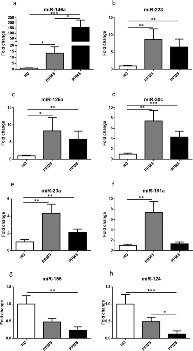
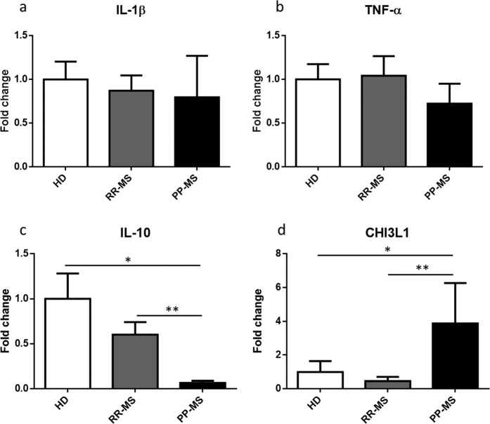
It is widely recognized that monocytes-macrophages adopt a wide variety of phenotypes, influencing the inflammatory activity and demyelination in Multiple Sclerosis (MS). However, how the phenotype of human monocytes evolves in the course of MS is largely unknown. The aim of our preliminary study was to analyse in monocytes of relapsing-remitting and progressive forms of MS patients the expression of a set of miRNAs which impact monocyte-macrophage immune function and their communication with brain cells. Quantitative PCR showed that miRNAs with anti-inflammatory functions, which promote pro-regenerative polarization, are increased in MS patients, while pro-inflammatory miR-155 is downregulated in the same patients. These changes may indicate the attempt of monocytes to counteract neuroinflammation. miR-124, an anti-inflammatory marker but also of myeloid cell quiescence was strongly downregulated, especially in progressive MS patients, suggesting complete loss of homeostatic monocyte function in the progressive disease phase. Profiling of miRNAs that control monocyte polarization may help to define not only the activation state of monocytes in the course of the disease but also novel pathogenic mechanisms.
Overview
The claims and statistics of this paper haven’t been checked yet.
Run the check — free, no account needed. It reads the open-access text and checks the sources it cites.
Integrity signals
Registry
Cites
Statistics
Figures
Verification
Claims the paper makes
The claims and statistics of this paper haven’t been checked yet.
Run the check — free, no account needed. It reads the open-access text and checks the sources it cites.
Claims other papers make about this one
Each is checked against this paper’s own text: does it say what the citing paper says it does?
In the citing paper
“Finally, miR-124, a marker of anti-inflammation and immune cell quiescence, was strongly decreased in progressive MS patients, suggesting the therapeutic prospect of MS by targeting inflammatory responses (Amoruso et al. 2020) (Fig. 3).”
From MiR-124 and MiR-155 as Therapeutic Targets in Microglia-Mediated Inflammation in Multiple Sclerosis (2025) · cites this paper as Amoruso et al. 2020
Self-check
Percentages, sums and reported differences re-computed from the paper’s own numbers.
The self-check needs the paper’s full text.
Citations
Cites
Defining the clinical course of multiple sclerosis: The 2013 revisions
Lublin, F. D., Reingold, S. C., Cohen, J. A., Cutter, G. R., Sørensen, P. S., Thompson, A. J., Wolinsky, J. S., Balcer, L. J., Banwell, B., Barkhof, F., et al. (2014) · Neurology · doi:10.1212/wnl.0000000000000560 · Cited by 3,177
Immunology of multiple sclerosis
Sospedra, M., & Martin, R. (2005) · Annual Review of Immunology · doi:10.1146/annurev.immunol.23.021704.115707 · Cited by 2,149
Macrophage Polarization Comes of Age
Mantovani, Sica, & Locati, M. (2005) · Immunity · doi:10.1016/j.immuni.2005.10.001 · Cited by 1,259
Ridder, K., Keller, S., Dams, M., Rupp, A.-K., Schlaudraff, J., Del Turco, D., Starmann, J., Macas, J., Karpova, D., Devraj, K., et al. (2014) · PLoS Biology · doi:10.1371/journal.pbio.1001874 · Cited by 412
Prada, I., Gabrielli, M., Turola, E., Iorio, A., D’Arrigo, G., Parolisi, R., De Luca, M., Pacifici, M., Bastoni, M., Lombardi, M., et al. (2018) · Acta Neuropathologica · doi:10.1007/s00401-017-1803-x · Cited by 312
Self-citationmiR-125a-5p Regulates Differential Activation of Macrophages and Inflammation
Banerjee, S., Cui, H., Xie, N., Tan, Z., Yang, S., Icyuz, M., Thannickal, V. J., Abraham, E., & Liu, G. (2013) · Journal of Biological Chemistry · doi:10.1074/jbc.m112.426866 · Cited by 258
Jovičić, A., Roshan, R., Moisoi, N., Pradervand, S., Moser, R., Pillai, B., & Luthi-Carter, R. (2013) · The Journal of Neuroscience · doi:10.1523/jneurosci.0600-12.2013 · Cited by 251
miR‐155 as a multiple sclerosis–relevant regulator of myeloid cell polarization
Moore, C. S., Rao, V. T. S., Durafourt, B. A., Bedell, B. J., Ludwin, S. K., Bar‐Or, A., & Antel, J. P. (2013) · Annals of Neurology · doi:10.1002/ana.23967 · Cited by 219
Reijerkerk, A., Lopez-Ramirez, M. A., van het Hof, B., Drexhage, J. A. R., Kamphuis, W. W., Kooij, G., Vos, J. B., van der Pouw Kraan, T. C. T. M., van Zonneveld, A. J., Horrevoets, A. J., et al. (2013) · The Journal of Neuroscience · doi:10.1523/jneurosci.3965-12.2013 · Cited by 138
Research works printed but not linked: 51
- Compston, A. & Coles, A. Multiple sclerosis. Lancet 372, 1502–1517 (2008). · DOI
- Sospedra, M. & Martin, R. Immunology of Multiple Sclerosis. Semin Neurol 36, 115–127 (2016). · DOI
- Milo, R. & Kahana, E. Multiple sclerosis: geoepidemiology, genetics and the environment. Autoimmun Rev 9, A387–394 (2010). · DOI
- Hemmer, B., Kerschensteiner, M. & Korn, T. Role of the innate and adaptive immune responses in the course of multiple sclerosis. Lancet Neurol 14, 406–419 (2015). · DOI
- Henderson, A. P. D., Barnett, M. H., Parratt, J. D. E. & Prineas, J. W. Multiple sclerosis: distribution of inflammatory cells in newly forming lesions. Ann. Neurol. 66, 739–753 (2009). · DOI
- Lucchinetti, C. et al. Heterogeneity of multiple sclerosis lesions: implications for the pathogenesis of demyelination. Ann. Neurol. 47, 707–717 (2000). · DOI
- Wang, N., Liang, H. & Zen, K. Molecular mechanisms that influence the macrophage m1-m2 polarization balance. Front Immunol 5, 614 (2014).
- Martinez, F. O., Sica, A., Mantovani, A. & Locati, M. Macrophage activation and polarization. Front. Biosci. 13, 453–461 (2008). · DOI · Paper page
- Jakobsson, J. et al. Monocyte and microglial activation in patients with mood-stabilized bipolar disorder. J Psychiatry Neurosci 40, 250–258 (2015). · DOI
- Locatelli, G. et al. Mononuclear phagocytes locally specify and adapt their phenotype in a multiple sclerosis model. Nat. Neurosci. 21, 1196–1208 (2018). · DOI · Paper page
- Filipowicz, W., Bhattacharyya, S. N. & Sonenberg, N. Mechanisms of post-transcriptional regulation by microRNAs: are the answers in sight? Nat. Rev. Genet. 9, 102–114 (2008). · DOI
- Essandoh, K., Li, Y., Huo, J. & Fan, G.-C. MiRNA-Mediated Macrophage Polarization and its Potential Role in the Regulation of Inflammatory Response. Shock 46, 122–131 (2016). · DOI
- Vella, L. J., Hill, A. F. & Cheng, L. Focus on Extracellular Vesicles: Exosomes and Their Role in Protein Trafficking and Biomarker Potential in Alzheimer’s and Parkinson’s Disease. Int J Mol Sci 17, 173 (2016). · DOI
- Selmaj, I. et al. Global exosome transcriptome profiling reveals biomarkers for multiple sclerosis. Ann. Neurol. 81, 703–717 (2017). · DOI
- Kimura, K. et al. Circulating exosomes suppress the induction of regulatory T cells via let-7i in multiple sclerosis. Nat Commun 9, 17 (2018). · DOI
- Jagot, F. & Davoust, N. Is It worth Considering Circulating microRNAs in Multiple Sclerosis? Front Immunol 7, 129 (2016). · DOI · Paper page
- Keller, A. et al. Multiple sclerosis: microRNA expression profiles accurately differentiate patients with relapsing-remitting disease from healthy controls. PLoS ONE 4, e7440 (2009). · DOI
- Junker, A. et al. MicroRNA profiling of multiple sclerosis lesions identifies modulators of the regulatory protein CD47. Brain 132, 3342–3352 (2009). · DOI · Paper page
- Ridolfi, E. et al. Expression and Genetic Analysis of MicroRNAs Involved in Multiple Sclerosis. Int J Mol Sci 14, 4375–4384 (2013). · DOI
- Fenoglio, C. et al. Expression and genetic analysis of miRNAs involved in CD4+ cell activation in patients with multiple sclerosis. Neurosci. Lett. 504, 9–12 (2011). · DOI
- Ghorbani, S. et al. MicroRNA-181 Variants Regulate T Cell Phenotype in the Context of Autoimmune Neuroinflammation. Front Immunol 8, 758 (2017). · DOI
- Fenoglio, C. et al. Effect of fingolimod treatment on circulating miR-15b, miR23a and miR-223 levels in patients with multiple sclerosis. J. Neuroimmunol. 299, 81–83 (2016). · DOI
- Ghadiri, N. et al. Analysis of the expression of mir-34a, mir-199a, mir-30c and mir-19a in peripheral blood CD4+T lymphocytes of relapsing-remitting multiple sclerosis patients. Gene 659, 109–117 (2018). · DOI
- Lu, L. et al. Time Series miRNA-mRNA integrated analysis reveals critical miRNAs and targets in macrophage polarization. Sci Rep 6, 37446 (2016). · DOI
- Xie, W. et al. MiR-181a regulates inflammation responses in monocytes and macrophages. PLoS ONE 8, e58639 (2013). · DOI
- Ceolotto, G. et al. miR-30c-5p regulates macrophage-mediated inflammation and pro-atherosclerosis pathways. Cardiovasc. Res. 113, 1627–1638 (2017). · DOI
- Ponomarev, E. D., Veremeyko, T. & Weiner, H. L. MicroRNAs are universal regulators of differentiation, activation, and polarization of microglia and macrophages in normal and diseased CNS. Glia 61, 91–103 (2013). · DOI · Paper page
- Harraz, M. M., Xu, J.-C., Guiberson, N., Dawson, T. M. & Dawson, V. L. MiR-223 regulates the differentiation of immature neurons. Mol Cell Ther 2 (2014). · DOI
- Blonda, M., Amoruso, A., Grasso, R., Di Francescantonio, V. & Avolio, C. Multiple Sclerosis Treatments Affect Monocyte-Derived Microvesicle Production. Front Neurol 8 (2017). · DOI
- Chuluundorj, D., Harding, S. A., Abernethy, D. & La Flamme, A. C. Expansion and preferential activation of the CD14(+)CD16(+) monocyte subset during multiple sclerosis. Immunol. Cell Biol. 92, 509–517 (2014). · DOI
- Waschbisch, A. et al. Pivotal Role for CD16+ Monocytes in Immune Surveillance of the Central Nervous System. J. Immunol. 196, 1558–1567 (2016). · DOI
- Wu, D. et al. Brain 491 endothelial miR-146a negatively modulates T-cell adhesion through repressing multiple 492 targets to inhibit NF-κB activation. J Cereb Blood Flow Metab. 35, 412–23 (2015). · DOI
- Wu, H. et al. Extracellular vesicles containing miR-146a attenuate experimental colitis by targeting TRAF6 and IRAK1. Int. Immunopharmacol. 68, 204–212 (2019). · DOI
- Fumagalli, M., Lombardi, M., Gressens, P. & Verderio, C. How to reprogram microglia toward 497 beneficial functions. Glia 66, 2531–49 (2018). · DOI
- Galloway, D. A. et al. miR-223 promotes regenerative myeloid cell phenotype and function in the demyelinated central nervous system. Glia. 67, 857–869 (2018). · DOI · Paper page
- Bartel, D. P. MicroRNAs: genomics, biogenesis, mechanism, and function. Cell. 116, 281–97 (2004). · DOI
- Ying, W. et al. MicroRNA-223 is a crucial mediator of PPARγ-regulated alternative macrophage activation. J. Clin. Invest. 125, 4149–4159 (2015). · DOI · Paper page
- Ismail, N. et al. Macrophage microvesicles induce macrophage differentiation and miR-223 transfer. Blood 121, 984–995 (2013). · DOI
- Seoudi, A. M., Lashine, Y. A. & Abdelaziz, A. I. MicroRNA-181a - a tale of discrepancies. Expert Rev Mol Med 14, e5 (2012). · DOI
- Sarchielli, P. et al. Fibroblast growth factor-2 levels are elevated in the cerebrospinal fluid of multiple sclerosis patients. Neurosci. Lett. 435, 223–228 (2008). · DOI
- Varendi, K., Kumar, A., Härma, M.-A. & Andressoo, J.-O. miR-1, miR-10b, miR-155, and miR-191 are novel regulators of BDNF. Cell. Mol. Life Sci. 71, 4443–4456 (2014). · DOI · Paper page
- Gaudet, A. D. et al. miR-155 Deletion in Mice Overcomes Neuron-Intrinsic and Neuron-Extrinsic Barriers to Spinal Cord Repair. J. Neurosci. 36, 8516–8532 (2016). · DOI
- Slota, J. A. & Booth, S. A. MicroRNAs in Neuroinflammation: Implications in Disease Pathogenesis, Biomarker Discovery and Therapeutic Applications. Noncoding RNA 5 (2019). · DOI
- Dutta, R. et al. Hippocampal demyelination and memory dysfunction are associated with increased levels of the neuronal microRNA miR-124 and reduced AMPA receptors. Ann. Neurol. 73, 637–645 (2013). · DOI · Paper page
- Kemanetzoglou, E. & Andreadou, E. CNS Demyelination with TNF-α Blockers. Curr Neurol Neurosci Rep 17, 36 (2017). · DOI
- O’Garra, A. & Vieira, P. T(H)1 cells control themselves by producing interleukin-10. Nat. Rev. Immunol. 7, 425–428 (2007). · DOI
- Novakova, L. et al. Cerebrospinal fluid biomarkers as a measure of disease activity and treatment efficacy in relapsing-remitting multiple sclerosis. J. Neurochem. 141, 296–304 (2017). · DOI
- Burman, J. et al. YKL-40 is a CSF biomarker of intrathecal inflammation in secondary progressive multiple sclerosis. J. Neuroimmunol. 292, 52–57 (2016). · DOI
- Hinsinger, G. et al. Chitinase 3-like proteins as diagnostic and prognostic biomarkers of multiple sclerosis. Mult. Scler. 21, 1251–1261 (2015). · DOI
- Rathcke, C. N. & Vestergaard, H. YKL-40, a new inflammatory marker with relation to insulin resistance and with a role in endothelial dysfunction and atherosclerosis. Inflamm. Res. 55, 221–227 (2006). · DOI
- Amoruso, A. et al. Effect of fingolimod action on the release of monocyte-derived microvesicles in multiple sclerosis patients. J. Neuroimmunol. 323, 43–48 (2018). · DOI
Cited by
Sorted by citations
MicroRNA-124: A Key Player in Microglia-Mediated Inflammation in Neurological Diseases
Zhao, J., He, Z., & Wang, J. (2021) · Frontiers in Cellular Neuroscience · doi:10.3389/fncel.2021.771898 · Cited by 66
miR-124: A Promising Therapeutic Target for Central Nervous System Injuries and Diseases
Xu, J., Zheng, Y., Wang, L., Liu, Y., Wang, X., Li, Y., & Chi, G. (2022) · Cellular and Molecular Neurobiology · doi:10.1007/s10571-021-01091-6 · Cited by 55
MiR-124 and MiR-155 as Therapeutic Targets in Microglia-Mediated Inflammation in Multiple Sclerosis
Asl, E. R., Hosseini, S. E., Tahmasebi, F., Bolandi, N., & Barati, S. (2025) · Cellular and Molecular Neurobiology · doi:10.1007/s10571-025-01578-6 · Cited by 19
Metadata
- Authors
- Amoruso, A., Blonda, M., Gironi, M., Grasso, R., Di Francescantonio, V., Scaroni, F., Furlan, R., Verderio, C., & Avolio, C.
- Venue
- Scientific Reports
- Published
- 2020-04-09
- Volume
- 10
- Issue
- 1
- ISSN
- 2045-2322
- PMC
- PMC7145856
- Cited by
- 46
- References
- 9
- Language
- en
About these checks
Registry, retraction, Cites and statistics results are computed, not judged, with one exception: a printed reference that no index lists is read once by a model before it counts as a citation error. Claims are extracted by a model and kept only when their quote appears verbatim in the text; each cited source is then checked, with a model’s help, for whether it says what the claim relies on. Those verdicts can be wrong — read the claim’s quote and the cited source before relying on one.
Statistics and arithmetic are recomputed, not judged.
Confidence bands are shown only for fields calibrated on our annotated test set. None is calibrated yet, so no band is shown.
What counts as a claim ›How we measure confidence ›How we check this ›
Are you an author, or do you see a problem with this page? Request a review
