

The DOI matches its Crossref record; 4 of the 4 references it cites were found in a registry or an index.
Structural changes in the oral microbiome of the adolescent patients with moderate or severe dental fluorosis
Abstract
Dental fluorosis is a very prevalent endemic disease. Although oral microbiome has been reported to correlate with different oral diseases, there appears to be an absence of research recognizing any relationship between the severity of dental fluorosis and the oral microbiome. To this end, we investigated the changes in oral microbial community structure and identified bacterial species associated with moderate and severe dental fluorosis. Salivary samples of 42 individuals, assigned into Healthy (N = 9), Mild (N = 14) and Moderate/Severe (M&S, N = 19), were investigated using the V4 region of 16S rRNA gene. The oral microbial community structure based on Bray Curtis and Weighted Unifrac were significantly changed in the M&S group compared with both of Healthy and Mild. As the predominant phyla, Firmicutes and Bacteroidetes showed variation in the relative abundance among groups. The Firmicutes/Bacteroidetes (F/B) ratio was significantly higher in the M&S group. LEfSe analysis was used to identify differentially represented taxa at the species level. Several genera such as Streptococcus mitis, Gemella parahaemolysans, Lactococcus lactis, and Fusobacterium nucleatum , were significantly more abundant in patients with moderate/severe dental fluorosis, while Prevotella melaninogenica and Schaalia odontolytica were enriched in the Healthy group. In conclusion, our study indicates oral microbiome shift in patients with moderate/severe dental fluorosis. We identified several differentially represented bacterial species enriched in moderate and severe fluorosis. Findings from this study suggests that the roles of these bacteria in oral health and related diseases warrant more consideration in patients with moderate and severe fluorosis.
Overview
The claims and statistics of this paper haven’t been checked yet.
Run the check — free, no account needed. It reads the open-access text and checks the sources it cites.
Integrity signals
Registry
Cites
Statistics
Figures
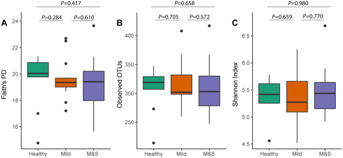
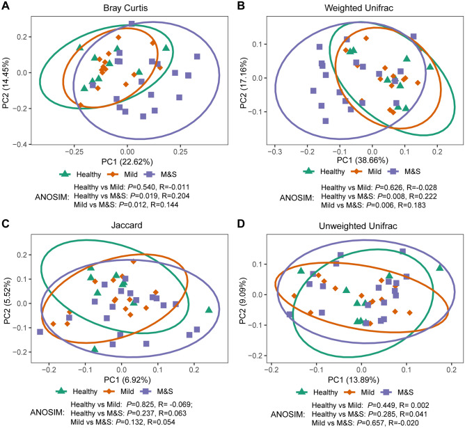
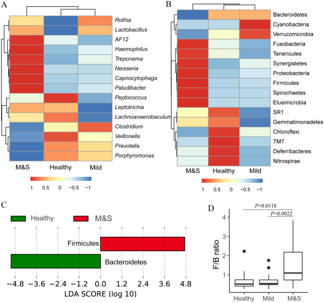
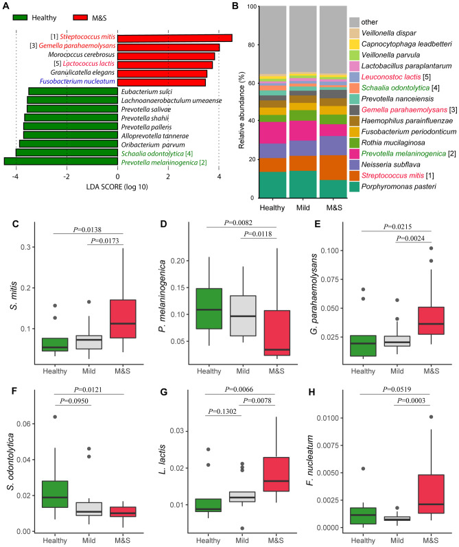
Verification
Claims the paper makes
The claims and statistics of this paper haven’t been checked yet.
Run the check — free, no account needed. It reads the open-access text and checks the sources it cites.
Claims other papers make about this one
Each is checked against this paper’s own text: does it say what the citing paper says it does?
The source backs part of the claim, not all of it — or backs it for a different population, condition or comparison.
In the citing paper
“For instance, Wang et al. demonstrated significant alterations in the oral microbial community structure among adolescents with severe dental fluorosis, compared to those without oral health issues [10].”
From Severity-dependent alterations in oral microbiota and antibiotic resistance in children with dental fluorosis (2026) · cites this paper as [10]
Experimental — accuracy not yet measuredThe cited source says what the claim attributes to it.
In the citing paper
“The specific differences in bacterial species observed in this study are inconsistent with those in the article published by Wang et al [23].”
From Saliva microbiome alterations in dental fluorosis population (2023) · cites this paper as [23]
In the citing paper
“Wang et al. previously analyzed saliva samples from 42 adolescents with moderate or severe dental fluorosis, revealing a unique microbial profile in these patients [23].”
From Saliva microbiome alterations in dental fluorosis population (2023) · cites this paper as [23]
In the citing paper
“Few studies to date have examined dental fluorosis-related changes in the oral microbiota [23].”
From Saliva microbiome alterations in dental fluorosis population (2023) · cites this paper as [23]
In the citing paper
“Owing to limitations in the sample size and disease severity in that study, however, the applicability of these results to other dental fluorosis patients such as those with mild disease remains unclear.”
From Saliva microbiome alterations in dental fluorosis population (2023) · cites this paper as [23]
Self-check
Percentages, sums and reported differences re-computed from the paper’s own numbers.
The self-check needs the paper’s full text.
Citations
Cites
Deblur Rapidly Resolves Single-Nucleotide Community Sequence Patterns
Amir, A., McDonald, D., Navas-Molina, J. A., Kopylova, E., Morton, J. T., Zech Xu, Z., Kightley, E. P., Thompson, L. R., Hyde, E. R., Gonzalez, A., et al. (2017) · mSystems · doi:10.1128/msystems.00191-16 · Cited by 1,881
Ectopic colonization of oral bacteria in the intestine drives T H 1 cell induction and inflammation
Atarashi, K., Suda, W., Luo, C., Kawaguchi, T., Motoo, I., Narushima, S., Kiguchi, Y., Yasuma, K., Watanabe, E., Tanoue, T., et al. (2017) · Science · doi:10.1126/science.aan4526 · Cited by 957
Human Microbiome Fusobacterium Nucleatum in Esophageal Cancer Tissue Is Associated with Prognosis
Yamamura, K., Baba, Y., Nakagawa, S., Mima, K., Miyake, K., Nakamura, K., Sawayama, H., Kinoshita, K., Ishimoto, T., Iwatsuki, M., et al. (2016) · Clinical Cancer Research · doi:10.1158/1078-0432.ccr-16-1786 · Cited by 457
Hsieh, Y.-Y., Tung, S.-Y., Pan, H.-Y., Yen, C.-W., Xu, H.-W., Lin, Y.-J., Deng, Y.-F., Hsu, W.-T., Wu, C.-S., & Li, C. (2018) · Scientific Reports · doi:10.1038/s41598-017-18596-0 · Cited by 269
Research works printed but not linked: 40
- Zhou, Y. et al. The prevalence and associated risk indicators of dental fluorosis in China: Findings from the 4th national oral health survey. Chin. J. Dent. Res. 21, 205–211 (2018).
- Verma, A., Shetty, B. K., Guddattu, V., Chourasia, M. K. & Pundir, P. High prevalence of dental fluorosis among adolescents is a growing concern: A school based cross-sectional study from Southern India. Environ. Health Prev. Med. 22, 17 (2017). · DOI
- Chankanka, O., Levy, S. M., Warren, J. J. & Chalmers, J. M. A literature review of aesthetic perceptions of dental fluorosis and relationships with psychosocial aspects/oral health-related quality of life. Commun. Dent. Oral Epidemiol. 38, 97–109 (2010). · DOI
- Vora, K. S. & Vora, P. K. Assessment of periodontal status of the patients with dental fluorosis in area with natural high levels of fluoride: A cross-sectional survey. Dental Hypotheses 4, 127 (2013). · DOI
- Costa, S. d. M. et al. Dental caries and endemic dental fluorosis in rural communities, Minas Gerais, Brazil. Revista Brasileira de Epidemiologia 16, 1021–1028 (2013). · DOI
- Menya, D. et al. Dental fluorosis and oral health in the African Esophageal Cancer Corridor: Findings from the Kenya ESCCAPE case–control study and a pan-African perspective. Int. J. Cancer 145, 99–109 (2019). · DOI
- Dewhirst, F. E. et al. The human oral microbiome. J. Bacteriol. 192, 5002–5017 (2010). · DOI · Paper page
- Ito, K. et al. Crystal structure of glucansucrase from the dental caries pathogen Streptococcus mutans. J. Mol. Biol. 408, 177–186 (2011). · DOI
- Takahashi, N. & Nyvad, B. The role of bacteria in the caries process: ecological perspectives. J. Dent. Res. 90, 294–303 (2011). · DOI
- Yang, S.-F. et al. Compositional and functional variations of oral microbiota associated with the mutational changes in oral cancer. Oral. Oncol. 77, 1–8 (2018). · DOI
- Fan, X. et al. Human oral microbiome and prospective risk for pancreatic cancer: A population-based nested case-control study. Gut 67, 120–127 (2018). · DOI · Paper page
- Farrell, J. J. et al. Variations of oral microbiota are associated with pancreatic diseases including pancreatic cancer. Gut 61, 582–588 (2012). · DOI · Paper page
- Flemer, B. et al. The oral microbiota in colorectal cancer is distinctive and predictive. Gut 67, 1454–1463 (2018). · DOI · Paper page
- Medina-Solis, C. E. et al. Dental fluorosis prevalence and severity using Dean’s index based on six teeth and on 28 teeth. Clin. Oral Invest. 12, 197–202 (2008). · DOI
- Dean, H., Arnold, F. & Elvolve, E. Additional studies of the relation of fluoride domestic waters to dental caries experience in 4425 white children aged 12–14 years in 13 cities in 4 states. Public Health Rep. 65, 1403–1408 (1942). · DOI
- Magoč, T. & Salzberg, S. L. FLASH: Fast length adjustment of short reads to improve genome assemblies. Bioinformatics 27, 2957–2963 (2011). · DOI · Paper page
- Martin, M. Cutadapt removes adapter sequences from high-throughput sequencing reads. EMBnet. J. 17, 10–12 (2011). · DOI · Paper page
- Bolyen, E. et al. Reproducible, interactive, scalable and extensible microbiome data science using QIIME 2. Nat. Biotechnol. 37, 852–857 (2019). · DOI · Paper page
- Deng, F. et al. The vaginal and fecal microbiomes are related to pregnancy status in beef heifers. J. Anim. Sci. Biotechnol. 10, 1–13 (2019). · DOI
- Pedregosa, F. et al. Scikit-learn: Machine learning in python. J. Mach. Learn. Res. 12, 2825–2830 (2011).
- McDonald, D. et al. An improved Greengenes taxonomy with explicit ranks for ecological and evolutionary analyses of bacteria and archaea. ISME J. 6, 610 (2012). · DOI
- Camacho, C. et al. BLAST+: Architecture and applications. BMC Bioinformatics 10, 421 (2009). · DOI · Paper page
- O’Leary, N. A. et al. Reference sequence (RefSeq) database at NCBI: current status, taxonomic expansion, and functional annotation. Nucleic Acids Res. 44, D733–D745 (2016). · DOI · Paper page
- Wickham, H. ggplot2: elegant graphics for data analysis (Springer, New York, 2016). · DOI
- Segata, N. et al. Metagenomic biomarker discovery and explanation. Genome Biol. 12, R60 (2011). · DOI · Paper page
- Brennan, C. A. & Garrett, W. S. Fusobacterium nucleatum—symbiont, opportunist and oncobacterium. Nat. Rev. Microbiol. 17, 156–166 (2019). · DOI · Paper page
- Fernandes, J., Su, W., Rahat-Rozenbloom, S., Wolever, T. & Comelli, E. Adiposity, gut microbiota and faecal short chain fatty acids are linked in adult humans. Nutr. Diab. 4, e121–e121 (2014). · DOI · Paper page
- Delzenne, N. M. & Cani, P. D. Interaction between obesity and the gut microbiota: relevance in nutrition. Annu. Rev. Nutr. 31, 15–31 (2011). · DOI
- Yuan, X. et al. Green tea liquid consumption alters the human intestinal and oral microbiome. Mol. Nutr. Food Res. 62, 1800178 (2018). · DOI
- Engström, K., Petersson, L., Sjöström, I. & Twetman, S. Composition of the salivary microflora during habitual consumption of fluoridated milk. Acta Odontol. Scand. 62, 143–146 (2004). · DOI
- Koopman, J. E. et al. The effect of fixed orthodontic appliances and fluoride mouthwash on the oral microbiome of adolescents: a randomized controlled clinical trial. PLoS ONE 10, e0137318 (2015). · DOI · Paper page
- Michel-Crosato, E., Biazevic, M. G. H. & Crosato, E. Relationship between dental fluorosis and quality of life: A population based study. Braz. Oral Res. 19, 150–155 (2005). · DOI
- Almerich-Silla, J. M., Montiel-Company, J. M. & Ruiz-Miravet, A. Caries and dental fluorosis in a western Saharan population of refugee children. Eur. J. Oral Sci. 116, 512–517 (2008). · DOI
- Aoba, T. Solubility properties of human tooth mineral and pathogenesis of dental caries. Oral Dis. 10, 249–257 (2004). · DOI
- Mitchell, J. Streptococcus mitis: walking the line between commensalism and pathogenesis. Mol. Oral Microbiol. 26, 89–98 (2011). · DOI · Paper page
- Abranches, J. et al. Biology of oral streptococci. Gram‐Positive Pathogens, 426–434 (2019). · DOI
- De Soet, J., Nyvad, B. & Kilian, M. Strain-related acid production by oral Streptococci. Caries Res. 34, 486–490 (2000). · DOI
- Gross, E. L. et al. Bacterial 16S sequence analysis of severe caries in young permanent teeth. J. Clin. Microbiol. 48, 4121–4128 (2010). · DOI
- Gross, E. L. et al. Beyond Streptococcus mutans: dental caries onset linked to multiple species by 16S rRNA community analysis. PLoS ONE 7, e47722 (2012). · DOI
- Zhao, H. et al. Variations in oral microbiota associated with oral cancer. Sci. Rep. 7, 1–10 (2017). · DOI · Paper page
Cited by
Sorted by citations
Saliva microbiome alterations in dental fluorosis population
Liu, S., Song, Q., Zhang, C., Li, M., Li, Z., Liu, Y., Xu, L., Xie, X., Zhao, L., Zhang, R., et al. (2023) · Journal of Oral Microbiology · doi:10.1080/20002297.2023.2180927 · Cited by 24
Self-citationDoes fluoride exposure impact on the human microbiome?
Moran, G. P., Zgaga, L., Daly, B., Harding, M., & Montgomery, T. (2023) · Toxicology Letters · doi:10.1016/j.toxlet.2023.03.001 · Cited by 18
Xia, Y., Peng, S., Yu, J., Wang, Y., Ye, Y., Liu, M., Shang, L., Cui, X., Wang, P., & Ding, Z. (2026) · Journal of Oral Microbiology · doi:10.1080/20002297.2026.2714622
Metadata
- Authors
- Wang, Q., Chen, X., Hu, H., Wei, X., Wang, X., Peng, Z., Ma, R., Zhao, Q., Zhao, J., Liu, J., & Deng, F.
- Venue
- Scientific Reports
- Published
- 2021-02-03
- Volume
- 11
- Issue
- 1
- ISSN
- 2045-2322
- PMC
- PMC7859183
- Cited by
- 22
- References
- 4
- Language
- en
- Funders
- Science and Technology Plan of Guizhou Province (QianKeHe[2018]5772-029)
- Outstanding Young Talent Project of Zunyi Medical University (17zy-002)
- Science and Technology fund of Guizhou Health Commission (gzwjkj2020-1-240)
- Top Science and Technology Talent Project in Higher Education Institution of Guizhou Province (Qian-Jiao-He KY Zi [2016]080)
About these checks
Registry, retraction, Cites and statistics results are computed, not judged, with one exception: a printed reference that no index lists is read once by a model before it counts as a citation error. Claims are extracted by a model and kept only when their quote appears verbatim in the text; each cited source is then checked, with a model’s help, for whether it says what the claim relies on. Those verdicts can be wrong — read the claim’s quote and the cited source before relying on one.
Statistics and arithmetic are recomputed, not judged.
Confidence bands are shown only for fields calibrated on our annotated test set. None is calibrated yet, so no band is shown.
What counts as a claim ›How we measure confidence ›How we check this ›
Are you an author, or do you see a problem with this page? Request a review
How to cite
Cite CoChat for the checks on this page, and the paper for its own findings.
- This page
- CoChat. (2026). Citation check: Structural changes in the oral microbiome of the adolescent patients with moderate or severe dental fluorosis. https://app.cochat.ai/papers/10.1038/s41598-021-82709-z
- The paper
- Wang, Q., Chen, X., Hu, H., Wei, X., Wang, X., Peng, Z., Ma, R., Zhao, Q., Zhao, J., Liu, J., & Deng, F. (2021). Structural changes in the oral microbiome of the adolescent patients with moderate or severe dental fluorosis. Scientific Reports. https://doi.org/10.1038/s41598-021-82709-z
