

The DOI matches its Crossref record; 1 of the 1 references it cites were found in a registry or an index.
MicroRNA profiles in hippocampal granule cells and plasma of rats with pilocarpine-induced epilepsy - comparison with human epileptic samples
Abstract
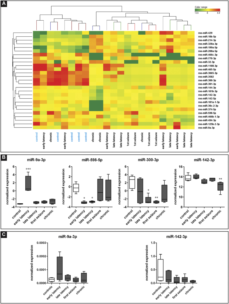
The identification of biomarkers of the transformation of normal to epileptic tissue would help to stratify patients at risk of epilepsy following brain injury and inform new treatment strategies. MicroRNAs (miRNAs) are an attractive option in this direction. In this study, miRNA microarrays were performed on laser-microdissected hippocampal granule cell layer (GCL) and on plasma, at different time points in the development of pilocarpine-induced epilepsy in the rat: latency, first spontaneous seizure and chronic epileptic phase. Sixty-three miRNAs were differentially expressed in the GCL when considering all time points. Three main clusters were identified that separated the control and chronic phase groups from the latency group and from the first spontaneous seizure group. MiRNAs from rats in the chronic phase were compared to those obtained from the laser-microdissected GCL of epileptic patients, identifying several miRNAs (miR-21-5p, miR-23a-5p, miR-146a-5p and miR-181c-5p) that were up-regulated in both human and rat epileptic tissue. Analysis of plasma samples revealed different levels between control and pilocarpine-treated animals for 27 miRNAs. Two main clusters were identified that segregated controls from all other groups. Those miRNAs that are altered in plasma before the first spontaneous seizure, like miR-9a-3p, may be proposed as putative biomarkers of epileptogenesis.
Overview
The claims and statistics of this paper haven’t been checked yet.
Run the check — free, no account needed. It reads the open-access text and checks the sources it cites.
Integrity signals
Registry
Cites
Statistics
Figures
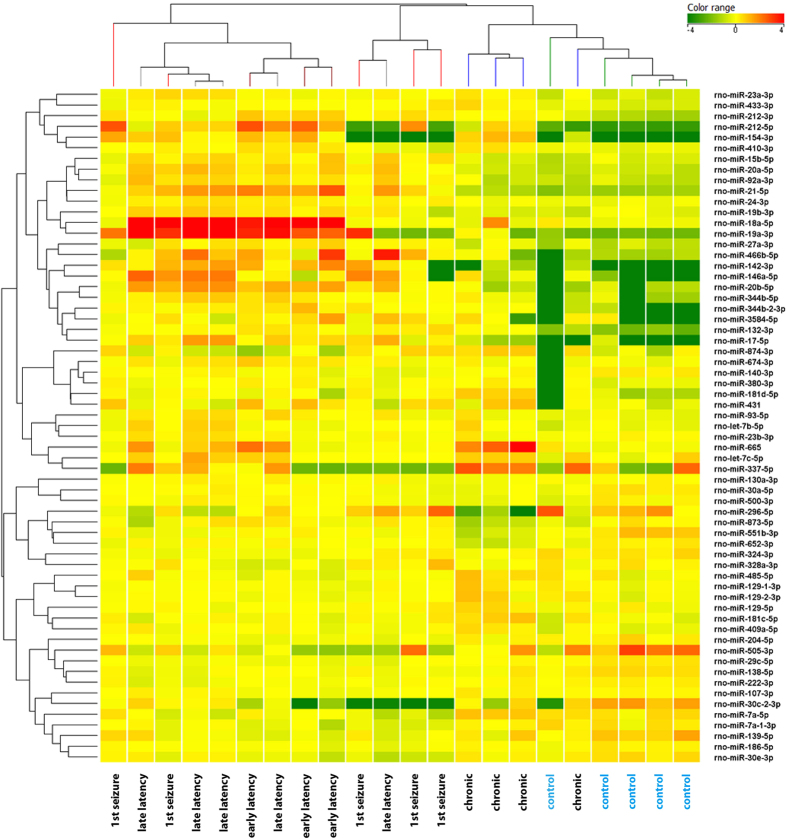
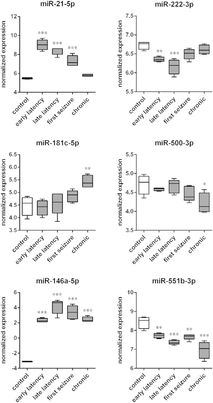
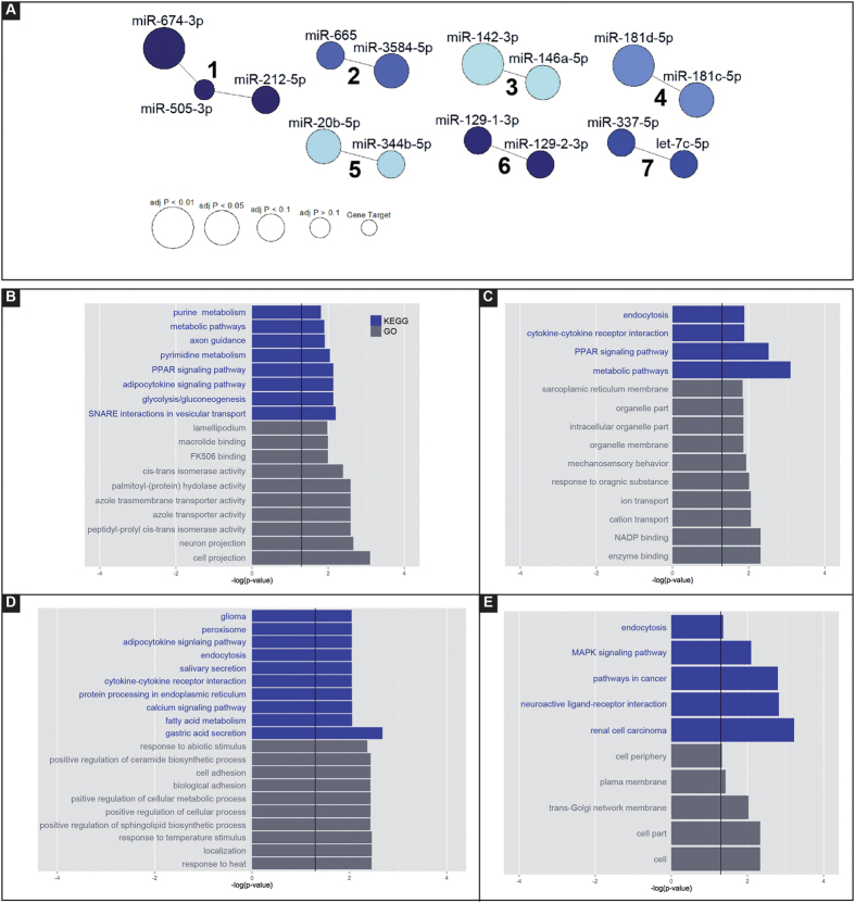
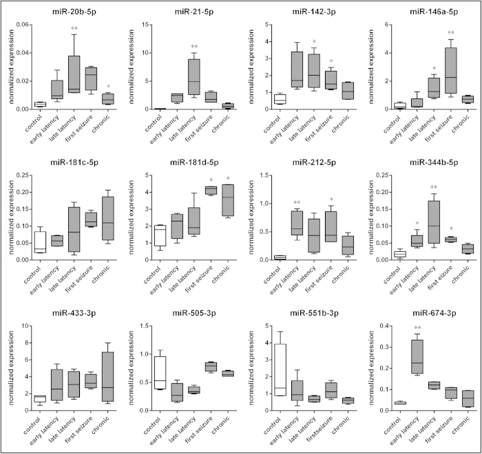
Verification
Claims the paper makes
The claims and statistics of this paper haven’t been checked yet.
Run the check — free, no account needed. It reads the open-access text and checks the sources it cites.
Claims other papers make about this one
Each is checked against this paper’s own text: does it say what the citing paper says it does?
In the citing paper
“Plasma from rodents subject to evoked seizures and animals with chronic epilepsy display elevations in brain-enriched and inflammation-associated miRNAs (Gorter et al., 2014; Roncon et al., 2015; Brennan et al., 2020).”
From Brain cell-specific origin of circulating microRNA biomarkers in experimental temporal lobe epilepsy (2023) · cites this paper as Roncon et al., 2015
Self-check
Percentages, sums and reported differences re-computed from the paper’s own numbers.
The self-check needs the paper’s full text.
Citations
Cites
MicroRNA-146a: A Key Regulator of Astrocyte-Mediated Inflammatory Response
Iyer, A., Zurolo, E., Prabowo, A., Fluiter, K., Spliet, W. G. M., van Rijen, P. C., Gorter, J. A., & Aronica, E. (2012) · PLoS ONE · doi:10.1371/journal.pone.0044789 · Cited by 330
Research works printed but not linked: 58
- Simonato, M. et al. The challenge and promise of anti-epileptic therapy development in animal models. Lancet Neurol 13, 949–60 (2014). · DOI
- Simonato, M. et al. Finding a better drug for epilepsy: preclinical screening strategies and experimental trial design. Epilepsia 53, 1860–7 (2012). · DOI
- Simonato, M., French, J. A., Galanopoulou, A. S. & O’Brien, T. J. Issues for new antiepilepsy drug development. Curr Opin Neurol 26, 195–200 (2013). · DOI
- Galanopoulou, A. S. et al. Identification of new epilepsy treatments: issues in preclinical methodology. Epilepsia 53, 571–82 (2012). · DOI
- Engel, J. Jr. et al. Epilepsy biomarkers. Epilepsia 4, 61–9 (2013). · DOI · Paper page
- Bartel, D. P. MicroRNAs: genomics, biogenesis, mechanism and function. Cell 116, 281–97 (2004). · DOI
- Im, H. I. & Kenny, P. J. MicroRNAs in neuronal function and dysfunction. Trends Neurosci 35, 325–34 (2012). · DOI · Paper page
- McNeill, E. & Van Vactor, D. MicroRNAs shape the neuronal landscape. Neuron 75, 363–79 (2012). · DOI · Paper page
- Vezzani, A., Aronica, E., Mazarati, A. & Pittman, Q. J. Epilepsy and brain inflammation. Exp Neurol 244, 11–21 (2013). · DOI · Paper page
- Mikkonen, M. et al. Remodeling of neuronal circuitries in human temporal lobe epilepsy: increased expression of highly polysialylated neural cell adhesion molecule in the hippocampus and the entorhinal cortex. Ann Neurol 44, 923–34 (1998). · DOI
- Blümcke, I. et al. International consensus classification of hippocampal sclerosis in temporal lobe epilepsy: a Task Force report from the ILAE Commission on Diagnostic Methods. Epilepsia 54, 1315–29 (2013). · DOI
- Wu, J. et al. MicroRNA-421 is a new potential diagnosis biomarker with higher sensitivity and specificity than carcinoembryonic antigen and cancer antigen 125 in gastric cancer. Biomarkers 16, 1–6 (2014).
- Sepramanian, S. et al. Circulating microRNA as biomarkers of acute stroke. Int J Mol Sci 15, 1418–32 (2014). · DOI
- Tan, L. et al. Genome-wide serum microRNA expression profiling identifies serum biomarkers for Alzheimer’s disease. J Alzheimers Dis 40, 1017–27 (2014). · DOI
- Cloutier, F., Marrero, A., O’Connell, C. & Morin, P. J. MicroRNAs as Potential Circulating Biomarkers for Amyotrophic Lateral Sclerosis. J Mol Neurosci 56, 102–112 (2014). · DOI
- Jin, X. F., Wu, N., Wang, L. & Li, J. Circulating microRNAs: a novel class of potential biomarkers for diagnosing and prognosing central nervous system diseases. Cell Mol Neurobiol 33, 601–13 (2013). · DOI
- Zhang, Y. et al. Altered expression levels of miRNAs in serum as sensitive biomarkers for early diagnosis of traumatic injury. J Cell Biochem 112, 2435–42 (2011). · DOI
- Kan, A. A. et al. Genome-wide microRNA profiling of human temporal lobe epilepsy identifies modulators of the immune response. Cell Mol Life Sci 69, 3127–45 (2012). · DOI
- Zucchini, S. et al. Identification of miRNAs differentially expressed in human epilepsy with or without granule cell pathology. PLoS One 9, e105521. 10.1371/journal.pone.0105521 (2014). · DOI
- Risbud, R. M. & Porter, B. E. Changes in microRNA expression in the whole hippocampus and hippocampal synaptoneurosome fraction following pilocarpine induced status epilepticus. PLoS One 8, e53464. 10.1371/journal.pone.0053464 (2013). · DOI · Paper page
- Hu, K. et al. MicroRNA expression profile of the hippocampus in a rat model of temporal lobe epilepsy and miR-34a-targeted neuroprotection against hippocampal neurone cell apoptosis post-status epilepticus. BMC Neurosci 13, 115 (2012). · DOI
- Jimenez-Mateos, E. M. et al. miRNA Expression profile after status epilepticus and hippocampal neuroprotection by targeting miR-132. Am J Pathol 179, 2519–32 (2011). · DOI
- Bot, A. M., Dębski, K. J. & Lukasiuk, K. Alterations in miRNA levels in the dentate gyrus in epileptic rats. PLoS One 8, e76051. 10.1371/journal.pone.0076051 (2013). · DOI
- Gorter, J. A. et al. Hippocampal subregion-specific microRNA expression during epileptogenesis in experimental temporal lobe epilepsy. Neurobiol Dis 62, 508–20 (2013). · DOI · Paper page
- Lucherini, O. M. et al. First report of circulating microRNAs in tumour necrosis factor receptor-associated periodic syndrome (TRAPS). PLoS One 8, e73443. 10.1371/journal.pone.0073443 (2013). · DOI
- Meng, F. et al. Neuronal calcium signaling pathways are associated with the development of epilepsy. Mol Med Rep 11, 196–202 (2015). · DOI
- Yelamanchili, S. V. & Fox, H. S. Defining larger roles for “tiny” RNA molecules: role of miRNAs in neurodegeneration research. J Neuroimmune Pharmacol 5, 63–9 (2010). · DOI
- Bienvenu, T., Diebold, B., Chelly, J. & Isidor, B. Refining the phenotype associated with MEF2C point mutations. Neurogenetics 14, 71–5 (2013). · DOI
- Johnson, M. R. et al. Systems genetics identifies Sestrin 3 as a regulator of a proconvulsant gene network in human epileptic hippocampus. Nat Commun 6, 6031 (2015). · DOI
- Pitkänen, A. & Sutula, T. P. Is epilepsy a progressive disorder? Prospects for new therapeutic approaches in temporal-lobe epilepsy. Lancet Neurol 1, 173–81 (2002). · DOI
- Curia, G., Longo, D., Biagini, G., Jones, R. S. & Avoli, M. The pilocarpine model of temporal lobe epilepsy. J Neurosci Methods 172, 143–57 (2008). · DOI
- Paradiso, B. et al. Localized overexpression of FGF-2 and BDNF in hippocampus reduced mossy fibers sprouting and spontaneous seizures up to 4 weeks after pilocarpine-induced status epilepticus. Epilepsia 52, 572–78 (2011). · DOI
- Wang, X. et al. M129V polymorphism in the prion protein gene is not associated with mesial temporal lobe epilepsy in a Han Chinese population. Eur J Neurol 15, 827–30 (2008). · DOI
- Schauwecker, P. E. Galanin receptor 1 deletion exacerbates hippocampal neuronal loss after systemic kainate administration in mice. PLoS One 5, e15657. 10.1371/journal.pone.0015657 (2010). · DOI
- McColl, C. D., Jacoby, A. S., Shine, J., Iismaa, T. P. & Bekkers, J. M. Galanin receptor-1 knockout mice exhibit spontaneous epilepsy, abnormal EEGs and altered inhibition in the hippocampus. Neuropharmacology 50, 209–18 (2006). · DOI
- Szczurowska, E. & Mareš, P. NMDA and AMPA receptors: development and status epilepticus. Physiol Res 62, S21–38 (2013). · DOI
- Pitkänen, A. et al. Issues related to development of antiepileptogenic therapies. Epilepsia 4, 35–43 (2013). · DOI
- Aronica, E. et al. Expression pattern of miR-146a, an inflammation-associated microRNA, in experimental and human temporal lobe epilepsy. Eur J Neurosci 31, 1100–7 (2010). · DOI · Paper page
- Song, Y. J. et al. Temporal lobe epilepsy induces differential expression of hippocampal miRNAs including let-7e and miR-23a/b. Brain Res 1387, 134–40 (2011). · DOI · Paper page
- Cheng, A. M., Byrom, M. V., Shelton, J. & Ford, L. P. Antisense inhibition of human miRNAs and indications for an involvement of miRNA in cell growth and apoptosis. Nucleic Acids Res 33, 1290–7 (2005). · DOI
- Song, Y. J. et al. miR-23 regulation of lamin B1 is crucial for oligodendrocyte development and myelination. Dis Model Mech 2, 178–88 (2009). · DOI
- Valadi, H. et al. Exosome-mediated transfer of mRNAs and microRNAs is a novel mechanism of genetic exchange between cells. Nat Cell Biol 9, 654–9 (2007). · DOI · Paper page
- Hunter, M. P. et al. Detection of microRNA expression in human peripheral blood microvesicles. PLoS One 3, e3694. 10.1371/journal.pone.0003694 (2008). · DOI
- Liu, D. Z. et al. Brain and blood microRNA expression profiling of ischemic stroke, intracerebral hemorrhage and kainate seizures. J Cereb Blood Flow Metab 30, 92–101 (2010). · DOI
- Kilkenny, C. et al. Animal research: reporting in vivo experiments: the ARRIVE guidelines. J Cereb Blood Flow Metab 31, 991–3 (2011). · DOI · Paper page
- Pellegrino, L. J., Pellegrino, A. S. & Cushman, A. J. A stereotaxic atlas of the rat brain. Plenum Press, New York and London (1979).
- Paradiso, B. et al. Localized delivery of fibroblast growth factor-2 and brain-derived neurotrophic factor reduces spontaneous seizures in an epilepsy model. Proc Natl Acad Sci USA 106, 7191–6 (2009). · DOI
- Mazzuferi, M. et al. Enhancement of GABA(A)-current run-down in the hippocampus occurs at the first spontaneous seizure in a model of temporal lobe epilepsy. Proc Natl Acad Sci USA 107, 3180–5 (2010). · DOI
- Soukupova, M. et al. Impairment of GABA release in the hippocampus at the time of the first spontaneous seizure in the pilocarpine model of temporal lobe epilepsy. Exp Neurol 257, 39–49 (2014). · DOI
- Racine, R. J. Modification of seizure activity by electrical stimulation. II. Motor seizure. Electroencephalogr Clin Neurophysiol 32, 281–94 (1972). · DOI
- Cheng, H. H. et al. Plasma processing conditions substantially influence circulating microRNA biomarkers levels. PLoS One 8, e64795. 10.1371/journal.pone.0064795 (2013). · DOI · Paper page
- Goldsworthy, S. M., Stocktown, P. S., Trempus, C. S., Foley, J. F. & Maronpot, R. R. Effects of fixation on RNA extraction and amplification from laser capture microdissected tissue. Mol Carcinog 25, 86–91 (1999). · DOI
- Burbach, G. J., Dehn, D., Del Turco, D. & Deher, T. Quantification of layer-specific gene expression in the hippocampus: effective use of laser microdissection in combination with quantitative RT-PCR. J of Neurosci Methods 131, 83–91(2003). · DOI
- Baj, G. et al. Regulation of the spatial code for BDNF mRNA isoform in the rat hippocampus following pilocarpine-treatment: a systematic analysis using laser microdissection and quantitative real-time PCR. Hippocampus 23, 413–23 (2013). · DOI
- Kroh, E. M., Parkin, R. K., Mitchell, P. S. & Tewari, M. Analysis of circulating microRNA biomarkers in plasma and serum using quantitative reverse transcription-PCR (qRT-PCR). Methods 50, 298–301 (2010). · DOI
- Dweep, H., Sticht, C., Pandey, P. & Gretz, N. miRWalk-database: prediction of possible miRNA binding sites by “walking” the genes of 3 genomes. J Neurosci Methods 208, 44–7 (2011).
- Livak, K. J. & Schmittgen, T. D. Analysis of relative gene expression data using real-time quantitative PCR and the 2−ΔΔCT method. Methods 25, 402–8 (2001). · DOI · Paper page
- de Araújo, M. A. et al. Identification of endogenous reference genes for the analysis of microRNA expression in the hippocampus of the pilocarpine-induced model of mesial temporal lobe epilepsy. PLoS One 9, e100529. 10.1371/journal.pone.0100529 (2014). · DOI
Cited by
Sorted by citations
Tang, C., Gu, Y., Wang, H., Wu, H., Wang, Y., Meng, Y., Han, Z., Gu, Y., Ma, W., Jiang, Z., et al. (2018) · Epilepsy Research · doi:10.1016/j.eplepsyres.2018.05.001 · Cited by 50
Brain cell-specific origin of circulating microRNA biomarkers in experimental temporal lobe epilepsy
Brindley, E., Heiland, M., Mooney, C., Diviney, M., Mamad, O., Hill, T. D. M., Yan, Y., Venø, M. T., Reschke, C. R., Batool, A., et al. (2023) · Frontiers in Molecular Neuroscience · doi:10.3389/fnmol.2023.1230942 · Cited by 21
Metadata
- Authors
- Roncon, P., Soukupovà, M., Binaschi, A., Falcicchia, C., Zucchini, S., Ferracin, M., Langley, S. R., Petretto, E., Johnson, M. R., Marucci, G., Michelucci, R., Rubboli, G., & Simonato, M.
- Venue
- Scientific Reports
- Published
- 2015-09-18
- Volume
- 5
- Issue
- 1
- ISSN
- 2045-2322
- PMC
- PMC4585664
- Cited by
- 98
- References
- 1
- Language
- en
About these checks
Registry, retraction, Cites and statistics results are computed, not judged, with one exception: a printed reference that no index lists is read once by a model before it counts as a citation error. Claims are extracted by a model and kept only when their quote appears verbatim in the text; each cited source is then checked, with a model’s help, for whether it says what the claim relies on. Those verdicts can be wrong — read the claim’s quote and the cited source before relying on one.
Statistics and arithmetic are recomputed, not judged.
Confidence bands are shown only for fields calibrated on our annotated test set. None is calibrated yet, so no band is shown.
What counts as a claim ›How we measure confidence ›How we check this ›
Are you an author, or do you see a problem with this page? Request a review
