

The DOI matches its Crossref record; 77 of the 77 references it cites were found in a registry or an index.
Systematic exploration of different E3 ubiquitin ligases: an approach towards potent and selective CDK6 degraders
Open accessAbstract
Cyclin-dependent kinase 6 (CDK6) is an important regulator of the cell cycle. Together with CDK4, it phosphorylates and inactivates retinoblastoma (Rb) protein.
Overview
The claims and statistics of this paper haven’t been checked yet.
Run the check — free, no account needed. It reads the open-access text and checks the sources it cites.
Integrity signals
Registry
Cites
Statistics
Figures
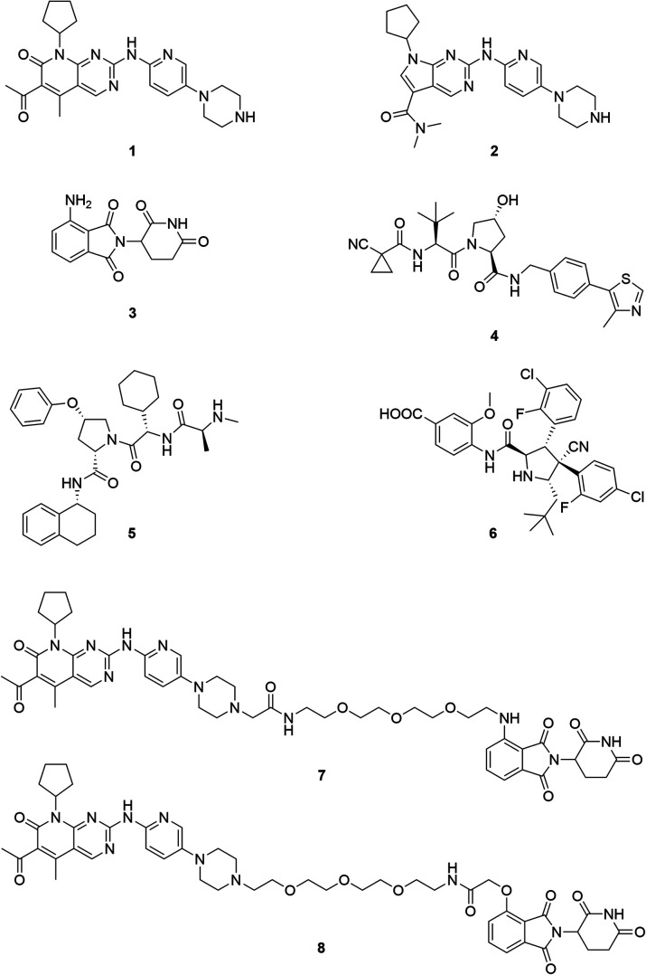
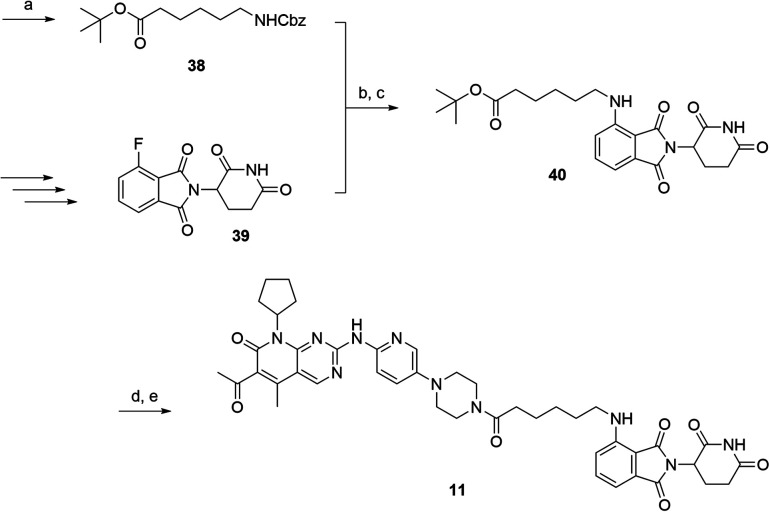
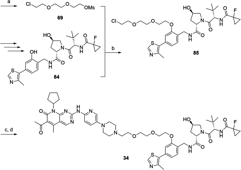
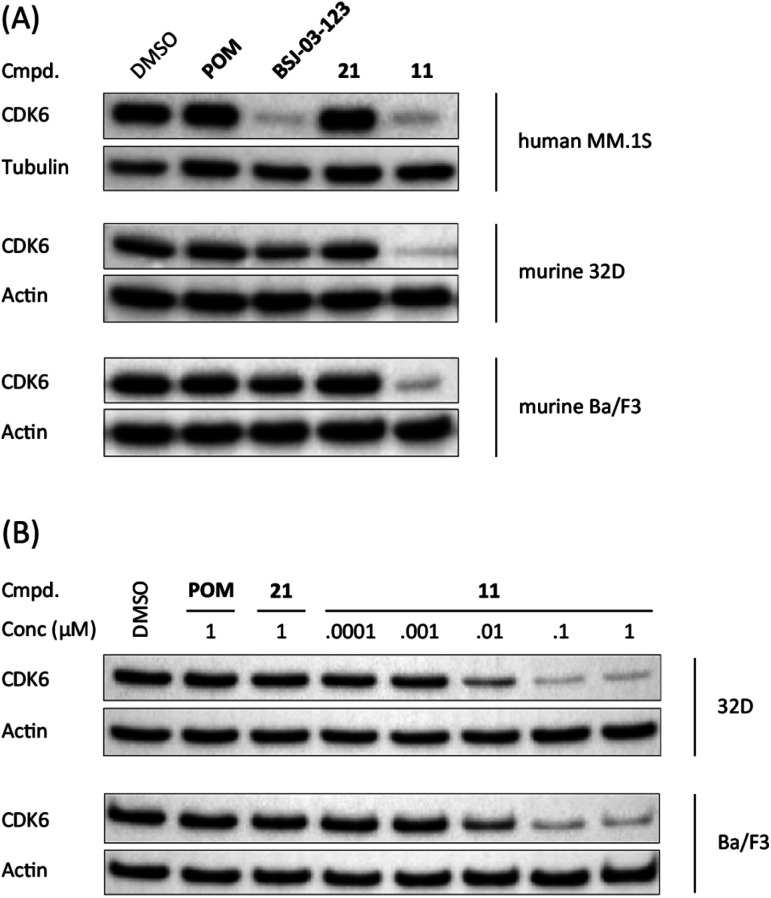
Verification
Claims the paper makes
The claims and statistics of this paper haven’t been checked yet.
Run the check — free, no account needed. It reads the open-access text and checks the sources it cites.
Claims other papers make about this one
Each is checked against this paper’s own text: does it say what the citing paper says it does?
In the citing paper
“Whereas multiple studies have described numerous CRBN-recruiting dual-selective degraders and CDK6-selective degraders 70–73, none report CDK4-selective degraders; the only reported CDK4-selective degraders are built on a slightly different warhead, ribociclib 74.”
From Rationalizing PROTAC-Mediated Ternary Complex Formation Using Rosetta (2021) · cites this paper as [72]
Self-check
Percentages, sums and reported differences re-computed from the paper’s own numbers.
The self-check needs the paper’s full text.
Citations
Cites
Cell cycle, CDKs and cancer: a changing paradigm
Malumbres, M., & Barbacid, M. (2009) · Nature Reviews Cancer · doi:10.1038/nrc2602 · Cited by 3,957
Palbociclib and letrozole in advanced breast cancer
Finn, R. S., Martin, M., Rugo, H. S., Jones, S., Im, S.-A., Gelmon, K., Harbeck, N., Lipatov, O. N., Walshe, J. M., Moulder, S., et al. (2016) · New England Journal of Medicine · doi:10.1056/nejmoa1607303 · Cited by 3,007
Meyers, R. M., Bryan, J. G., McFarland, J. M., Weir, B. A., Sizemore, A. E., Xu, H., Dharia, N. V., Montgomery, P. G., Cowley, G. S., Pantel, S., et al. (2017) · Nature Genetics · doi:10.1038/ng.3984 · Cited by 1,952
Lenalidomide Causes Selective Degradation of IKZF1 and IKZF3 in Multiple Myeloma Cells
Krönke, J., Udeshi, N. D., Narla, A., Grauman, P., Hurst, S. N., McConkey, M., Svinkina, T., Heckl, D., Comer, E., Li, X., et al. (2014) · Science · doi:10.1126/science.1244851 · Cited by 1,879
Self-citationCristofanilli, M., Turner, N. C., Bondarenko, I., Ro, J., Im, S.-A., Masuda, N., Colleoni, M., DeMichele, A., Loi, S., Verma, S., et al. (2016) · The Lancet Oncology · doi:10.1016/s1470-2045(15)00613-0 · Cited by 1,827
Phthalimide conjugation as a strategy for in vivo target protein degradation
Winter, G. E., Buckley, D. L., Paulk, J., Roberts, J. M., Souza, A., Dhe-Paganon, S., & Bradner, J. E. (2015) · Science · doi:10.1126/science.aab1433 · Cited by 1,760
The Myeloma Drug Lenalidomide Promotes the Cereblon-Dependent Destruction of Ikaros Proteins
Lu, G., Middleton, R. E., Sun, H., Naniong, M., Ott, C. J., Mitsiades, C. S., Wong, K.-K., Bradner, J. E., & Kaelin, W. G. (2014) · Science · doi:10.1126/science.1244917 · Cited by 1,583
New functional activities for the p21 family of CDK inhibitors.
LaBaer, J., Garrett, M. D., Stevenson, L. F., Slingerland, J. M., Sandhu, C., Chou, H. S., Fattaey, A., & Harlow, E. (1997) · Genes & Development · doi:10.1101/gad.11.7.847 · Cited by 1,402
Finn, R. S., Dering, J., Conklin, D., Kalous, O., Cohen, D. J., Desai, A. J., Ginther, C., Atefi, M., Chen, I., Fowst, C., et al. (2009) · Breast Cancer Research · doi:10.1186/bcr2419 · Cited by 1,398
Treating cancer with selective CDK4/6 inhibitors
O'Leary, B., Finn, R. S., & Turner, N. C. (2016) · Nature Reviews Clinical Oncology · doi:10.1038/nrclinonc.2016.26 · Cited by 1,137
Research works printed but not linked: 1
- Schumacher, 1965, Br. J. Pharmacol., 25, 324
Cited by
Sorted by citations
Current strategies for the design of PROTAC linkers: a critical review
Troup, R. I., Fallan, C., & Baud, M. G. J. (2020) · Exploration of Targeted Anti-tumor Therapy · doi:10.37349/etat.2020.00018 · Cited by 424
Proteolysis-targeting chimeras (PROTACs) in cancer therapy
Li, X., Pu, W., Zheng, Q., Ai, M., Chen, S., & Peng, Y. (2022) · Molecular Cancer · doi:10.1186/s12943-021-01434-3 · Cited by 389
Self-citationTarget and tissue selectivity of PROTAC degraders
Guenette, R. G., Yang, S. W., Min, J., Pei, B., & Potts, P. R. (2022) · Chemical Society Reviews · doi:10.1039/d2cs00200k · Cited by 324
PROTACs: great opportunities for academia and industry (an update from 2020 to 2021)
He, M., Cao, C., Ni, Z., Liu, Y., Song, P., Hao, S., He, Y., Sun, X., & Rao, Y. (2022) · Signal Transduction and Targeted Therapy · doi:10.1038/s41392-022-00999-9 · Cited by 283
Zeng, S., Huang, W., Zheng, X., Cheng, L., Zhang, Z., Wang, J., & Shen, Z. (2020) · European Journal of Medicinal Chemistry · doi:10.1016/j.ejmech.2020.112981 · Cited by 264
Diehl, C. J., & Ciulli, A. (2022) · Chemical Society Reviews · doi:10.1039/d2cs00387b · Cited by 269
Unraveling the Role of Linker Design in Proteolysis Targeting Chimeras
Bemis, T. A., Clair, J. J. L., & Burkart, M. D. (2021) · Journal of Medicinal Chemistry · doi:10.1021/acs.jmedchem.1c00482 · Cited by 250
Bond, M. J., & Crews, C. M. (2021) · RSC Chemical Biology · doi:10.1039/d1cb00011j · Cited by 232
Recent Developments in PROTAC‐Mediated Protein Degradation: From Bench to Clinic
Hu, Z., & Crews, C. M. (2022) · ChemBioChem · doi:10.1002/cbic.202100270 · Cited by 231
E3 Ligase Ligands in Successful PROTACs: An Overview of Syntheses and Linker Attachment Points
Bricelj, A., Steinebach, C., Kuchta, R. D., Gütschow, M., & Sosič, I. (2021) · Frontiers in Chemistry · doi:10.3389/fchem.2021.707317 · Cited by 232
Self-citationCecchini, C., Pannilunghi, S., Tardy, S., & Scapozza, L. (2021) · Frontiers in Chemistry · doi:10.3389/fchem.2021.672267 · Cited by 201
The roles of E3 ubiquitin ligases in cancer progression and targeted therapy
Sampson, C., Wang, Q., Otkur, W., Zhao, H., Lu, Y., Liu, X., & Piao, H. (2023) · Clinical and Translational Medicine · doi:10.1002/ctm2.1204 · Cited by 193
E3 ligase ligand chemistries: from building blocks to protein degraders
Sosič, I., Bricelj, A., & Steinebach, C. (2022) · Chemical Society Reviews · doi:10.1039/d2cs00148a · Cited by 176
Self-citationGoracci, L., Desantis, J., Valeri, A., Castellani, B., Eleuteri, M., & Cruciani, G. (2020) · Journal of Medicinal Chemistry · doi:10.1021/acs.jmedchem.0c00793 · Cited by 161
Rationalizing PROTAC-Mediated Ternary Complex Formation Using Rosetta
Bai, N., Miller, S. A., Andrianov, G. V., Yates, M., Kirubakaran, P., & Karanicolas, J. (2021) · Journal of Chemical Information and Modeling · doi:10.1021/acs.jcim.0c01451 · Cited by 157
Developments of CRBN-based PROTACs as potential therapeutic agents
Wang, C., Zhang, Y., Wu, Y., & Xing, D. (2021) · European Journal of Medicinal Chemistry · doi:10.1016/j.ejmech.2021.113749 · Cited by 158
Expanding the arsenal of E3 ubiquitin ligases for proximity-induced protein degradation
Kannt, A., & Đikić, I. (2021) · Cell chemical biology · doi:10.1016/j.chembiol.2021.04.007 · Cited by 153
An E3 ligase guide to the galaxy of small-molecule-induced protein degradation
Jevtić, P., Haakonsen, D. L., & Rapé, M. (2021) · Cell chemical biology · doi:10.1016/j.chembiol.2021.04.002 · Cited by 149
A bibliometric analysis of PROTAC from 2001 to 2021
Li, D., Yu, D., Li, Y., & Yang, R. (2022) · European Journal of Medicinal Chemistry · doi:10.1016/j.ejmech.2022.114838 · Cited by 125
Yang, L., Yang, Y., Zhang, J., Li, M., Yang, L., Wang, X., Chen, M., Zhang, H., He, B., Wang, X., et al. (2024) · Signal Transduction and Targeted Therapy · doi:10.1038/s41392-024-01983-1 · Cited by 131
CDK4: a master regulator of the cell cycle and its role in cancer
Baker, S. J., Poulikakos, P. I., Irie, H. Y., Parekh, S. S., & Reddy, E. P. (2022) · Genes & Cancer · doi:10.18632/genesandcancer.221 · Cited by 84
Peng, X., Hu, Z., Zeng, L., Zhang, M., Xu, C., Lu, B., Tao, C., Chen, W., Hou, W., Cheng, K., et al. (2023) · Acta Pharmaceutica Sinica B · doi:10.1016/j.apsb.2023.09.003 · Cited by 87
Bricelj, A., Ng, Y. L. D., Ferber, D., Kuchta, R. D., Müller, S., Monschke, M., Wagner, K. G., Krönke, J., Sosič, I., Gütschow, M., et al. (2021) · ACS Medicinal Chemistry Letters · doi:10.1021/acsmedchemlett.1c00368 · Cited by 71
Self-citationThe Renaissance of Cyclin Dependent Kinase Inhibitors
Ettl, T., Schulz, D., & Bauer, R. J. (2022) · Cancers · doi:10.3390/cancers14020293 · Cited by 71
VHL-based PROTACs as potential therapeutic agents: Recent progress and perspectives
Wang, C., Zhang, Y., Wang, J., & Xing, D. (2021) · European Journal of Medicinal Chemistry · doi:10.1016/j.ejmech.2021.113906 · Cited by 73
Ng, Y. L. D., Ramberger, E., Bohl, S., Dolnik, A., Steinebach, C., Conrad, T., Müller, S., Popp, O., Kull, M., Haji, M., et al. (2022) · Nature Communications · doi:10.1038/s41467-022-28515-1 · Cited by 68
Self-citationRecent Advances of Degradation Technologies Based on PROTAC Mechanism
Xiao, M., Zhao, J., Wang, Q., Liu, J., & Ma, L. (2022) · Biomolecules · doi:10.3390/biom12091257 · Cited by 69
Targeted protein degradation in cancers: Orthodox PROTACs and beyond
Li, J., Chen, X., Lü, A., & Liang, C. (2023) · The Innovation · doi:10.1016/j.xinn.2023.100413 · Cited by 68
A beginner’s guide to current synthetic linker strategies towards VHL-recruiting PROTACs
Zografou-Barredo, N. A., Hallatt, A. J., Goujon-Ricci, J., & Cano, C. (2023) · Bioorganic & Medicinal Chemistry · doi:10.1016/j.bmc.2023.117334 · Cited by 62
Resistance mechanisms and therapeutic strategies of CDK4 and CDK6 kinase targeting in cancer
Asciolla, J. J., Wu, X., Adamopoulos, C., Gavathiotis, E., & Poulikakos, P. I. (2025) · Nature Cancer · doi:10.1038/s43018-024-00893-z · Cited by 66
Optimization of PROTAC Ternary Complex Using DNA Encoded Library Approach
Chen, Q., Liu, C., Wang, W., Meng, X., Cheng, X., Li, X.-Y., Cai, L., Luo, L., He, X., Qu, H., et al. (2023) · ACS Chemical Biology · doi:10.1021/acschembio.2c00797 · Cited by 55
Current advances of small molecule E3 ligands for proteolysis-targeting chimeras design
Mi, D., Li, Y., Gu, H., Li, Y., & Chen, Y. (2023) · European Journal of Medicinal Chemistry · doi:10.1016/j.ejmech.2023.115444 · Cited by 58
DNA framework-engineered chimeras platform enables selectively targeted protein degradation
Zhou, L.-M., Yu, B., Gao, M., Chen, R., Li, Z., Gu, Y., Bian, J., & Ma, Y. (2023) · Nature Communications · doi:10.1038/s41467-023-40244-7 · Cited by 61
Enriching Proteolysis Targeting Chimeras with a Second Modality: When Two Are Better Than One
Salerno, A., Seghetti, F., Caciolla, J., Uliassi, E., Testi, E., Guardigni, M., Roberti, M., Milelli, A., & Bolognesi, M. L. (2022) · Journal of Medicinal Chemistry · doi:10.1021/acs.jmedchem.2c00302 · Cited by 58
Targeting Protein Kinases Degradation by PROTACs
Yu, F., Cai, M., Shao, L., & Zhang, J. (2021) · Frontiers in Chemistry · doi:10.3389/fchem.2021.679120 · Cited by 55
Steinebach, C., Bricelj, A., Murgai, A., Sosič, I., Bischof, L., Ng, Y. L. D., Heim, C., Maiwald, S. A., Proj, M., Voget, R., et al. (2023) · Journal of Medicinal Chemistry · doi:10.1021/acs.jmedchem.3c00851 · Cited by 57
Self-citationHu, R., Wang, W., Yang, Y.-Y., Hu, X.-T., Wang, Q., Zuo, W., Xu, Y., Feng, Q., & Wang, N. (2021) · European Journal of Medicinal Chemistry · doi:10.1016/j.ejmech.2021.113922 · Cited by 53
Vicente, A. T. S., & Salvador, J. A. R. (2022) · International Journal of Molecular Sciences · doi:10.3390/ijms231911068 · Cited by 56
Maity, P., Chatterjee, J., Patil, K. T., Arora, S., Katiyar, M. K., Kumar, M., Samarbakhsh, A., Joshi, G., Bhutani, P., Chugh, M., et al. (2023) · Journal of Medicinal Chemistry · doi:10.1021/acs.jmedchem.2c01242 · Cited by 51
Recent advances in IAP-based PROTACs (SNIPERs) as potential therapeutic agents
Wang, C., Zhang, Y., Shi, L., Yang, S., Chang, J., Zhong, Y., Li, Q., & Xing, D. (2022) · Journal of Enzyme Inhibition and Medicinal Chemistry · doi:10.1080/14756366.2022.2074414 · Cited by 50
Targeting CDK4/6 for Anticancer Therapy
Qi, J., & Ouyang, Z. (2022) · Biomedicines · doi:10.3390/biomedicines10030685 · Cited by 49
The impact of E3 ligase choice on PROTAC effectiveness in protein kinase degradation
Sobierajski, T., Małolepsza, J., Pichlak, M., Gendaszewska‐Darmach, E., & Błażewska, K. M. (2024) · Drug Discovery Today · doi:10.1016/j.drudis.2024.104032 · Cited by 48
PROTACs bearing piperazine-containing linkers: what effect on their protonation state?
Desantis, J., Mammoli, A., Eleuteri, M., Coletti, A., Croci, F., Macchiarulo, A., & Goracci, L. (2022) · RSC Advances · doi:10.1039/d2ra03761k · Cited by 46
Development of CDK4/6 Inhibitors: A Five Years Update
Ammazzalorso, A., Agamennone, M., Filippis, B. D., & Fantacuzzi, M. (2021) · Molecules · doi:10.3390/molecules26051488 · Cited by 48
Rana, S., Mallareddy, J. R., Singh, S., Boghean, L., & Natarajan, A. (2021) · Cancers · doi:10.3390/cancers13215506 · Cited by 43
Recent advances in targeted protein degraders as potential therapeutic agents
Yang, N., Kong, B., Zhu, Z., Huang, F., Zhang, L., Lu, T., Chen, Y., Zhang, Y., & Jiang, Y. (2023) · Molecular Diversity · doi:10.1007/s11030-023-10606-w · Cited by 44
Solubility Enhanced Formulation Approaches to Overcome Oral Delivery Obstacles of PROTACs
Pöstges, F., Kayser, K., Appelhaus, J., Monschke, M., Gütschow, M., Steinebach, C., & Wagner, K. G. (2023) · Pharmaceutics · doi:10.3390/pharmaceutics15010156 · Cited by 40
Self-citationYang, N., Fan, Z., Sun, S., Hu, X., Mao, Y., Jia, C., Cai, X., Xu, T., Li, B., Li, Y., et al. (2023) · European Journal of Medicinal Chemistry · doi:10.1016/j.ejmech.2023.115857 · Cited by 40
Hung, C.-L., Liu, H.-H., Fu, C., Yeh, H.-H., Hu, T.-L., Kuo, Z., Lin, Y.-C., Jhang, M.-R., Hwang, C.-S., Hsu, H., et al. (2023) · EBioMedicine · doi:10.1016/j.ebiom.2023.104500 · Cited by 40
Designing HDAC-PROTACs: Lessons Learned So Far
Fischer, F., Avelar, L. A. A., Murray, L., & Kurz, T. (2021) · Future Medicinal Chemistry · doi:10.4155/fmc-2021-0206 · Cited by 38
PROTAC: targeted drug strategy. Principles and limitations
Koroleva, O. A., Dutikova, Y. V., Trubnikov, A. V., Zenov, F. A., Manasova, E. V., Shtil, A. A., & Kurkin, A. V. (2022) · Russian Chemical Bulletin · doi:10.1007/s11172-022-3659-z · Cited by 38
CRL4CRBN E3 Ligase Complex as a Therapeutic Target in Multiple Myeloma
Barankiewicz, J., Salomon‐Perzyński, A., Misiewicz-Krzemińska, I., & Lech‐Marańda, E. (2022) · Cancers · doi:10.3390/cancers14184492 · Cited by 38
Ubiquitination and Ubiquitin-Like Modifications in Multiple Myeloma: Biology and Therapy
Wirth, M., Schick, M., Keller, U. B., & Krönke, J. (2020) · Cancers · doi:10.3390/cancers12123764 · Cited by 35
Self-citationSchiedel, M., Lehotzky, A., Szunyogh, S., Oláh, J., Hammelmann, S., Wössner, N., Robaa, D., Einsle, O., Sippl, W., Ovádi, J., et al. (2020) · ChemBioChem · doi:10.1002/cbic.202000351 · Cited by 30
Recent Progress in CDK4/6 Inhibitors and PROTACs
Wang, H., Ba, J., Kang, Y., Gong, Z., Liang, T., Zhang, Y., Qi, J., & Wang, J. (2023) · Molecules · doi:10.3390/molecules28248060 · Cited by 33
Progress of small molecules for targeted protein degradation: PROTACs and other technologies
Zhao, H., Xin, M., & Zhang, S. (2023) · Drug Development Research · doi:10.1002/ddr.22026 · Cited by 31
Current strategies for improving limitations of proteolysis targeting chimeras
Pu, C., Wang, S., Liu, L., Feng, Z., Zhang, H., Gong, Q., Sun, Y., Guo, Y., & Li, R. (2022) · Chinese Chemical Letters · doi:10.1016/j.cclet.2022.107927 · Cited by 32
Therapeutic potential of CDK4/6 inhibitors in renal cell carcinoma
Sager, R., Backe, S. J., Ahanin, E. F., Smith, G., Nsouli, I. S., Woodford, M. R., Bratslavsky, G., Bourboulia, D., & Mollapour, M. (2022) · Nature Reviews Urology · doi:10.1038/s41585-022-00571-8 · Cited by 29
Molecular glues: enhanced protein-protein interactions and cell proteome editing
Weagel, E. G., Foulks, J. M., Siddiqui, A. Q., & Warner, S. L. (2022) · Medicinal Chemistry Research · doi:10.1007/s00044-022-02882-2 · Cited by 31
Targeting CDKs in cancer therapy: advances in PROTACs and molecular glues
Marei, H. E. S., Bedair, K., Hasan, A., Al-Mansoori, L., Gaiba, A., Morrione, A., Cenciarelli, C., & Giordano, A. (2025) · npj Precision Oncology · doi:10.1038/s41698-025-00931-8 · Cited by 31
Sweet and Blind Spots in E3 Ligase Ligand Space Revealed by a Thermophoresis-Based Assay
Maiwald, S. A., Heim, C., Alvarez, B. H., & Hartmann, M. D. (2020) · ACS Medicinal Chemistry Letters · doi:10.1021/acsmedchemlett.0c00440 · Cited by 26
Riching, K. M., Vasta, J. D., Hughes, S. J., Zoppi, V., Maniaci, C., Testa, A., Urh, M., Ciulli, A., & Daniels, D. L. (2021) · Current Research in Chemical Biology · doi:10.1016/j.crchbi.2021.100009 · Cited by 26
PROTAC: Novel degradable approach for different targets to treat breast cancer
Wang, Z., Che, S., & Yu, Z. (2024) · European Journal of Pharmaceutical Sciences · doi:10.1016/j.ejps.2024.106793 · Cited by 25
Gockel, L. M., Pfeifer, V., Baltes, F., Bachmaier, R. D., Wagner, K. G., Bendas, G., Gütschow, M., Sosič, I., & Steinebach, C. (2022) · Archiv der Pharmazie · doi:10.1002/ardp.202100467 · Cited by 24
Self-citationSynthesis, biological evaluation and clinical trials of Cereblon-based PROTACs
Vicente, A. T. S., Moura, S. P. S. P., & Salvador, J. A. R. (2025) · Communications Chemistry · doi:10.1038/s42004-025-01598-9 · Cited by 23
3-Aminophthalic acid, a new cereblon ligand for targeted protein degradation by O’PROTAC
Yan, Y., Shao, J., Ding, D., Pan, Y., Tran, P., Yan, W., Wang, Z., Li, H., & Huang, H. (2022) · Chemical Communications · doi:10.1039/d1cc06525d · Cited by 22
Vu, L. P., Diehl, C. J., Casement, R., Bond, A. G., Steinebach, C., Strašek, N., Bricelj, A., Perdih, A., Schnakenburg, G., Sosič, I., et al. (2023) · Journal of Medicinal Chemistry · doi:10.1021/acs.jmedchem.3c00434 · Cited by 20
Self-citationProteolysis Targeting Chimeras (PROTACs) in Breast Cancer Therapy
Jin, Y., & Lee, Y. (2024) · ChemMedChem · doi:10.1002/cmdc.202400267 · Cited by 21
Targeting micro-environmental pathways by PROTACs as a therapeutic strategy
Liu, J., Peng, Y., Inuzuka, H., & Wei, W. (2022) · Seminars in Cancer Biology · doi:10.1016/j.semcancer.2022.07.001 · Cited by 20
Heim, C., Maiwald, S. A., Steinebach, C., Collins, M. K., Strope, J. D., Chau, C. H., Figg, W. D., Gütschow, M., & Hartmann, M. D. (2020) · Biochemical and Biophysical Research Communications · doi:10.1016/j.bbrc.2020.11.117 · Cited by 19
Self-citationDevelopment of Degraders of Cyclin-Dependent Kinases 4 and 6 Based on Rational Drug Design
He, H., Zhang, X., Wang, J., Liu, Q., Zhang, L., Lu, C., Yuan, Y., Zhao, Z., Li, H., & Chen, Z. (2024) · Journal of Medicinal Chemistry · doi:10.1021/acs.jmedchem.4c00965 · Cited by 18
Proteolysis-targeting chimeras in biotherapeutics: Current trends and future applications
Li, Q., Zhou, L., Qin, S., Huang, Z., Li, B., Li, B., Yang, M., Nice, E. C., Zhu, H., & Huang, C. (2023) · European Journal of Medicinal Chemistry · doi:10.1016/j.ejmech.2023.115447 · Cited by 17
Development of PROTACS degrading KRAS and SOS1
Hamilton, G., Eggerstorfer, M.-T., & Stickler, S. (2024) · Oncology Research Featuring Preclinical and Clinical Cancer Therapeutics · doi:10.32604/or.2024.051653 · Cited by 16
Discovery of HyT‐Based Degraders of CDK9‐Cyclin T1 Complex
Lin, R., Yang, J., Liu, T., Wang, M., Ke, C., Luo, C., Lin, J. H., Li, J., & Lin, H. (2023) · Chemistry & Biodiversity · doi:10.1002/cbdv.202300769 · Cited by 15
PROteolysis‐Targeting Chimeras (PROTACs) in leukemia: overview and future perspectives
Vicente, A. T. S., & Salvador, J. A. R. (2024) · MedComm · doi:10.1002/mco2.575 · Cited by 14
Small-Molecule Degraders of Cyclin-Dependent Kinase Protein: A Review
Yu, B., Du, Z., Zhang, Y., Li, Z., & Bian, J. (2021) · Future Medicinal Chemistry · doi:10.4155/fmc-2021-0154 · Cited by 14
PROTACs: The Future of Leukemia Therapeutics
Anwar, Z., Ali, M. S., Galvano, A., Perez, A., Mantia, M. L., Bukhari, I., & Świątczak, B. (2022) · Frontiers in Cell and Developmental Biology · doi:10.3389/fcell.2022.851087 · Cited by 13
Rothweiler, E. M., Brennan, P. E., & Huber, K. (2022) · Biological Chemistry · doi:10.1515/hsz-2021-0396 · Cited by 12
Development and Characterization of the First Selective Class IIb Histone Deacetylase Degraders
Zhai, S., Honin, I., Schäker‐Hübner, L., Hanl, M., Jacobi, L., Dressler, F., Pieńkowska, D. E., König, P., Gerhartz, J., Voget, R., et al. (2025) · Journal of Medicinal Chemistry · doi:10.1021/acs.jmedchem.5c00674 · Cited by 10
Self-citationEncoding BRAF inhibitor functions in protein degraders
Miller, D. S. J., Voell, S. A., Sosič, I., Proj, M., Rossanese, O. W., Schnakenburg, G., Gütschow, M., Collins, I., & Steinebach, C. (2022) · RSC Medicinal Chemistry · doi:10.1039/d2md00064d · Cited by 12
Self-citationHeterobifunctional Ligase Recruiters Enable pan-Degradation of Inhibitor of Apoptosis Proteins
Ng, Y. L. D., Bricelj, A., Jansen, J. A., Murgai, A., Peter, K., Donovan, K. A., Gütschow, M., Krönke, J., Steinebach, C., & Sosič, I. (2023) · Journal of Medicinal Chemistry · doi:10.1021/acs.jmedchem.2c01817 · Cited by 10
Self-citationTargeting the cell-cycle machinery for cancer therapy
Peng, Y., Liu, J., Inuzuka, H., Liu, J., & Wei, W. (2025) · Trends in Cell Biology · doi:10.1016/j.tcb.2025.08.008 · Cited by 12
Targeting the ubiquitin pathway in lymphoid malignancies
Li, B., Eichhorn, P. J. A., & Chng, W. J. (2024) · Cancer Letters · doi:10.1016/j.canlet.2024.216978 · Cited by 10
Zheng, S., Chen, Y., Zhu, Z., Li, N., He, C., Koeffler, H. P., Han, X., Wei, Q., & Xu, L. (2025) · Journal of Zhejiang University SCIENCE B · doi:10.1631/jzus.b2500021 · Cited by 9
Chen, X., Haishan, S., Shao, Y., Ma, Q., Niu, Y., & Shang, Z. (2021) · Translational Andrology and Urology · doi:10.21037/tau-20-1357 · Cited by 9
Kuchta, R. D., Heim, C., Herrmann, A., Maiwald, S. A., Ng, Y. L. D., Sosič, I., Keuler, T., Krönke, J., Gütschow, M., Hartmann, M. D., et al. (2023) · RSC Chemical Biology · doi:10.1039/d2cb00223j · Cited by 9
Self-citationFDA-approved kinase inhibitors in PROTAC design, development and synthesis
Kossakowski, K., Cherniienko, A., Zaprutko, L., & Pawełczyk, A. (2025) · Journal of Enzyme Inhibition and Medicinal Chemistry · doi:10.1080/14756366.2025.2542357 · Cited by 12
Enhancing Solubility in VHL-Based PROTACs: Optimized USP7 Degraders for Improved Developability
Wittenburg, S., Zuleeg, M. R., Peter, K., Lemnitzer, P., Voget, R., Bricelj, A., Gobec, M., Dierlamm, N., Braun, M. B., Geiger, T. M., et al. (2025) · Journal of Medicinal Chemistry · doi:10.1021/acs.jmedchem.5c00718 · Cited by 9
Self-citationZheng, M., Zhang, X., Chen, W., Xia, F., Yang, H., Yuan, K., & Yang, P. (2024) · Future Medicinal Chemistry · doi:10.4155/fmc-2023-0259 · Cited by 7
He, J., Liu, S., Zhang, S., Gao, Q., Zhu, L., Xu, N., Hu, Z., Zhang, X., Ma, S., Wang, X., et al. (2025) · Scientific Reports · doi:10.1038/s41598-025-92494-8 · Cited by 7
Guo, S., Liu, J., Zheng, H., & Wu, X. (2026) · Biomedicines · doi:10.3390/biomedicines14040835 · Cited by 5
Targeted protein degraders from an oncologist point of view: The Holy Grail of cancer therapy?
Ambrosini, M., Fucà, G., Duca, M., Damian, S., Santis, F. D., Corti, F., Cresta, S., de Braud, F., & Nicola, M. D. (2021) · Critical Reviews in Oncology/Hematology · doi:10.1016/j.critrevonc.2021.103532 · Cited by 5
Selective CDK6 Degradation via the KLHDC2 E3 Ubiquitin Ligase
Jeon, E., Kim, Y., Ahn, H., Martinez, M. J., Hwang, K., Cho, S., Dwyer, B. G., Romero, B. A., Hinshaw, S. M., Gray, N. S., et al. (2025) · Journal of Medicinal Chemistry · doi:10.1021/acs.jmedchem.5c02713 · Cited by 4
Emerging Strategies to Inhibit the G1/S Transition for Cancer Therapy
Rubin, S. M., Sage, J., & Skotheim, J. M. (2026) · Cancer Research · doi:10.1158/0008-5472.can-25-0916 · Cited by 5
Design and synthesis of JNK1-targeted PROTACs and research on the activity
Guo, Y., Liu, F., Chi, M., Qian, H., Zhang, Y., Yuan, Y., Hou, S., Chen, X., & Ma, L. (2024) · Bioorganic Chemistry · doi:10.1016/j.bioorg.2024.108044 · Cited by 4
PROTAC-Mediated GSPT1 Degradation Impairs the Expression of Fusion Genes in Acute Myeloid Leukemia
Perzolli, A., Steinebach, C., Krönke, J., Gütschow, M., Zwaan, C. M., Barneh, F., & Heidenreich, O. T. (2025) · Cancers · doi:10.3390/cancers17020211 · Cited by 4
Self-citationAdhikari, B., Schneider, K., Diebold, M., Sotriffer, C., & Wolf, E. (2024) · eLife · doi:10.7554/elife.98450 · Cited by 4
Palbociclib as an Antitumor Drug: A License to Kill
Łupicka‐Słowik, A., Cossu, F., & Sieńczyk, M. (2024) · Molecules · doi:10.3390/molecules29225334 · Cited by 5
PROTACs improve selectivity for targeted proteins
Liu, L., Guan, X., Zhuo, J., Wu, X., & Han, X. (2025) · Acta Materia Medica · doi:10.15212/amm-2025-0015 · Cited by 3
Zhai, S., Kemkes, M., Kponomaizoun, C.-E., Gerhartz, J., Feller, F., Tu, J., Pieńkowska, D. E., Schliehe‐Diecks, J., Dressel, I., Gütschow, M., et al. (2026) · Journal of Medicinal Chemistry · doi:10.1021/acs.jmedchem.5c00939 · Cited by 3
Self-citation
Metadata
- Authors
- Steinebach, C., Ng, Y. L. D., Sosič, I., Lee, C.-S., Chen, S., Lindner, S., Vu, L. P., Bricelj, A., Haschemi, R., Monschke, M., Steinwarz, E., Wagner, K. G., Bendas, G., Luo, J., Gütschow, M., & Krönke, J.
- Affiliations
- University of Bonn (DE)
- Universität Ulm (DE)
- University of Ljubljana (SI)
- National Cancer Institute (US)
- Corresponding author
- Gütschow, Michael
- Krönke, Jan
- ORCID
- Venue
- Chemical Science
- Publisher
- Royal Society of Chemistry (RSC)
- Published
- 2020
- Volume
- 11
- Issue
- 13
- Pages
- 3474-3486
- ISSN
- 2041-6520 (ISSN-L)
- 2041-6539
- PMC
- PMC7552917
- OpenAlex
- W3009753365
- Cited by
- 148
- References
- 77
- Citations by year
- 2026: 20 · 2025: 24 · 2024: 27 · 2023: 20 · 2022: 27 · 2021: 22 · 2020: 8
- FWCI
- 6.56
- Type
- journal article
- Language
- en
- Open access
- diamond
- doi.org
- Licence
- Locations
- Chemical Science (publishedVersion, cc-by)
- Europe PMC (PubMed Central) (publishedVersion, cc-by)
- PubMed Central (publishedVersion, cc-by)
- PubMed (publishedVersion)
- Funders
- Deutsche Forschungsgemeinschaft (FOR2372, Kr-3886/2-1, SFB1074)
- Javna Agencija za Raziskovalno Dejavnost RS (P1-0208)
- Topics
- Life Sciences › Biochemistry, Genetics and Molecular Biology › Molecular Biology › Protein Degradation and Inhibitors
- Life Sciences › Biochemistry, Genetics and Molecular Biology › Molecular Biology › Ubiquitin and proteasome pathways
- Physical Sciences › Chemistry › Organic Chemistry › Chemical Synthesis and Analysis
- Keywords
- PROTACs, CDK4/6, E3 ubiquitin ligase, VHL, cIAP1, targeted protein degradation, tumor cell proliferation, leukemia, multiple myeloma, breast cancer
About these checks
Registry, retraction, Cites and statistics results are computed, not judged, with one exception: a printed reference that no index lists is read once by a model before it counts as a citation error. Claims are extracted by a model and kept only when their quote appears verbatim in the text; each cited source is then checked, with a model’s help, for whether it says what the claim relies on. Those verdicts can be wrong — read the claim’s quote and the cited source before relying on one.
Statistics and arithmetic are recomputed, not judged.
Confidence bands are shown only for fields calibrated on our annotated test set. None is calibrated yet, so no band is shown.
What counts as a claim ›How we measure confidence ›How we check this ›
Are you an author, or do you see a problem with this page? Request a review
