

The DOI matches its Crossref record; 66 of the 66 references it cites were found in a registry or an index.
Permanent neuroglial remodeling of the retina following infiltration of CSF1R inhibition-resistant peripheral monocytes
Published version of a preprint
Abstract
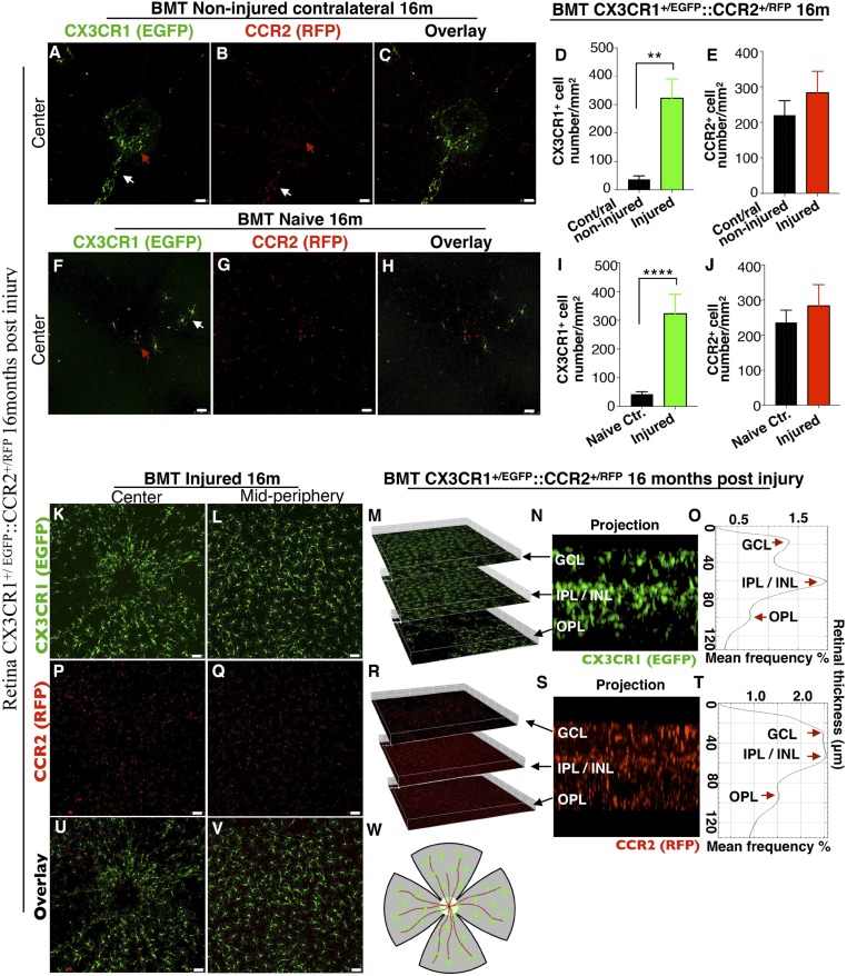
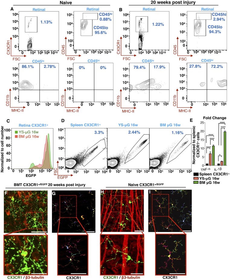
Significance: This work contributes to the understanding of the enigmatic progressive retinal damage following acute ocular surface injury. Clinical findings in patients suggest that such injuries can adversely affect the retina. This study demonstrates that corneal injury leads to rapid infiltration of blood-derived monocytes into the retina and to subsequent remodeling of the neuroglial system. In contrast to previously held belief, this study shows that the blood-derived monocytes engraft permanently into the retina and differentiate into microglia-like cells. Although these cells are morphologically indistinguishable from native microglia, they retain a distinct signature and insensitivity to CSF1R inhibition and exhibit a reactive phenotype which persists long after the noxious stimuli is removed, ultimately contributing to progressive neuroretinal degeneration.
Overview
The claims and statistics of this paper haven’t been checked yet.
Run the check — free, no account needed. It reads the open-access text and checks the sources it cites.
Integrity signals
Registry
Cites
Statistics
Figures
Verification
Claims the paper makes
The claims and statistics of this paper haven’t been checked yet.
Run the check — free, no account needed. It reads the open-access text and checks the sources it cites.
Claims other papers make about this one
Each is checked against this paper’s own text: does it say what the citing paper says it does?
In the citing paper
“Given that peripheral monocytes have been shown to participate in CNS disease via both infiltration and repopulation of neuroglia following microglia depletion (4–10), it is important to determine if CSF1R inhibition causes functional changes in peripheral immune cells that can become part of the CNS (3, 4, 10–14).”
From CSF1R inhibition by a small-molecule inhibitor is not microglia specific; affecting hematopoiesis and the function of macrophages (2020) · cites this paper as [8]
In the citing paper
“Considering that BMDMs permanently engraft into diseased CNS tissue (8, 10), this work suggests that small-molecule inhibition is not restricted to microglia but additionally affects the turnover and function of bone marrow-derived, circulating, and tissue-resident macrophages.”
From CSF1R inhibition by a small-molecule inhibitor is not microglia specific; affecting hematopoiesis and the function of macrophages (2020) · cites this paper as [8]
Self-check
Percentages, sums and reported differences re-computed from the paper’s own numbers.
The self-check needs the paper’s full text.
Citations
Cites
1 retracted
Stutchfield, B. M., Antoine, D. J., MacKinnon, A. C., Gow, D., Bain, C. C., Hawley, C. A., Hughes, M. J., Francis, B., Wojtacha, D., Man, T. Y., et al. (2015) · Gastroenterology · doi:10.1053/j.gastro.2015.08.053 · Cited by 178
RetractedA robust and high-throughput Cre reporting and characterization system for the whole mouse brain
Madisen, L., Zwingman, T. A., Sunkin, S. M., Oh, S. W., Zariwala, H. A., Gu, H., Ng, L. L., Palmiter, R. D., Hawrylycz, M. J., Jones, A. R., et al. (2010) · Nature Neuroscience · doi:10.1038/nn.2467 · Cited by 6,907
Macrophages in Tissue Repair, Regeneration, and Fibrosis
Wynn, T. A., & Vannella, K. M. (2016) · Immunity · doi:10.1016/j.immuni.2016.02.015 · Cited by 4,481
Kettenmann, H., Hanisch, U.-K., Noda, M., & Verkhratsky, A. (2011) · Physiological Reviews · doi:10.1152/physrev.00011.2010 · Cited by 3,547
Fate Mapping Reveals Origins and Dynamics of Monocytes and Tissue Macrophages under Homeostasis
Yona, S., Kim, K.-W., Wolf, Y., Mildner, A., Varol, D., Breker, M., Strauss-Ayali, D., Viukov, S., Guilliams, M., Misharin, A., et al. (2013) · Immunity · doi:10.1016/j.immuni.2012.12.001 · Cited by 3,245
Jung, S., Aliberti, J., Graemmel, P., Sunshine, M. J., Kreutzberg, G. W., Sher, A., & Littman, D. R. (2000) · Molecular and Cellular Biology · doi:10.1128/mcb.20.11.4106-4114.2000 · Cited by 2,632
Microglia promote learning-dependent synapse formation through brain-derived neurotrophic factor
Parkhurst, C. N., Yang, G., Ninan, I., Savas, J. N., Yates, J. R., Lafaille, J. J., Hempstead, B. L., Littman, D. R., & Gan, W.-B. (2013) · Cell · doi:10.1016/j.cell.2013.11.030 · Cited by 2,599
Identification of a unique TGF-β–dependent molecular and functional signature in microglia
Butovsky, O., Jedrychowski, M. P., Moore, C. S., Cialic, R., Lanser, A. J., Gabriely, G., Koeglsperger, T., Dake, B., Wu, P. M., Doykan, C. E., et al. (2014) · Nature Neuroscience · doi:10.1038/nn.3599 · Cited by 2,543
Hashimoto, D., Chow, A., Noizat, C., Teo, P., Beasley, M. B., Leboeuf, M., Becker, C. D., See, P., Price, J., Lucas, D., et al. (2013) · Immunity · doi:10.1016/j.immuni.2013.04.004 · Cited by 2,311 · cited 13×
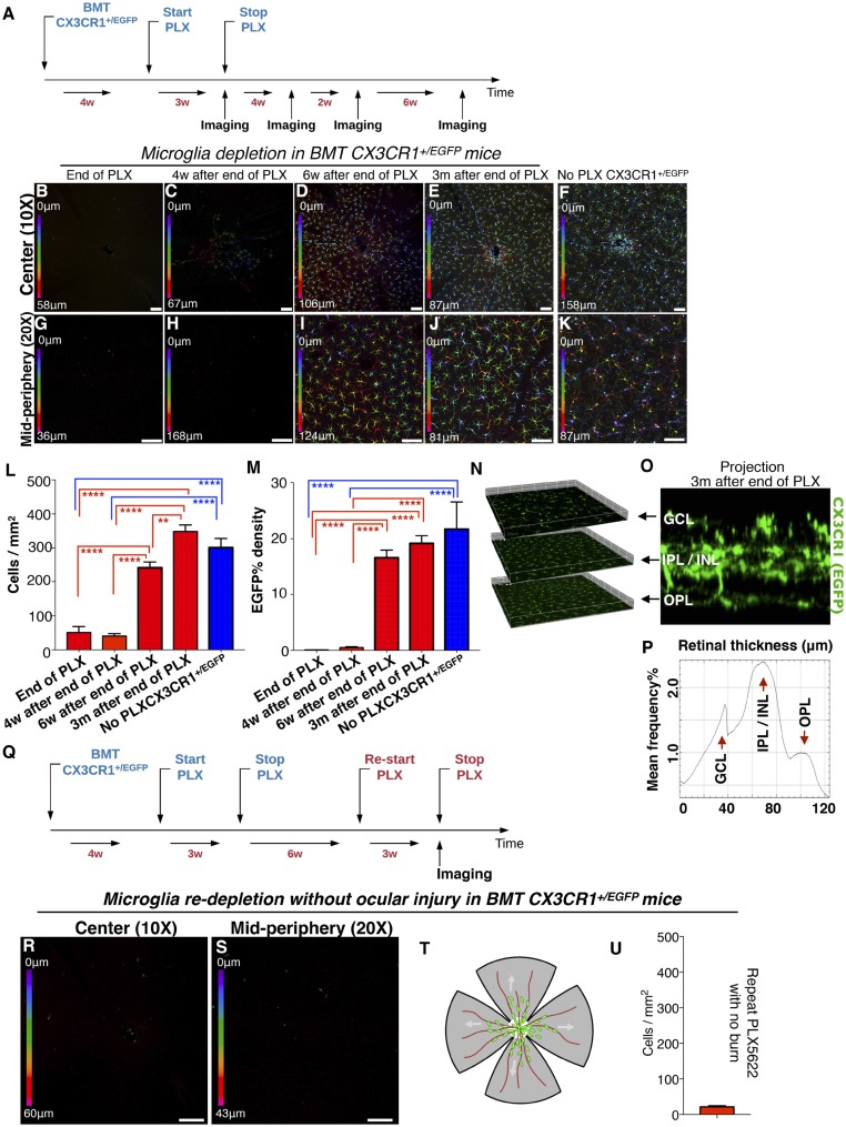
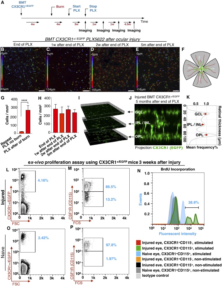
“Using a small molecule inhibitor (PLX5622) of CSF1R (an essential receptor for microglia survival) we show that engrafted peripheral CX3CR1 + monocytes after injury differ from the yolk-sac derived microglia, and cannot be ablated using CSF1R inhibition.”
BackgroundInfluentialTissue-Resident Macrophage Enhancer Landscapes Are Shaped by the Local Microenvironment
Lavin, Y., Winter, D., Blecher-Gonen, R., David, E., Keren-Shaul, H., Merad, M., Jung, S., & Amit, I. (2014) · Cell · doi:10.1016/j.cell.2014.11.018 · Cited by 2,167
Elmore, M. R. P., Najafi, A. R., Koike, M. A., Dagher, N. N., Spangenberg, E. E., Rice, R. A., Kitazawa, M., Matusow, B., Nguyen, H., West, B. L., et al. (2014) · Neuron · doi:10.1016/j.neuron.2014.02.040 · Cited by 1,968
Dendritic cells, monocytes and macrophages: a unified nomenclature based on ontogeny
Guilliams, M., Ginhoux, F., Jakubzick, C., Naik, S. H., Onai, N., Schraml, B. U., Segura, É., Tussiwand, R., & Yona, S. (2014) · Nature reviews. Immunology · doi:10.1038/nri3712 · Cited by 1,944
Tissue-Resident Macrophage Ontogeny and Homeostasis
Ginhoux, F., & Guilliams, M. (2016) · Immunity · doi:10.1016/j.immuni.2016.02.024 · Cited by 1,884
New tools for studying microglia in the mouse and human CNS
Bennett, M. L., Bennett, F. C., Liddelow, S. A., Ajami, B., Zamanian, J. L., Fernhoff, N. B., Mulinyawe, S. B., Bohlen, C. J., Adil, A., Tucker, A., et al. (2016) · Proceedings of the National Academy of Sciences · doi:10.1073/pnas.1525528113 · Cited by 1,804
The role of microglia and macrophages in glioma maintenance and progression
Hambardzumyan, D., Gutmann, D. H., & Kettenmann, H. (2016) · Nature Neuroscience · doi:10.1038/nn.4185 · Cited by 1,683
Local self-renewal can sustain CNS microglia maintenance and function throughout adult life
Ajami, B., Bennett, J. L., Krieger, C., Tetzlaff, W., & Rossi, F. M. V. (2007) · Nature Neuroscience · doi:10.1038/nn2014 · Cited by 1,643
THE ORIGIN AND KINETICS OF MONONUCLEAR PHAGOCYTES
van Furth, R., & Cohn, Z. A. (1968) · The Journal of Experimental Medicine · doi:10.1084/jem.128.3.415 · Cited by 1,564
Gosselin, D., Link, V. M., Romanoski, C. E., Fonseca, G. J., Eichenfield, D. Z., Spann, N. J., Stender, J. D., Chun, H. B., Garner, H., Geissmann, F., et al. (2014) · Cell · doi:10.1016/j.cell.2014.11.023 · Cited by 1,307
Hoeffel, G., Chen, J., Lavin, Y., Low, D., Almeida, F. F., See, P., Beaudin, A. E., Lum, J., Low, I., Forsberg, E. C., et al. (2015) · Immunity · doi:10.1016/j.immuni.2015.03.011 · Cited by 1,135 · cited 2×
“The ability of CSF1R-independent embryonic precursors to functionally replace yolk-sac–derived microglia during development has been demonstrated (30), suggesting that other tissue macrophages may assume the role of microglia under certain conditions.”
BackgroundMicroglia in the adult brain arise from Ly-6ChiCCR2+ monocytes only under defined host conditions
Mildner, A., Schmidt, H., Nitsche, M., Merkler, D., Hanisch, U.-K., Mack, M., Heikenwalder, M., Brück, W., Priller, J., & Prinz, M. (2007) · Nature Neuroscience · doi:10.1038/nn2015 · Cited by 1,089
Innate immune memory in the brain shapes neurological disease hallmarks
Wendeln, A.-C., Degenhardt, K., Kaurani, L., Gertig, M., Ulas, T., Jain, G., Wagner, J., Häsler, L. M., Wild, K., Skodras, A., et al. (2018) · Nature · doi:10.1038/s41586-018-0023-4 · Cited by 956 · cited 2×
“6 altering microglia epigenetic reprogramming through cytokine inhibition may be a potential therapeutic avenue for retina and brain neuropathology (15, 18).”
BackgroundMicroglia as neuroprotective, immunocompetent cells of the CNS
Streit, W. J. (2002) · Glia · doi:10.1002/glia.10154 · Cited by 919
Schlitzer, A., McGovern, N. N., Teo, P., Zelante, T., Atarashi, K., Low, D., Ho, A. W. S., See, P., Shin, A., Wasan, P. S., et al. (2013) · Immunity · doi:10.1016/j.immuni.2013.04.011 · Cited by 886
Differential roles of microglia and monocytes in the inflamed central nervous system
Yamasaki, R., Lu, H., Butovsky, O., Ohno, N., Rietsch, A. M., Cialic, R., Wu, P. M., Doykan, C. E., Lin, J., Cotleur, A. C., et al. (2014) · Journal of Experimental Medicine · doi:10.1084/jem.20132477 · Cited by 856
Imamura, K., Hishikawa, N., Sawada, M., Nagatsu, T., Yoshida, M., & Hashizume, Y. (2003) · Acta Neuropathologica · doi:10.1007/s00401-003-0766-2 · Cited by 791
Turnover of resident microglia in the normal adult mouse brain
Lawson, L. J., Perry, V. H., & Gordon, S. (1992) · Neuroscience · doi:10.1016/0306-4522(92)90500-2 · Cited by 709
van de Laar, L., Saelens, W., De Prijck, S., Martens, L., Scott, C. L., Van Isterdael, G., Hoffmann, E., Beyaert, R., Saeys, Y., Lambrecht, B. N., et al. (2016) · Immunity · doi:10.1016/j.immuni.2016.02.017 · Cited by 648
Bruttger, J., Karram, K., Wörtge, S., Regen, T., Marini, F., Hoppmann, N., Klein, M., Blank, T., Yona, S., Wolf, Y., et al. (2015) · Immunity · doi:10.1016/j.immuni.2015.06.012 · Cited by 634
Saederup, N., Cardona, A. E., Croft, K., Mizutani, M., Cotleur, A. C., Tsou, C.-L., Ransohoff, R. M., & Charo, I. F. (2010) · PLoS ONE · doi:10.1371/journal.pone.0013693 · Cited by 599 · cited 1×
“129(Cg)-Ccr2/J (CCR2) (Stock# 017586),(47) B6.”
Most Tissue-Resident Macrophages Except Microglia Are Derived from Fetal Hematopoietic Stem Cells
Sheng, J., Ruedl, C., & Karjalainen, K. (2015) · Immunity · doi:10.1016/j.immuni.2015.07.016 · Cited by 524
Dagher, N. N., Najafi, A. R., Kayala, K. M. N., Elmore, M. R. P., White, T. E., Medeiros, R., West, B. L., & Green, K. N. (2015) · Journal of Neuroinflammation · doi:10.1186/s12974-015-0366-9 · Cited by 505
Jenkins, S. J., Rückerl, D., Thomas, G. D., Hewitson, J. P., Duncan, S., Brombacher, F., Maizels, R. M., Hume, D. A., & Allen, J. E. (2013) · The Journal of Experimental Medicine · doi:10.1084/jem.20121999 · Cited by 417
Regulation of Microglial Proliferation during Chronic Neurodegeneration
Gómez-Nicola, D., Fransen, N. L., Suzzi, S., & Perry, V. H. (2013) · The Journal of Neuroscience · doi:10.1523/jneurosci.4440-12.2013 · Cited by 407
Mizutani, M., Pino, P. A., Saederup, N., Charo, I. F., Ransohoff, R. M., & Cardona, A. E. (2012) · The Journal of Immunology · doi:10.4049/jimmunol.1100421 · Cited by 346
The Many Alternative Faces of Macrophage Activation
Hume, D. A. (2015) · Frontiers in Immunology · doi:10.3389/fimmu.2015.00370 · Cited by 320
Microglia are the major cell type expressing MHC class II in human white matter
Hayes, G. M., Woodroofe, M. N., & Cuzner, M. L. (1987) · Journal of the Neurological Sciences · doi:10.1016/0022-510x(87)90218-8 · Cited by 306
Malm, T. M., Koistinaho, M., Pärepalo, M., Vatanen, T., Ooka, A., Karlsson, S., & Koistinaho, J. (2005) · Neurobiology of Disease · doi:10.1016/j.nbd.2004.09.009 · Cited by 301
Commensal microflora-induced T cell responses mediate progressive neurodegeneration in glaucoma
Chen, H., Cho, K., Vu, T. H. K., Shen, C.-H., Kaur, M., Chen, G., Mathew, R., McHam, M. L., Fazelat, A., Lashkari, K., et al. (2018) · Nature Communications · doi:10.1038/s41467-018-05681-9 · Cited by 297
Greenhalgh, A. D., & David, S. (2014) · The Journal of Neuroscience · doi:10.1523/jneurosci.4912-13.2014 · Cited by 296
Intestinal CD103− dendritic cells migrate in lymph and prime effector T cells
Cerovic, V., Houston, S. A., Scott, C. L., Aumeunier, A., Yrlid, U., Mowat, A. M., & Milling, S. W. F. (2012) · Mucosal Immunology · doi:10.1038/mi.2012.53 · Cited by 286
Gibbings, S. L., Goyal, R., Desch, A. N., Leach, S. M., Prabagar, M., Atif, S. M., Bratton, D. L., Janssen, W., & Jakubzick, C. V. (2015) · Blood · doi:10.1182/blood-2015-01-624809 · Cited by 240
Microglial Expression of MHC Class II Increases in Normal Aging of Nonhuman Primates
Sheffield, L. G., & Berman, N. E. J. (1998) · Neurobiology of Aging · doi:10.1016/s0197-4580(97)00168-1 · Cited by 237
MHC class II‐positive microglia in human brain: Association with alzheimer lesions
Perlmutter, L. S., Scott, S. A., Barrón, E., & Chui, H. C. (1992) · Journal of Neuroscience Research · doi:10.1002/jnr.490330407 · Cited by 228
The role of CSF1R-dependent macrophages in control of the intestinal stem-cell niche
Sehgal, A., Donaldson, D. S., Pridans, C., Sauter, K. A. D., Hume, D. A., & Mabbott, N. A. (2018) · Nature Communications · doi:10.1038/s41467-018-03638-6 · Cited by 221
The role of cytokines in the pathogenesis of inflammatory eye disease
Wakefield, D. (1992) · Cytokine · doi:10.1016/1043-4666(92)90028-p · Cited by 219
Reactive nonproliferative gliosis predominates in a chronic mouse model of glaucoma
Inman, D. M., & Horner, P. J. (2007) · Glia · doi:10.1002/glia.20516 · Cited by 205
Djukic, M. (2006) · Brain · doi:10.1093/brain/awl206 · Cited by 197
Evans, T. A., Barkauskas, D. S., Myers, J. T., Hare, E. G., You, J. Q., Ransohoff, R. M., Huang, A. Y., & Silver, J. (2014) · Experimental Neurology · doi:10.1016/j.expneurol.2014.01.013 · Cited by 181
Bone Marrow Cell Recruitment to the Brain in the Absence of Irradiation or Parabiosis Bias
Kierdorf, K., Katzmarski, N., Haas, C. A., & Prinz, M. (2013) · PLoS ONE · doi:10.1371/journal.pone.0058544 · Cited by 172
Microglia plasticity during health and disease: An immunological perspective
Shemer, A., Erny, D., Jung, S., & Prinz, M. (2015) · Trends in Immunology · doi:10.1016/j.it.2015.08.003 · Cited by 168
Incidence of Glaucoma in Patients with Uveitis
Neri, P., Azuara‐Blanco, A., & Forrester, J. V. (2004) · Journal of Glaucoma · doi:10.1097/01.ijg.0000146391.77618.d0 · Cited by 153
Zhang, Y., Zhao, L., Wang, X., Ma, W., Lazere, A., Qian, H., Zhang, J., Abu-Asab, M. S., Fariss, R. N., Roger, J. E., et al. (2018) · Science Advances · doi:10.1126/sciadv.aap8492 · Cited by 148
Guadagno, J., Xu, X., Karajgikar, M., Brown, A. M., & Cregan, S. P. (2013) · Cell Death and Disease · doi:10.1038/cddis.2013.59 · Cited by 146
Glaucoma Progression and Role of Glaucoma Surgery in Patients With Boston Keratoprosthesis
Črnej, A., Paschalis, E. I., Salvador‐Culla, B., Tauber, A., Drnovšek‐Olup, B., Shen, L. Q., & Dohlman, C. H. (2014) · Cornea · doi:10.1097/ico.0000000000000067 · Cited by 124
Self-citationSolomon, J. N., Lewis, C. B., Ajami, B., Corbel, S. Y., Rossi, F. M. V., & Krieger, C. (2006) · Glia · doi:10.1002/glia.20331 · Cited by 105
Hawley, C. A., Rojo, R., Raper, A., Sauter, K. A. D., Lisowski, Z. M., Grabert, K., Bain, C. C., Davis, G. M., Louwe, P. A., Ostrowski, M. C., et al. (2018) · The Journal of Immunology · doi:10.4049/jimmunol.1701488 · Cited by 91
Early and Rapid Engraftment of Bone Marrow-Derived Microglia in Scrapie
Priller, J., Prinz, M., Heikenwälder, M. F., Zeller, N., Schwarz, P., Heppner, F. L., & Aguzzi, A. (2006) · Journal of Neuroscience · doi:10.1523/jneurosci.2275-06.2006 · Cited by 89
Mechanisms of Retinal Damage after Ocular Alkali Burns
Paschalis, E. I., Zhou, C., Lei, F., Scott, N. L., Kapoulea, V., Robert, M.-C., Vavvas, D. G., Dana, R., Chodosh, J., & Dohlman, C. H. (2017) · American Journal Of Pathology · doi:10.1016/j.ajpath.2017.02.005 · Cited by 85
Self-citationGlaucoma in Eyes With Severe Chemical Burn, Before and After Keratoprosthesis
Cade, F., Grosskreutz, C. L., Tauber, A., & Dohlman, C. H. (2011) · Cornea · doi:10.1097/ico.0b013e31821eead6 · Cited by 74
Self-citationZhou, C., Robert, M.-C., Kapoulea, V., Lei, F., Stagner, A. M., Jakobiec, F. A., Dohlman, C. H., & Paschalis, E. I. (2017) · Investigative Ophthalmology & Visual Science · doi:10.1167/iovs.16-20339 · Cited by 72
Self-citationThe Role of Microglia and Peripheral Monocytes in Retinal Damage after Corneal Chemical Injury
Paschalis, E. I., Lei, F., Zhou, C., Kapoulea, V., Thanos, A., Dana, R., Vavvas, D. G., Chodosh, J., & Dohlman, C. H. (2018) · The American Journal of Pathology · doi:10.1016/j.ajpath.2018.03.005 · Cited by 68
Self-citationBerman, N. E., Marcario, J., Yong, C., Raghavan, R., Raymond, L., Joag, S. V., Narayan, O., & Cheney, P. (1999) · Neurobiology of Disease · doi:10.1006/nbdi.1999.0261 · Cited by 64
Sauter, K. A. D., Waddell, L. A., Lisowski, Z. M., Young, R. A., Lefèvre, L., Davis, G. M., Clohisey, S., McCulloch, M. E. B., Magowan, E. A., Mabbott, N. A., et al. (2016) · American Journal of Physiology-Gastrointestinal and Liver Physiology · doi:10.1152/ajpgi.00116.2016 · Cited by 54
Microglia Regulate Neuroglia Remodeling in Various Ocular and Retinal Injuries
Paschalis, E. I., Lei, F., Zhou, C., Chen, X. N., Kapoulea, V., Hui, P.-C., Dana, R., Chodosh, J., Vavvas, D. G., & Dohlman, C. H. (2019) · The Journal of Immunology · doi:10.4049/jimmunol.1800982 · Cited by 50
Self-citationMittal, R., González-Gómez, I., Panigrahy, A., Goth, K., Bonnet, R., & Prasadarao, N. V. (2010) · The Journal of Experimental Medicine · doi:10.1084/jem.20092265 · Cited by 47
Chemical Burns of the Eye: The Role of Retinal Injury and New Therapeutic Possibilities
Dohlman, C. H., Cade, F., Regatieri, C. V., Zhou, C., Lei, F., Črnej, A., Harissi‐Dagher, M., Robert, M.-C., Papaliodis, G. N., Chen, D., et al. (2017) · Cornea · doi:10.1097/ico.0000000000001438 · Cited by 40
Self-citation
Cited by
Lei, F., Cui, N., Zhou, C., Chodosh, J., Vavvas, D. G., & Paschalis, E. I. (2020) · Proceedings of the National Academy of Sciences · doi:10.1073/pnas.1922788117 · Cited by 322
Self-citationTarale, P., & Alam, M. M. (2022) · Inflammopharmacology · doi:10.1007/s10787-022-00958-4 · Cited by 26
Metadata
- Authors
- Paschalis, E. I., Lei, F., Zhou, C., Kapoulea, V., Dana, R., Chodosh, J., Vavvas, D. G., & Dohlman, C. H.
- Affiliations
- Department of Ophthalmology, Massachusetts Eye and Ear Infirmary, Harvard Medical School, Boston, MA 02114;
- Venue
- Proceedings of the National Academy of Sciences
- Publisher
- National Academy of Sciences
- Published
- 2018-11-15
- Volume
- 115
- Issue
- 48
- ISSN
- 0027-8424
- 1091-6490
- PMC
- PMC6275537
- Cited by
- 72
- References
- 66
- Language
- en
- Funders
- Massachusetts Eye and Ear (Boston Keratoprosthesis)
- Massachusetts Lions Eye Research Fund (MEEI fund)
- HHS | NIH | National Eye Institute (P30EY003790)
About these checks
Registry, retraction, Cites and statistics results are computed, not judged, with one exception: a printed reference that no index lists is read once by a model before it counts as a citation error. Claims are extracted by a model and kept only when their quote appears verbatim in the text; each cited source is then checked, with a model’s help, for whether it says what the claim relies on. Those verdicts can be wrong — read the claim’s quote and the cited source before relying on one.
Statistics and arithmetic are recomputed, not judged.
Confidence bands are shown only for fields calibrated on our annotated test set. None is calibrated yet, so no band is shown.
What counts as a claim ›How we measure confidence ›How we check this ›
Are you an author, or do you see a problem with this page? Request a review
Data sources
Bibliographic data: Semantic Scholar, OpenAlex, Crossref.
Contains information from the Semantic Scholar Academic Graph Datasets, made available under the ODC Attribution License (ODC-BY 1.0).
Semantic Scholar’s data is described in: Kinney, R., et al. (2023). The Semantic Scholar Open Data Platform. arXiv:2301.10140.
