

The DOI matches its Crossref record; 58 of the 58 references it cites were found in a registry or an index.
Structure and Biosynthesis of Two Exopolysaccharides Produced by Lactobacillus johnsonii FI9785
Open accessAbstract
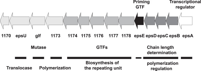
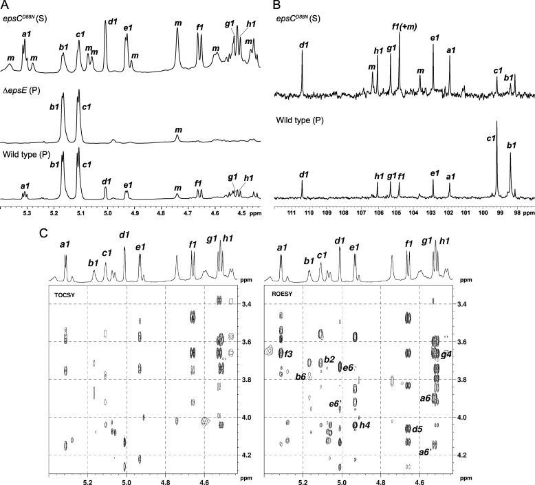
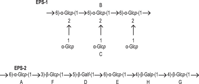
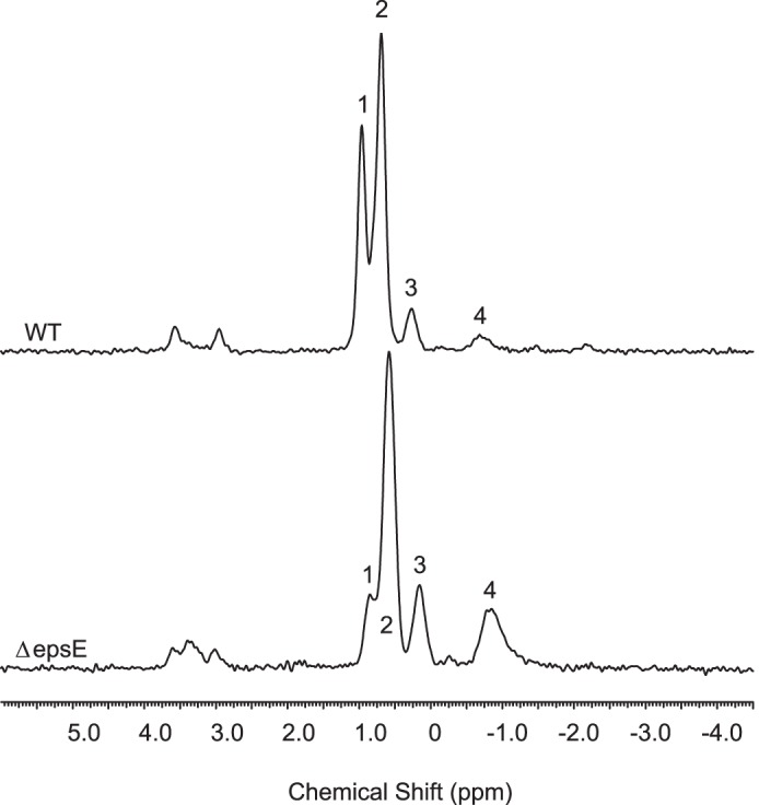
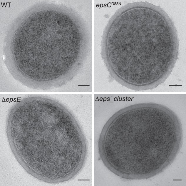
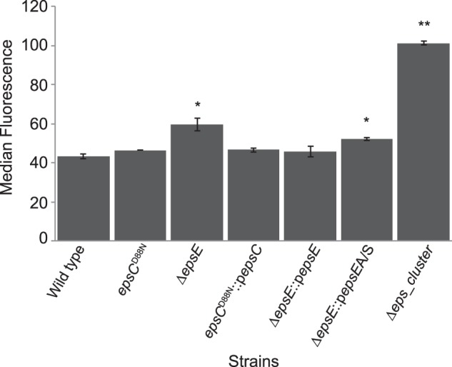
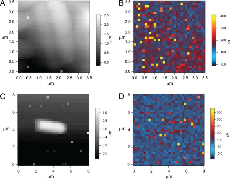
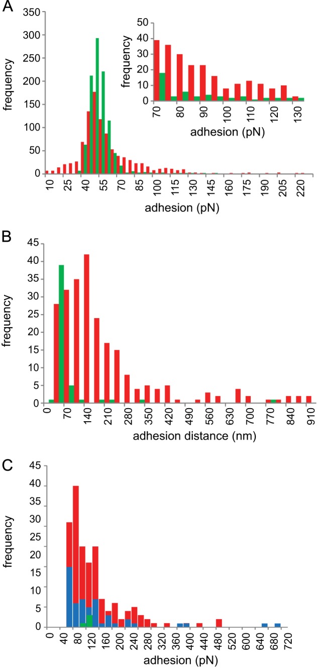
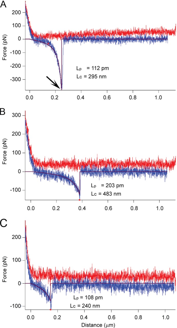
Exopolysaccharides were isolated and purified from Lactobacillus johnsonii FI9785, which has previously been shown to act as a competitive exclusion agent to control Clostridium perfringens in poultry. Structural analysis by NMR spectroscopy revealed that L. johnsonii FI9785 can produce two types of exopolysaccharide: EPS-1 is a branched dextran with the unusual feature that every backbone residue is substituted with a 2-linked glucose unit, and EPS-2 was shown to have a repeating unit with the following structure: -6)-α-Glc p -(1–3)-β-Glc p -(1–5)-β-Gal f -(1–6)-α-Glc p -(1–4)-β-Gal p -(1–4)-β-Glc p -(1-. Sites on both polysaccharides were partially occupied by substituent groups: 1-phosphoglycerol and O -acetyl groups in EPS-1 and a single O -acetyl group in EPS-2. Analysis of a deletion mutant (Δ epsE ) lacking the putative priming glycosyltransferase gene located within a predicted eps gene cluster revealed that the mutant could produce EPS-1 but not EPS-2, indicating that epsE is essential for the biosynthesis of EPS-2. Atomic force microscopy confirmed the localization of galactose residues on the exterior of wild type cells and their absence in the Δ epsE mutant. EPS2 was found to adopt a random coil structural conformation. Deletion of the entire 14-kb eps cluster resulted in an acapsular mutant phenotype that was not able to produce either EPS-2 or EPS-1. Alterations in the cell surface properties of the EPS-specific mutants were demonstrated by differences in binding of an anti-wild type L. johnsonii antibody. These findings provide insights into the biosynthesis and structures of novel exopolysaccharides produced by L. johnsonii FI9785, which are likely to play an important role in biofilm formation, protection against harsh environment of the gut, and colonization of the host. Background: Bacterial cell surface polysaccharides are important in pathogenesis, cell adhesion, and protection against harsh environments. Results: Two novel exopolysaccharide (EPS) structures were identified in Lactobacillus johnsonii . Conclusion: The eps cluster is essential for production of both EPS, but epsE is required only for the heteropolymer. Significance: This study will guide functional analysis of EPS in survival and colonization of gut commensals.
Overview
The claims and statistics of this paper haven’t been checked yet.
Run the check — free, no account needed. It reads the open-access text and checks the sources it cites.
Integrity signals
Registry
Cites
Statistics
Figures
Verification
Claims the paper makes
The claims and statistics of this paper haven’t been checked yet.
Run the check — free, no account needed. It reads the open-access text and checks the sources it cites.
Claims other papers make about this one
Each is checked against this paper’s own text: does it say what the citing paper says it does?
The source backs part of the claim, not all of it — or backs it for a different population, condition or comparison.
In the citing paper
“EPS produced by Lb. johnsonii FI9785 may favor the colonization of probiotic bacteria in the host gastrointestinal tract by replacing pathogenic bacteria in a competitive inhibition mode [145].”
From Lactic Acid Bacteria Exopolysaccharides Producers: A Sustainable Tool for Functional Foods (2021) · cites this paper as [145]
Experimental — accuracy not yet measured
Self-check
Percentages, sums and reported differences re-computed from the paper’s own numbers.
The self-check needs the paper’s full text.
Citations
Cites
Statistical mechanics of chain molecules
Flory, P. J., & Volkenstein, M. V. (1969) · Biopolymers · doi:10.1002/bip.1969.360080514 · Cited by 6,449
Calibration of atomic-force microscope tips
Hutter, J. L., & Bechhoefer, J. (1993) · Review of Scientific Instruments · doi:10.1063/1.1143970 · Cited by 4,205
Röntgenuntersuchung gelöster Fadenmoleküle
Kratky, O., & Porod, G. (1949) · Recueil des Travaux Chimiques des Pays-Bas · doi:10.1002/recl.19490681203 · Cited by 1,994
Heteropolysaccharides from lactic acid bacteria
Vuyst, L. D., & Degeest, B. (1999) · FEMS Microbiology Reviews · doi:10.1111/j.1574-6976.1999.tb00395.x · Cited by 840
Genetic Analysis of the Capsular Biosynthetic Locus from All 90 Pneumococcal Serotypes
Bentley, S. D., Aanensen, D. M., Mavroidi, A., Saunders, D., Rabbinowitsch, E., Collins, M. D., Donohoe, K., Harris, D. J., Murphy, L., Quail, M. A., et al. (2006) · PLoS Genetics · doi:10.1371/journal.pgen.0020031 · Cited by 759
Fanning, S., Hall, L. J., Cronin, M. A., Zomer, A., MacSharry, J. A., Goulding, D., Motherway, M. O., Shanahan, F., Nally, K., Dougan, G., et al. (2012) · Proceedings of the National Academy of Sciences · doi:10.1073/pnas.1115621109 · Cited by 563
New Perspectives for Lactobacilli Exopolysaccharides
Badel, S., Bernardi, T., & Michaud, P. (2011) · Biotechnology Advances · doi:10.1016/j.biotechadv.2010.08.011 · Cited by 497
Efficient insertional mutagenesis in lactococci and other gram-positive bacteria
Maguin, E., Prévost, H., Ehrlich, S. D., & Gruss, A. (1996) · Journal of Bacteriology · doi:10.1128/jb.178.3.931-935.1996 · Cited by 486
van Hijum, S. A. F. T., Kralj, S., Ozimek, L. K., Dijkhuizen, L., & van Geel-Schutten, I. H. (2006) · Microbiology and Molecular Biology Reviews · doi:10.1128/mmbr.70.1.157-176.2006 · Cited by 466
Statistical mechanics of chain molecules
Volkenstein, M. (1970) · Biopolymers · doi:10.1002/bip.1970.360090311 · Cited by 446
Cited by
Sorted by citations
Polysaccharide Production by Lactic Acid Bacteria: From Genes to Industrial Applications
Zeidan, A. A., Poulsen, V. K., Janzen, T., Buldo, P., Derkx, P. M. F., Øregaard, G., & Neves, A. R. (2017) · FEMS Microbiology Reviews · doi:10.1093/femsre/fux017 · Cited by 326
Castro-Bravo, N., Wells, J. M., Margolles, A., & Ruas-Madiedo, P. (2018) · Frontiers in Microbiology · doi:10.3389/fmicb.2018.02426 · Cited by 284
The Critical Roles of Polysaccharides in Gut Microbial Ecology and Physiology
Porter, N. T., & Martens, E. C. (2017) · Annual Review of Microbiology · doi:10.1146/annurev-micro-102215-095316 · Cited by 251
Nanoencapsulation for Probiotic Delivery
Centurion, F., Basit, A. W., Liu, J., Gaisford, S., Rahim, M. A., & Kalantar‐Zadeh, K. (2021) · ACS Nano · doi:10.1021/acsnano.1c09951 · Cited by 225
Dertli̇, E., Mayer, M. J., & Narbad, A. (2015) · BMC Microbiology · doi:10.1186/s12866-015-0347-2 · Cited by 216
Liu, Z., Zhang, Z., Qiu, L., Zhang, F., Xu, X., Wei, H., & Tao, X. (2017) · Journal of Dairy Science · doi:10.3168/jds.2016-11944 · Cited by 205
Vandana, & Das, S. (2022) · Carbohydrate Polymers · doi:10.1016/j.carbpol.2022.119536 · Cited by 199
Lactic Acid Bacteria Exopolysaccharides Producers: A Sustainable Tool for Functional Foods
Prete, R., Alam, M. K., Perpetuini, G., Perla, C., Pittia, P., & Corsetti, A. (2021) · Foods · doi:10.3390/foods10071653 · Cited by 194
Bacteria Facilitate Enteric Virus Co-infection of Mammalian Cells and Promote Genetic Recombination
Erickson, A. K., Jesudhasan, P. R. R., Mayer, M. J., Narbad, A., Winter, S., & Pfeiffer, J. K. (2017) · Cell Host & Microbe · doi:10.1016/j.chom.2017.11.007 · Cited by 187
Xu, Y., Cui, Y., Yue, F., Liu, L., Shan, Y., Liu, B., Zhou, Y., & Lü, X. (2019) · Food Hydrocolloids · doi:10.1016/j.foodhyd.2019.03.032 · Cited by 189
Dertli, E., Mercan, E., Arıcı, M., Yılmaz, M. T., & Sağdıç, O. (2016) · LWT - Food Science and Technology · doi:10.1016/j.lwt.2016.03.030 · Cited by 168
Benefits of bacteria-derived exopolysaccharides on gastrointestinal microbiota, immunity and health
Oerlemans, M. M. P., Akkerman, R., Ferrari, M. D. L., Walvoort, M. T. C., & de Vos, P. (2020) · Journal of Functional Foods · doi:10.1016/j.jff.2020.104289 · Cited by 159
da Silva, L. A., Neto, J. H. P. L., & Cardarelli, H. R. (2019) · Annals of Microbiology · doi:10.1007/s13213-019-01456-9 · Cited by 152
Taylan, O., Yılmaz, M. T., & Dertli̇, E. (2019) · International Journal of Biological Macromolecules · doi:10.1016/j.ijbiomac.2019.06.078 · Cited by 126
Werning, M. L., Hernández-Alcántara, A. M., Ruiz, M. J., Soto, L. P., Dueñas, M. T., López, P., & Frizzo, L. S. (2022) · Foods · doi:10.3390/foods11091284 · Cited by 132
Protein-Polysaccharide Interactions to Alter Texture
van de Velde, F., de Hoog, E. H. A., Oosterveld, A., & Tromp, R. H. (2015) · Annual Review of Food Science and Technology · doi:10.1146/annurev-food-022814-015558 · Cited by 116
Hatziioanou, D., Gherghisan-Filip, C., Saalbach, G., Horn, N., Wegmann, U., Duncan, S. H., Flint, H. J., Mayer, M. J., & Narbad, A. (2017) · Microbiology · doi:10.1099/mic.0.000515 · Cited by 113
Bibi, A., Xiong, Y., Rajoka, M. S. R., Mehwish, H. M., Radicetti, E., Umair, M., Shoukat, M., Khan, M. K. I., & Aadil, R. M. (2021) · Sustainability · doi:10.3390/su132212429 · Cited by 108
Lactobacillus-produced exopolysaccharides and their potential health benefits: A review
Patten, D. A., & Laws, A. P. (2015) · Beneficial Microbes · doi:10.3920/bm2014.0117 · Cited by 100
Dertli̇, E., Toker, Ö. S., Durak, M. Z., Yılmaz, M. T., Tatlısu, N. B., Sağdıç, O., & Cankurt, H. (2015) · Carbohydrate Polymers · doi:10.1016/j.carbpol.2015.08.047 · Cited by 98
Yılmaz, M. T., Dertli̇, E., Toker, Ö. S., Tatlısu, N. B., Sağdıç, O., & Arıcı, M. (2014) · Journal of Dairy Science · doi:10.3168/jds.2014-8936 · Cited by 92
Deo, D., Davray, D., & Kulkarni, R. (2019) · Microorganisms · doi:10.3390/microorganisms7100444 · Cited by 90
Next-Generation Probiotics and Chronic Diseases: A Review of Current Research and Future Directions
Tiwari, A. K., Krisnawati, D. I., Susilowati, E., Mutalik, C., & Kuo, T. (2024) · Journal of Agricultural and Food Chemistry · doi:10.1021/acs.jafc.4c08702 · Cited by 91
Structural analysis of the α-d-glucan produced by the sourdough isolate Lactobacillus brevis E25
Dertli̇, E., Colquhoun, I. J., Côté, G. L., Gall, G. L., & Narbad, A. (2017) · Food Chemistry · doi:10.1016/j.foodchem.2017.09.017 · Cited by 80
İspirli, H., Sagdic, O., Yılmaz, M. T., & Dertli, E. (2019) · Process Biochemistry · doi:10.1016/j.procbio.2018.12.015 · Cited by 77
Exopolysaccharide (EPS) Synthesis by Oenococcus oeni: From Genes to Phenotypes
Dimopoulou, M., Vuillemin, M., Campbell-Sills, H., Lucas, P., Ballestra, P., Miot‐Sertier, C., Favier, M., Coulon, J., Moine, V., Doco, T., et al. (2014) · PLoS ONE · doi:10.1371/journal.pone.0098898 · Cited by 75
Tiwari, O. N., Sasmal, S., Kataria, A. K., & Devi, I. (2020) · 3 Biotech · doi:10.1007/s13205-020-02200-w · Cited by 75
İspirli, H., Özmen, D., Yılmaz, M. T., Sağdıç, O., & Dertli̇, E. (2019) · Food Control · doi:10.1016/j.foodcont.2019.106812 · Cited by 67
Deka, P. C., Goswami, G., Das, P., Gautom, T., Chowdhury, N., Boro, R. C., & Barooah, M. (2018) · Molecular Biology Reports · doi:10.1007/s11033-018-4566-0 · Cited by 66
Sengupta, D., Datta, S., & Biswas, D. (2018) · Applied Microbiology and Biotechnology · doi:10.1007/s00253-018-8745-7 · Cited by 60
Aburas, H. M., İspirli, H., Taylan, O., Yılmaz, M. T., & Dertli̇, E. (2020) · International Journal of Biological Macromolecules · doi:10.1016/j.ijbiomac.2020.06.030 · Cited by 59
Potential Health Benefits of Ropy Exopolysaccharides Produced by Lactobacillus plantarum
Yılmaz, T., & Şimşek, Ö. (2020) · Molecules · doi:10.3390/molecules25143293 · Cited by 54
Dertli̇, E., Yılmaz, M. T., Tatlısu, N. B., Toker, Ö. S., Cankurt, H., & Sağdıç, O. (2016) · Meat Science · doi:10.1016/j.meatsci.2016.06.008 · Cited by 54
Sran, K. S., Bisht, B., Mayilraj, S., & Choudhury, A. R. (2019) · International Journal of Biological Macromolecules · doi:10.1016/j.ijbiomac.2019.03.016 · Cited by 53
Debnath, S., Chakraborty, S., Langthasa, M., Choure, K., Agnihotri, V., Srivastava, A., Kant, P., Tilwari, A., Maheshwari, D. K., & Pandey, P. (2023) · Frontiers in Plant Science · doi:10.3389/fpls.2023.1152875 · Cited by 51
Richards, P. J., Lafontaine, G. M. F., Connerton, P. L., Liang, L., Asiani, K., Fish, N. M., & Connerton, I. F. (2020) · mSystems · doi:10.1128/msystems.00827-19 · Cited by 48
Özkan, E. R., Demirci, T., & Akın, N. (2021) · LWT · doi:10.1016/j.lwt.2021.110908 · Cited by 50
Immunomodulatory Roles of Polysaccharide Capsules in the Intestine
Hsieh, S. A., & Allen, P. M. (2020) · Frontiers in Immunology · doi:10.3389/fimmu.2020.00690 · Cited by 54
Liao, N., Pang, B., Han, J., Xu, X., Yan, L., Li, H., Shao, D., & Shi, J. (2020) · Journal of Controlled Release · doi:10.1016/j.jconrel.2020.04.022 · Cited by 48
Castro‐Alves, V., Gomes, D., Menolli, N., Sforça, M. L., & do Nascimento, J. R. O. (2016) · International Journal of Biological Macromolecules · doi:10.1016/j.ijbiomac.2016.11.059 · Cited by 47
Banerjee, A., Das, D., Rudra, S. G., Mazumder, K., Andler, R., & Bandopadhyay, R. (2019) · Journal of Polymers and the Environment · doi:10.1007/s10924-019-01602-z · Cited by 47
Johnson, C., Ridley, H., Marchetti, R., Silipo, A., Griffin, D. C., Crawford, L., Bonev, B. B., Molinaro, A., & Lakey, J. H. (2014) · Molecular Microbiology · doi:10.1111/mmi.12568 · Cited by 45
Zhai, Q., Tian, F., Wang, G., Zhao, J., Liu, X., Cross, K. L., Zhang, H., Narbad, A., & Chen, W. (2016) · RSC Advances · doi:10.1039/c5ra24843d · Cited by 43
Özkan, E. R., Demirci, T., Öztürk, H. İ., & Akın, N. (2020) · Journal of the Science of Food and Agriculture · doi:10.1002/jsfa.10909 · Cited by 44
Yalmancı, D., İspirli, H., & Dertli̇, E. (2022) · Food Bioscience · doi:10.1016/j.fbio.2022.102014 · Cited by 43
Laffargue, T., Moulis, C., & Remaud‐Siméon, M. (2023) · Biotechnology Advances · doi:10.1016/j.biotechadv.2023.108140 · Cited by 41
Structural and bioactive characteristics of a dextran produced by Lactobacillus kunkeei AK1
Yılmaz, M. T., İspirli, H., Taylan, O., Bilgrami, A. L., & Dertli̇, E. (2022) · International Journal of Biological Macromolecules · doi:10.1016/j.ijbiomac.2022.01.012 · Cited by 41
İspirli, H., Yüzer, M. O., Skory, C. D., Colquhoun, I. J., Sağdıç, O., & Dertli̇, E. (2019) · Biocatalysis and Biotransformation · doi:10.1080/10242422.2019.1593969 · Cited by 40
Bioactive and technological properties of an α-D-glucan synthesized by Weissella cibaria PDER21
Yılmaz, M. T., İspirli, H., Taylan, O., Alamoudi, M., & Dertli̇, E. (2022) · Carbohydrate Polymers · doi:10.1016/j.carbpol.2022.119227 · Cited by 40
Xiao, L., Xu, D., Tang, N., Rui, X., Zhang, Q., Chen, X., Dong, M., & Li, W. (2021) · Food Chemistry Molecular Sciences · doi:10.1016/j.fochms.2021.100054 · Cited by 42
Yılmaz, M. T., İspirli, H., Taylan, O., Taşdemir, V., Sağdıç, O., & Dertli̇, E. (2021) · Food Bioscience · doi:10.1016/j.fbio.2021.101330 · Cited by 39
EpsA is an essential gene in exopolysaccharide production in L actobacillus johnsonii FI9785
Dertli̇, E., Mayer, M. J., Colquhoun, I. J., & Narbad, A. (2015) · Microbial Biotechnology · doi:10.1111/1751-7915.12314 · Cited by 39
Živković, M., Hidalgo-Cantabrana, C., Kojić, M., Gueimonde, M., Golić, N., & Ruas‐Madiedo, P. (2015) · Food Research International · doi:10.1016/j.foodres.2015.05.012 · Cited by 39
Singh, S., Sran, K. S., Pinnaka, A. K., & Choudhury, A. R. (2019) · International Journal of Biological Macromolecules · doi:10.1016/j.ijbiomac.2019.06.080 · Cited by 36
Şentürk, D. Z., Dertli̇, E., Erten, H., & Şimşek, Ö. (2019) · Food Science and Biotechnology · doi:10.1007/s10068-019-00641-5 · Cited by 36
Yang, Y., Jiang, G., & Tian, Y. (2023) · World Journal of Microbiology and Biotechnology · doi:10.1007/s11274-023-03610-7 · Cited by 39
İspirli, H., Demirbaş, F., & Dertli̇, E. (2018) · Journal of Food Science and Technology · doi:10.1007/s13197-018-3181-3 · Cited by 35
Isolation and identification of exopolysaccharide producer lactic acid bacteria from Turkish yogurt
İspirli, H., & Dertli̇, E. (2017) · Journal of Food Processing and Preservation · doi:10.1111/jfpp.13351 · Cited by 34
Putative Adhesion Factors in Vaginal Lactobacillus gasseri DSM 14869: Functional Characterization
Zeng, Z., Zuo, F., & Marcotte, H. (2019) · Applied and Environmental Microbiology · doi:10.1128/aem.00800-19 · Cited by 35
Xiao, L., Yang, Y., Han, S., Rui, X., Ma, K., Zhang, C., Wang, G., & Li, W. (2022) · International Journal of Biological Macromolecules · doi:10.1016/j.ijbiomac.2022.10.124 · Cited by 32
Nakamura, S., Kurata, R., Tonozuka, T., Funane, K., Park, E. Y., & Miyazaki, T. (2023) · Journal of Biological Chemistry · doi:10.1016/j.jbc.2023.104885 · Cited by 31
Zhou, J., Yan, B., Wu, Y., Zhu, H., Lian, H., Zhao, J., Zhang, H., Chen, W., & Fan, D. (2021) · LWT · doi:10.1016/j.lwt.2021.111396 · Cited by 30
Özpınar, F. B., İspirli, H., Kayacan, S., Korkmaz, K., Dere, S., Sağdıç, O., Alkay, Z., Tunçil, Y. E., Ayyash, M. M., & Dertli̇, E. (2024) · International Journal of Biological Macromolecules · doi:10.1016/j.ijbiomac.2024.130507 · Cited by 29
Mayer, M. J., D’Amato, A., Colquhoun, I. J., Gall, G. L., & Narbad, A. (2020) · Applied and Environmental Microbiology · doi:10.1128/aem.02808-19 · Cited by 28
Food Supplements to Mitigate Detrimental Effects of Pelvic Radiotherapy
Segers, C., Verslegers, M., Baatout, S., Leys, N., Lebeer, S., & Mastroleo, F. (2019) · Microorganisms · doi:10.3390/microorganisms7040097 · Cited by 27
Revealing the Compact Structure of Lactic Acid Bacterial Heteroexopolysaccharides by SAXS and DLS
Khan, S., Birch, J., Harris, P., Calsteren, M. V., Ipsen, R., Peters, G. H. J., Svensson, B., & Almdal, K. (2017) · Biomacromolecules · doi:10.1021/acs.biomac.6b01597 · Cited by 26
Lactic acid bacteria - derived exopolysaccharide: Biosynthesis and antibacterial characterization
Zhang, M., Zhao, D., Yang, H., Jiao, X., Zhou, R., Zheng, J., & Wu, C. (2025) · Trends in Food Science & Technology · doi:10.1016/j.tifs.2025.105033 · Cited by 26
İspirli, H., Demirbaş, F., Yüzer, M. O., & Dertli̇, E. (2018) · Food Biotechnology · doi:10.1080/08905436.2018.1507913 · Cited by 24
He, Y., Chen, H., Ye, Z., Zhang, X., Ye, H., & Ye, M. (2022) · Process Biochemistry · doi:10.1016/j.procbio.2022.08.035 · Cited by 24
Debnath, S., Das, A., Maheshwari, D. K., & Pandey, P. (2022) · Microbiological Research · doi:10.1016/j.micres.2022.127255 · Cited by 24
Updates on Metabolism in Lactic Acid Bacteria in Light of “Omic” Technologies
Kowalczyk, M., Mayo, B., Fernández, M., & Aleksandrzak‐Piekarczyk, T. (2015) · Wiley eBooks · doi:10.1002/9781118868386.ch1 · Cited by 25
Isolation and characterisation of lactic acid bacteria from traditional koumiss and kurut
İspirli, H., & Dertli̇, E. (2017) · International Journal of Food Properties · doi:10.1080/10942912.2017.1372473 · Cited by 22
Le, T.-S., Nguyen, P.-T., Nguyen-Ho, S.-H., Nguyen, T.-P., Nguyen, T.-T., Thai, M.-N., Nguyen-Thi, T.-U., Nguyen, M.-C., Hoang, Q.-K., & Nguyen, H. (2021) · Archives of Microbiology · doi:10.1007/s00203-021-02479-0 · Cited by 21
Mercan, E., İspirli, H., Sert, D., Yılmaz, M. T., & Dertli̇, E. (2015) · Archives of Microbiology · doi:10.1007/s00203-015-1141-0 · Cited by 21
Yalmancı, D., Dertli̇, E., Tekin‐Cakmak, Z. H., & Karasu, S. (2023) · International Journal of Food Science & Technology · doi:10.1111/ijfs.16287 · Cited by 18
Ermiş, E., Poyraz, E., Dertli̇, E., & Yılmaz, M. T. (2020) · Sakarya University Journal of Science · doi:10.16984/saufenbilder.545929 · Cited by 18
Yalmancı, D., Dertli̇, E., Tekin‐Cakmak, Z. H., & Karasu, S. (2022) · International Journal of Biological Macromolecules · doi:10.1016/j.ijbiomac.2022.12.069 · Cited by 17
Do, T. B. T., Tran, B.-K., Trần, T. V. T., Hiếu, L. T., Cnockaert, M. C., Vandamme, P. A. R., Nguyen, T. H. C., Nguyen, C. C., Hong, S. H., Kim, S. Y., et al. (2020) · ACS Omega · doi:10.1021/acsomega.0c05256 · Cited by 16
Knirel, Y. A., & Calsteren, M. V. (2020) · Elsevier eBooks · doi:10.1016/b978-0-12-819475-1.00005-5 · Cited by 16
İspirli, H., Sağdıç, O., & Dertli̇, E. (2020) · Preparative Biochemistry & Biotechnology · doi:10.1080/10826068.2020.1795673 · Cited by 15
A contextualised protein language model reveals the functional syntax of bacterial evolutionPreprint
Wiatrak, M., Abelson, D. C., Mikhaylenko, R., Torné, R. V., Ntemourtsidou, M., Dinan, A. M., Arora, D., Horsfield, S., Lees, J. A., Brbić, M., et al. (2025) · bioRxiv (Cold Spring Harbor Laboratory) · doi:10.1101/2025.07.20.665723 · Cited by 19
Sample Collection and Preparation of Biofluids and Extracts for NMR Spectroscopy
Gall, G. L. (2015) · Methods in molecular biology · doi:10.1007/978-1-4939-2377-9_2 · Cited by 13
Himar1 Transposon for Efficient Random Mutagenesis in Aggregatibacter actinomycetemcomitans
Ding, Q., & Tan, K. S. (2017) · Frontiers in Microbiology · doi:10.3389/fmicb.2017.01842 · Cited by 14
Tho, N. P., Son, L. T., Tho, N. T., Cuong, B. D., Toan, H. P., Khánh, H. Q., & Thanh, N. H. (2021) · Microbiology · doi:10.1134/s0026261721040147 · Cited by 13
Xiao, L., Zhao, X., Lin, L., Mahsa, G. C., Ma, K., Zhang, C., Rui, X., & Li, W. (2024) · Journal of Agricultural and Food Chemistry · doi:10.1021/acs.jafc.4c04256 · Cited by 13
Ge, Z., Wang, D., Zhao, W., Wang, P., & Zhao, X. (2025) · Food Reviews International · doi:10.1080/87559129.2025.2467163 · Cited by 14
Novel imaging technologies for characterization of microbial extracellular polysaccharides
Lilledahl, M. B., & Stokke, B. T. (2015) · Frontiers in Microbiology · doi:10.3389/fmicb.2015.00525 · Cited by 13
Zhang, Y., Wu, Y., Zhao, X., Ye, Q., Cao, L., Liu, M., Gao, B., Niu, Q., Chen, N., Duan, Z., et al. (2025) · Acta Pharmaceutica Sinica B · doi:10.1016/j.apsb.2025.07.028 · Cited by 12
Cell surface polysaccharides in the gut microbiota: occurrence, structure and role
Laplanche, V., Speciale, I., Castro, C. D., & Juge, N. (2025) · Gut Microbes · doi:10.1080/19490976.2025.2536082 · Cited by 12
Wang, K., Wang, Y., Gu, L., Yu, J., Liu, Q., Zhang, R., Liang, G., Chen, H., Gu, F., Liu, H., et al. (2024) · Microorganisms · doi:10.3390/microorganisms12040672 · Cited by 15
Dev, M. J., Mahajan, G., Warke, R. G., Warke, G. M., Patil, T. A., Satardekar, M. R., Dalvi, R. C., & Singhal, R. S. (2023) · International Microbiology · doi:10.1007/s10123-023-00399-1 · Cited by 12
Llamas-Arriba, M. G., Pérez-Ramos, A., Puertas, A. I., López, P., Dueñas, M. T., & Prieto, A. (2018) · Frontiers in Microbiology · doi:10.3389/fmicb.2018.02041 · Cited by 13
Comparative genomics of Lactobacillus johnsonii reveals extensive intraspecific genetic variation
Chen, K., Zhou, X., Zhao, J., Ross, R. P., Stanton, C., Chen, W., & Yang, B. K. (2023) · Food Bioscience · doi:10.1016/j.fbio.2023.103190 · Cited by 11
Karasu, E. N., & Ermiş, E. (2018) · Journal of Food Engineering · doi:10.1016/j.jfoodeng.2018.08.026 · Cited by 11
Özkan, E. R., Öztürk, H. İ., Demirci, T., & Akın, N. (2021) · LWT · doi:10.1016/j.lwt.2021.112077 · Cited by 12
Sengupta, D., Datta, S., & Biswas, D. (2019) · Journal of Basic Microbiology · doi:10.1002/jobm.201900080 · Cited by 11
Woo, S., Sung, J.-M., Park, J., Park, E. Y., & Park, E. Y. (2025) · Food Research International · doi:10.1016/j.foodres.2025.117523 · Cited by 10
Meng, F., Lyu, Y., Chen, X., Lü, F., Zhao, H., Lu, Y., Zhao, M., & Lu, Z. (2023) · Journal of Agricultural and Food Chemistry · doi:10.1021/acs.jafc.2c07880 · Cited by 9
Yang, S., Liu, L., Wang, J., Guo, S., Liu, G., Chen, X., Deng, X., Tu, M., Tao, Y., & Rao, Y. (2022) · Archives of Microbiology · doi:10.1007/s00203-022-03232-x · Cited by 9
Characterisation of dextran AP-27 produced by bee pollen isolate Lactobacillus kunkeei AP-27
Yılmaz, M. T., İspirli, H., Alidrisi, H. M., Taylan, O., & Dertli̇, E. (2023) · Process Biochemistry · doi:10.1016/j.procbio.2023.03.007 · Cited by 7
Metadata
- Authors
- Dertli, E., Colquhoun, I. J., Gunning, A. P., Bongaerts, R. J., Le Gall, G., Bonev, B. B., Mayer, M. J., & Narbad, A.
- Affiliations
- Quadram Institute (GB)
- University of Nottingham (GB)
- Corresponding author
- Mayer, Melinda J.
- Venue
- Journal of Biological Chemistry
- Publisher
- Elsevier BV
- Published
- 2013-11
- Volume
- 288
- Issue
- 44
- Pages
- 31938-31951
- ISSN
- 0021-9258 (ISSN-L)
- PMC
- PMC3814790
- OpenAlex
- W1964087334
- Cited by
- 134
- References
- 58
- Citations by year
- 2026: 13 · 2025: 13 · 2024: 4 · 2023: 13 · 2022: 11 · 2021: 10 · 2020: 14 · 2019: 13 · 2018: 10 · 2017: 11 · 2016: 4 · 2015: 14 · 2014: 4
- FWCI
- 10.85
- Type
- journal article
- Language
- en
- Open access
- hybrid
- doi.org
- Licence
- Locations
- Journal of Biological Chemistry (publishedVersion, cc-by)
- Europe PMC (PubMed Central) (publishedVersion, cc-by)
- PubMed Central (publishedVersion, cc-by)
- PubMed (publishedVersion)
- DSpace@Bayburt (submittedVersion, cc-by-nc-nd)
- UEA Digital Repository (University of East Anglia) (submittedVersion)
- Funders
- Biotechnology and Biological Sciences Research Council ((BB/J004529/1, BBS/E/F/00044453, BB/J004545/1)
- Topics
- Life Sciences › Agricultural and Biological Sciences › Food Science › Polysaccharides Composition and Applications
- Life Sciences › Agricultural and Biological Sciences › Food Science › Probiotics and Fermented Foods
- Life Sciences › Immunology and Microbiology › Microbiology › Escherichia coli research studies
- Keywords
- exopolysaccharides, exopolysaccharide biosynthesis, heteropolymers, galactose substitution, O-acetylation, cell surface polysaccharides, biofilm formation
- MeSH terms
- Bacterial Proteins, Carbohydrate Conformation, Genes, Bacterial, Multigene Family, Glucosyltransferases, Lactobacillus, Mutation, Polysaccharides, Bacterial
About these checks
Registry, retraction, Cites and statistics results are computed, not judged, with one exception: a printed reference that no index lists is read once by a model before it counts as a citation error. Claims are extracted by a model and kept only when their quote appears verbatim in the text; each cited source is then checked, with a model’s help, for whether it says what the claim relies on. Those verdicts can be wrong — read the claim’s quote and the cited source before relying on one.
Statistics and arithmetic are recomputed, not judged.
Confidence bands are shown only for fields calibrated on our annotated test set. None is calibrated yet, so no band is shown.
What counts as a claim ›How we measure confidence ›How we check this ›
Are you an author, or do you see a problem with this page? Request a review
