

The DOI matches its Crossref record; 36 of the 36 references it cites were found in a registry or an index.
An acetylation–phosphorylation switch that regulates tau aggregation propensity and function
Open accessAbstract
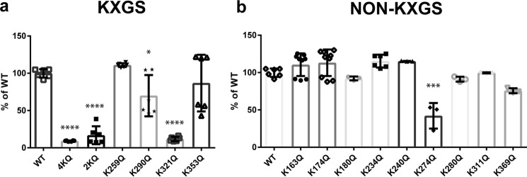
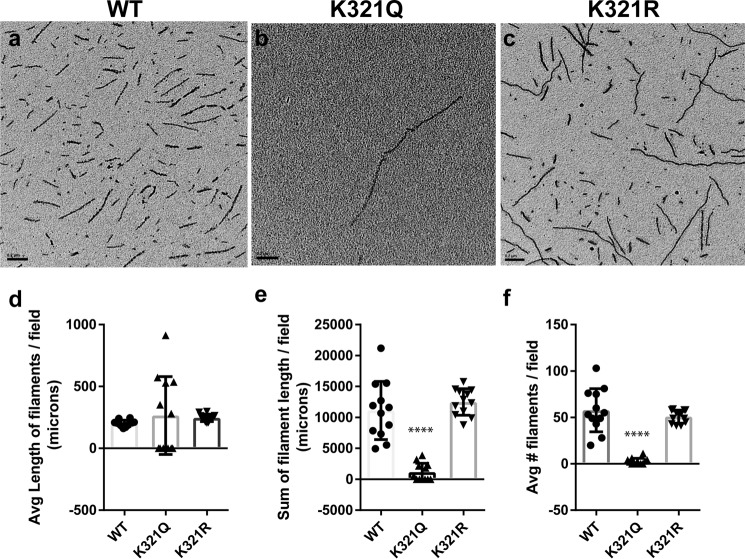
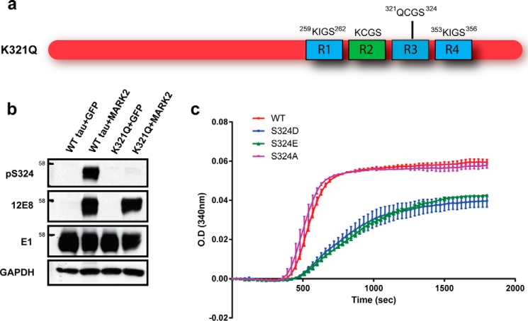
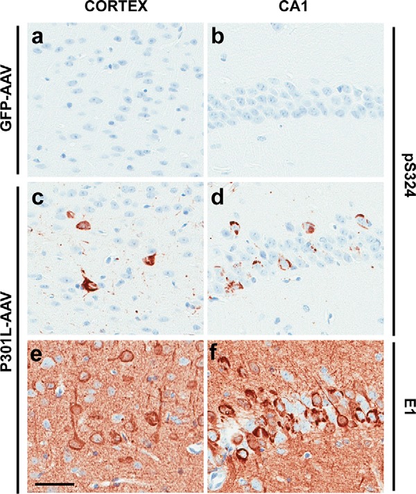
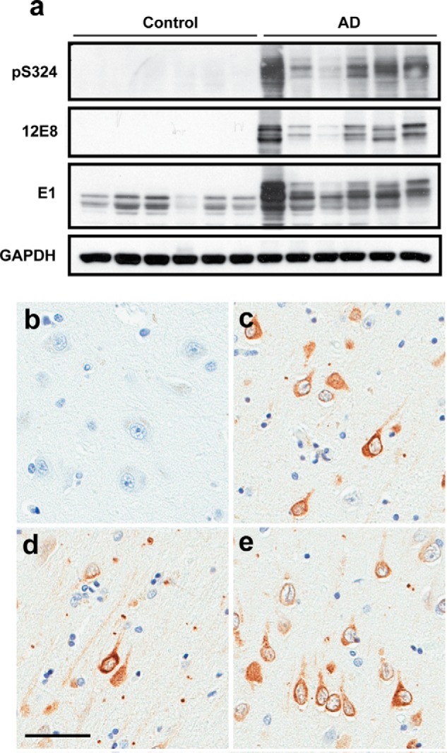
The aberrant accumulation of tau protein is a pathological hallmark of a class of neurodegenerative diseases known as tauopathies, including Alzheimer's disease and related dementias. On the basis of previous observations that tau is a direct substrate of histone deacetylase 6 (HDAC6), we sought to map all HDAC6-responsive sites in tau and determine how acetylation in a site-specific manner affects tau's biophysical properties in vitro . Our findings indicate that several acetylation sites in tau are responsive to HDAC6 and that acetylation on Lys-321 (within a KCGS motif) is both essential for acetylation-mediated inhibition of tau aggregation in vitro and a molecular tactic for preventing phosphorylation on the downstream Ser-324 residue. To determine the functional consequence of this HDAC6-regulated phosphorylation event, we examined tau's ability to promote microtubule assembly and found that phosphorylation of Ser-324 interferes with the normal microtubule-stabilizing function of tau. Tau phosphorylation of Ser-324 (pSer-324) has not previously been evaluated in the context of tauopathy, and here we observed increased deposition of pSer-324–positive tau both in mouse models of tauopathy and in patients with Alzheimer's disease. These findings uncover a novel acetylation–phosphorylation switch at Lys-321/Ser-324 that coordinately regulates tau polymerization and function. Because the disease relevance of this finding is evident, additional studies are needed to examine the role of pSer-324 in tau pathobiology and to determine whether therapeutically modulating this acetylation–phosphorylation switch affects disease progression in vivo .
Overview
The claims and statistics of this paper haven’t been checked yet.
Run the check — free, no account needed. It reads the open-access text and checks the sources it cites.
Integrity signals
Registry
Cites
Statistics
Figures
Verification
Claims the paper makes
The claims and statistics of this paper haven’t been checked yet.
Run the check — free, no account needed. It reads the open-access text and checks the sources it cites.
Claims other papers make about this one
Each is checked against this paper’s own text: does it say what the citing paper says it does?
The source backs part of the claim, not all of it — or backs it for a different population, condition or comparison.
In the citing paper
“while increased Tau acetylation reduces Tau phosphorylation [34, 42].”
From Elevated SGK1 increases Tau phosphorylation and microtubule instability in Alzheimer’s patient-derived cortical neurons (2026) · cites this paper as [42]
Experimental — accuracy not yet measured
Self-check
Percentages, sums and reported differences re-computed from the paper’s own numbers.
The self-check needs the paper’s full text.
Citations
Cites
Buée, L., Bussière, T., Buée‐Scherrer, V., Delacourte, A., & Hof, P. R. (2000) · Brain Research Reviews · doi:10.1016/s0165-0173(00)00019-9 · Cited by 2,142
Cryo-EM structures of tau filaments from Alzheimer’s disease
Fitzpatrick, A. W. P., Falcon, B., He, S., Murzin, A. G., Murshudov, G., Garringer, H. J., Crowther, R. A., Ghetti, B., Goedert, M., & Scheres, S. H. W. (2017) · Nature · doi:10.1038/nature23002 · Cited by 2,061
Alzheimer’s Disease: The Challenge of the Second Century
Holtzman, D. M., Morris, J. C., & Goate, A. M. (2011) · Science Translational Medicine · doi:10.1126/scitranslmed.3002369 · Cited by 1,424 · cited 1×
“Whereas the increasing burden of tau pathology correlates with cognitive dysfunction and continues to progress with disease severity (3), both the pathogenic species of tau and the mechanism of toxicity remain unclear.”
Augustinack, J. C., Schneider, A., Mandelkow, E.-M., & Hyman, B. T. (2002) · Acta Neuropathologica · doi:10.1007/s004010100423 · Cited by 998 · cited 2×
“Phosphorylation of tau on KIGS motifs (pSer-262/356) is observed at early stages of neurofibrillary tangle (NFT) formation in AD (14), but assessment of pSer-324 immunoreactivity in AD has not been performed.”
Acetylation of Tau Inhibits Its Degradation and Contributes to Tauopathy
Min, S.-W., Cho, S.-H., Zhou, Y., Schroeder, S., Haroutunian, V., Seeley, W. W., Huang, E. J., Shen, Y., Masliah, E., Mukherjee, C., et al. (2010) · Neuron · doi:10.1016/j.neuron.2010.08.044 · Cited by 949 · cited 1×
“which tau acetylation inhibits turnover and leads to accumulation (34), given that insoluble tau species purified from NFTs have been shown to be ubiquitinated (33), a lack of sufficient ubiquitination alone does not appear to be adequate to account for tau accumulation in disease.”
Biochemistry and Cell Biology of Tau Protein in Neurofibrillary DegenerationPreprint
Mandelkow, E., & Mandelkow, E. (2012) · Cold Spring Harbor Perspectives in Medicine · doi:10.1101/cshperspect.a006247 · Cited by 897 · cited 1×
“4–6).”
Biernat, J., Gustke, N., Drewes, G., Mandelkow, E., & Mandelkow, E. (1993) · Neuron · doi:10.1016/0896-6273(93)90279-z · Cited by 796 · cited 2×
“Given that phosphorylation of Ser-262 in tau’s first KIGS motif has also been shown to impair tau’s ability to promote microtubule assembly (11), the phosphorylation status of all K X GS motifs may act to regulate tau function.”
The acetylation of tau inhibits its function and promotes pathological tau aggregation
Cohen, T. J., Guo, J. L., Hurtado, D. E., Kwong, L. K., Mills, I. P., Trojanowski, J. Q., & Lee, V. M. Y. (2011) · Nature Communications · doi:10.1038/ncomms1255 · Cited by 724 · cited 6×
“However, because acetylation of Lys-321 was not detected following CBP-mediated acetylation when increased aggregation was observed (18) (see supplemental Table S1 for a comparison of results across studies) but was identified under conditions that reduced aggregation (10), this supports our finding that modification…”
InfluentialCHIP and Hsp70 regulate tau ubiquitination, degradation and aggregation
Petrucelli, L. (2004) · Human Molecular Genetics · doi:10.1093/hmg/ddh083 · Cited by 723
Self-citationDickey, C. A., Kamal, A., Lundgren, K., Klosak, N., Bailey, R. M., Dunmore, J., Ash, P., Shoraka, S., Zlatkovic, J., Eckman, C. B., et al. (2007) · Journal of Clinical Investigation · doi:10.1172/jci29715 · Cited by 642 · cited 1×
“In addition, tau species phosphorylated on Ser-262/356 are required for the synaptic toxicity of oligomeric A (25, 26), are unable to bind and stabilize microtubules (11, 27–29), and are also not recognized by the HSP chaperone network (30, 31).”
Self-citationCritical role of acetylation in tau-mediated neurodegeneration and cognitive deficits
Min, S.-W., Chen, X., Tracy, T. E., Li, Y., Zhou, Y., Wang, C., Shirakawa, K., Minami, S. S., Defensor, E., Mok, S. A., et al. (2015) · Nature Medicine · doi:10.1038/nm.3951 · Cited by 516 · cited 3×
“Giventhat HDAC6 also regulates acetylation of non-K X GS sites in tau, it is important to note that acetylation of some residues, in particular Lys-174 (19) and Lys-280 (18, 20), has been linked to neurodegeneration and increased tau toxicity.”
Morris, M., Knudsen, G. M., Maeda, S., Trinidad, J. C., Ioanoviciu, A., Burlingame, A. L., & Mucke, L. (2015) · Nature Neuroscience · doi:10.1038/nn.4067 · Cited by 472 · cited 7×
“Despite this, a recent report failed to detect acetylation of either Lys-174 or Lys-280 in nontransgenic mice (6), which may suggest that these residues are only modified by acetylation under pathological conditions in vivo.”
InfluentialMicrotubule-associated Protein/Microtubule Affinity-regulating Kinase (p110mark)
Meyer, H. E., Mandelkow, E.-M., Drewes, G., Trinczek, B., Illenberger, S., Biernat, J., Schmitt-Ulms, G., & Mandelkow, E. (1995) · Journal of Biological Chemistry · doi:10.1074/jbc.270.13.7679 · Cited by 388
Histone deacetylase 6 interacts with the microtubule‐associated protein tau
Ding, H., Dolan, P. J., & Johnson, G. V. W. (2008) · Journal of Neurochemistry · doi:10.1111/j.1471-4159.2008.05564.x · Cited by 387
Mairet-Coello, G., Courchet, J., Pieraut, S., Courchet, V., Maximov, A., & Polleux, F. (2013) · Neuron · doi:10.1016/j.neuron.2013.02.003 · Cited by 351
Cook, C. N., Carlomagno, Y., Gendron, T. F., Dunmore, J. H., Scheffel, K., Stetler, C. T., Davis, M., Dickson, D. W., Jarpe, M. B., Deture, M. A., et al. (2013) · Human Molecular Genetics · doi:10.1093/hmg/ddt402 · Cited by 325 · cited 18×
“Whereas deposition of tau species phosphorylated on KIGS motifs (pSer-262/356) increases with age in the well-characterized rTg4510 mouse model (7, 12), it is unclear whether pSer-324 accumulation follows a similar disease course because this phosphoepitope has not been evaluated in animal models of tauopathy.”
InfluentialSelf-citationNishimura, I., Yang, Y., & Lu, B. (2004) · Cell · doi:10.1016/s0092-8674(04)00170-9 · Cited by 324 · cited 1×
“Specifically, Par-1/MARK2–mediated phosphorylation of the KIGS motifs (pSer-262/356) has been shown to be required for tau toxicity in Drosophila (24), and detection of this phosphoepitope is observed at very early stages of NFT formation in AD brain (14).”
Cripps, D., Thomas, S. N., Jeng, Y., Yang, F., Davies, P., & Yang, A. J. (2006) · Journal of Biological Chemistry · doi:10.1074/jbc.m512786200 · Cited by 312 · cited 2×
“which tau acetylation inhibits turnover and leads to accumulation (34), given that insoluble tau species purified from NFTs have been shown to be ubiquitinated (33), a lack of sufficient ubiquitination alone does not appear to be adequate to account for tau accumulation in disease.”
Acetylated tau, a novel pathological signature in Alzheimer's disease and other tauopathies
Irwin, D. J., Cohen, T. J., Grossman, M., Arnold, S. E., Xie, S. X., Lee, V. M.-Y., & Trojanowski, J. Q. (2012) · Brain · doi:10.1093/brain/aws013 · Cited by 281 · cited 4×
“Giventhat HDAC6 also regulates acetylation of non-K X GS sites in tau, it is important to note that acetylation of some residues, in particular Lys-174 (19) and Lys-280 (18, 20), has been linked to neurodegeneration and increased tau toxicity.”
InfluentialNovel diffusion barrier for axonal retention of Tau in neurons and its failure in neurodegeneration
Li, X., Kumar, Y., Zempel, H., Mandelkow, E., Biernat, J., & Mandelkow, E. (2011) · The EMBO Journal · doi:10.1038/emboj.2011.376 · Cited by 232 · cited 1×
“In addition, tau species phosphorylated on Ser-262/356 are required for the synaptic toxicity of oligomeric A (cid:2) (25, 26), are unable to bind and stabilize microtubules (11, 27–29), and are also not recognized by the HSP chaperone network (30, 31).”
Akt and CHIP coregulate tau degradation through coordinated interactions
Dickey, C. A., Koren, J., Zhang, Y.-J., Xu, Y.-F., Jinwal, U. K., Birnbaum, M. J., Monks, B., Sun, M., Cheng, J. Q., Patterson, C., et al. (2008) · Proceedings of the National Academy of Sciences · doi:10.1073/pnas.0709180105 · Cited by 226
Self-citationDickey, C. A., Dunmore, J., Lu, B., Wang, J., Lee, W. C., Kamal, A., Burrows, F., Eckman, C., Hutton, M., & Petrucelli, L. (2006) · The FASEB Journal · doi:10.1096/fj.05-5343fje · Cited by 160 · cited 1×
“In addition, tau species phosphorylated on Ser-262/356 are required for the synaptic toxicity of oligomeric A (cid:2) (25, 26), are unable to bind and stabilize microtubules (11, 27–29), and are also not recognized by the HSP chaperone network (30, 31).”
Self-citationBiernat, J., & Mandelkow, E.-M. (1999) · Molecular Biology of the Cell · doi:10.1091/mbc.10.3.727 · Cited by 149 · cited 1×
“In addition, tau species phosphorylated on Ser-262/356 are required for the synaptic toxicity of oligomeric A (cid:2) (25, 26), are unable to bind and stabilize microtubules (11, 27–29), and are also not recognized by the HSP chaperone network (30, 31).”
The microtubule-associated tau protein has intrinsic acetyltransferase activity
Cohen, T. J., Friedmann, D., Hwang, A. W., Marmorstein, R., & Lee, V. M. Y. (2013) · Nature Structural & Molecular Biology · doi:10.1038/nsmb.2555 · Cited by 149 · cited 1×
“As an additional control, we included a reaction in which recombinant tau was incubated with acetyl-CoA in the absence of the p300 acetyltransferase enzyme to control for potential nonenzymatic acetylation and/or autoacetylation of tau that may occur (9, 10).”
Selenica, M. B., Benner, L., Housley, S. B., Manchec, B., Lee, D. C., Nash, K. R., Kalin, J. H., Bergman, J. A., Kozikowski, A. P., Gordon, M. N., et al. (2014) · Alzheimer s Research & Therapy · doi:10.1186/alzrt241 · Cited by 148 · cited 1×
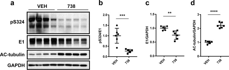
“Consistent with previous studies (7, 15, 16), HDAC6 inhibition leads to a significant reduction in tau levels as detected by the human tau-specific antibody E1 (Fig.”
Funk, K. E., Thomas, S. N., Schafer, K. N., Cooper, G. L., Liao, Z., Clark, D. J., Yang, A. J., & Kuret, J. (2014) · Biochemical Journal · doi:10.1042/bj20140372 · Cited by 142 · cited 1×
“Finally, considering that lysine residues within the KXGS motifs can also be modified by ubiquitination and methylation (6, 32, 33), it will be important in future studies to determine how these various posttranslational modifications differentially impact tau function and aggregation.”
Acetylation: a new key to unlock tau’s role in neurodegeneration
Cook, C. N., Stankowski, J. N., Carlomagno, Y., Stetler, C. T., & Petrucelli, L. (2014) · Alzheimer s Research & Therapy · doi:10.1186/alzrt259 · Cited by 140
Self-citationLoss of HDAC6, a novel CHIP substrate, alleviates abnormal tau accumulation
Cook, C. N., Gendron, T. F., Scheffel, K., Carlomagno, Y., Dunmore, J. H., Deture, M. A., & Petrucelli, L. (2012) · Human Molecular Genetics · doi:10.1093/hmg/dds125 · Cited by 132 · cited 2×
“E1 (human-specific tau antibody, rabbit host) was generated by our group against amino acid residues 19 –33 within exon 1 of human tau (15, 35, 36).”
Self-citationThe Role of HDAC6 in Alzheimer's Disease
Zhang, L., Sheng, S., & Qin, C. (2012) · Journal of Alzheimer's Disease · doi:10.3233/jad-2012-120727 · Cited by 127
Kim, C., Choi, H., Jung, E. S., Lee, W., Oh, S., Jeon, N. L., & Mook‐Jung, I. (2012) · PLoS ONE · doi:10.1371/journal.pone.0042983 · Cited by 115 · cited 2×
“…and/or activity is increased in primary hippocampal neurons treated with A (cid:2) (1–42) (21), as well as in animal models and patients with AD (21–23), may suggest that A (cid:2) -mediated stimulation of HDAC6 activity promotes K X GS deacetylation, leaving these motifs exposed and vulnerable…”
Yu, W., Polepalli, J., Wagh, D., Rajadas, J., Malenka, R., & Lu, B. (2012) · Human Molecular Genetics · doi:10.1093/hmg/ddr576 · Cited by 103
Dickey, C. A., Kraft, C., Jinwal, U. K., Koren, J., Johnson, A. G., Anderson, L. C., Lebson, L. A., Lee, D. C., Dickson, D. W., de Silva, R., et al. (2008) · American Journal Of Pathology · doi:10.2353/ajpath.2009.080764 · Cited by 86 · cited 1×
“Whereas deposition of tau species phosphorylated on KIGS motifs (pSer-262/356) increases with age in the well-characterized rTg4510 mouse model (7, 12), it is unclear whether pSer324 accumulation follows a similar disease course because this phosphoepitope has not been evaluated in animal models of tauopathy.”
Self-citationTau and Synuclein and Their Role in Neuropathology
Dickson, D. W. (1999) · Brain Pathology · doi:10.1111/j.1750-3639.1999.tb00548.x · Cited by 83
Self-citationCook, C., Kang, S. S., Carlomagno, Y., Lin, W.-L., Yue, M., Kurti, A., Shinohara, M., Jansen-West, K., Perkerson, E., Castanedes-Casey, M., et al. (2015) · Human Molecular Genetics · doi:10.1093/hmg/ddv336 · Cited by 82 · cited 2×
“S3), we assessed whether pSer-324 –positive tau species were detected along with other phospho-tau epitopes in our recently developed AAV model of tauopathy (13).”
Self-citationNuclear Magnetic Resonance Analysis of the Acetylation Pattern of the Neuronal Tau Protein
Kamah, A., Huvent, I., Cantrelle, F., Qi, H., Lippens, G. M., Landrieu, I., & Smet‐Nocca, C. (2014) · Biochemistry · doi:10.1021/bi500006v · Cited by 77 · cited 3×
“As an additional control, we included a reaction in which recombinant tau was incubated with acetyl-CoA in the absence of the p300 acetyltransferase enzyme to control for potential nonenzymatic acetylation and/or autoacetylation of tau that may occur (9, 10).”
Cited by
Sorted by citations
Novel tau filament fold in chronic traumatic encephalopathy encloses hydrophobic molecules
Falcon, B., Zivanov, J., Zhang, W., Murzin, A. G., Garringer, H. J., Vidal, R., Crowther, R. A., Newell, K. L., Ghetti, B. F., Goedert, M., et al. (2019) · Nature · doi:10.1038/s41586-019-1026-5 · Cited by 722
Nguyen, P. H., Ramamoorthy, A., Sahoo, B. R., Zheng, J., Faller, P., Straub, J. E., Domínguez, L., Shea, J., Dokholyan, N. V., Simone, A. D., et al. (2021) · Chemical Reviews · doi:10.1021/acs.chemrev.0c01122 · Cited by 680
Role of Tau as a Microtubule-Associated Protein: Structural and Functional Aspects
Barbier, P., Zejneli, O., Martinho, M., Lasorsa, A., Belle, V., Smet‐Nocca, C., Tsvetkov, P. O., Devred, F., & Landrieu, I. (2019) · Frontiers in Aging Neuroscience · doi:10.3389/fnagi.2019.00204 · Cited by 655
“In addition, cross-talks between acetylation and phosphorylation modifications of Tau interplay in their regulatory role of MT dynamics (Carlomagno et al., 2017).”
BackgroundInfluentialSavelieff, M. G., Nam, G., Kang, J., Lee, H. J., Lee, M., & Lim, M. H. (2018) · Chemical Reviews · doi:10.1021/acs.chemrev.8b00138 · Cited by 612
Phase separation and neurodegenerative diseases: a disturbance in the force
Zbinden, A., Pérez-Berlanga, M., De Rossi, P., & Polymenidou, M. (2020) · Developmental Cell · doi:10.1016/j.devcel.2020.09.014 · Cited by 500
Metadata
- Authors
- Carlomagno, Y., Chung, D.-E. C., Yue, M., Castanedes-Casey, M., Madden, B. J., Dunmore, J., Tong, J., DeTure, M., Dickson, D. W., Petrucelli, L., & Cook, C.
- Affiliations
- Mayo Clinic (US)
- Corresponding author
- Petrucelli, Leonard
- Cook, Casey
- ORCID
- Venue
- Journal of Biological Chemistry
- Publisher
- Elsevier BV
- Published
- 2017-09
- Volume
- 292
- Issue
- 37
- Pages
- 15277-15286
- ISSN
- 0021-9258 (ISSN-L)
- PMC
- PMC5602388
- OpenAlex
- W2741056309
- Cited by
- 143
- References
- 36
- Citations by year
- 2026: 19 · 2025: 14 · 2024: 14 · 2023: 8 · 2022: 11 · 2021: 24 · 2020: 17 · 2019: 19 · 2018: 17
- FWCI
- 10.73
- Type
- journal article
- Language
- en
- Open access
- hybrid
- doi.org
- Licence
- Locations
- Journal of Biological Chemistry (publishedVersion, cc-by)
- Europe PMC (PubMed Central) (publishedVersion, cc-by)
- PubMed Central (publishedVersion, cc-by)
- PubMed (publishedVersion)
- Funders
- BrightFocus Foundation (A2013546S)
- National Institute of Neurological Disorders and Stroke (U54NS100693, R35NS097273, R01NS089544, R21NS094489)
- National Institute on Aging (ADRC 2 P50 AG016574–16)
- Mayo Clinic
- Liston Family Foundation
- Topics
- Life Sciences › Neuroscience › Cellular and Molecular Neuroscience › Alzheimer's disease research and treatments
- Life Sciences › Neuroscience › Neurology › Neuroinflammation and Neurodegeneration Mechanisms
- Health Sciences › Medicine › Psychiatry and Mental health › Dementia and Cognitive Impairment Research
- Keywords
- tau protein, acetylation, phosphorylation, HDAC6, tau aggregation, microtubule stabilization, tauopathy, Alzheimer's disease
- MeSH terms
- Histone Deacetylase 6, Acetylation, Aged, Alzheimer Disease, Animals, Animals, Newborn, Cells, Cultured, Cerebral Cortex, Female, Histone Deacetylases, Humans, Lysine, Male, Mice, Transgenic, Mutation, Neurons, Phosphorylation, Protein Processing, Post-Translational, Serine, Tissue Banks, tau Proteins, Amino Acid Substitution, Tauopathies, Histone Deacetylase Inhibitors
About these checks
Registry, retraction, Cites and statistics results are computed, not judged, with one exception: a printed reference that no index lists is read once by a model before it counts as a citation error. Claims are extracted by a model and kept only when their quote appears verbatim in the text; each cited source is then checked, with a model’s help, for whether it says what the claim relies on. Those verdicts can be wrong — read the claim’s quote and the cited source before relying on one.
Statistics and arithmetic are recomputed, not judged.
Confidence bands are shown only for fields calibrated on our annotated test set. None is calibrated yet, so no band is shown.
What counts as a claim ›How we measure confidence ›How we check this ›
Are you an author, or do you see a problem with this page? Request a review
