

The DOI matches its Crossref record; 34 of the 34 references it cites were found in a registry or an index; 1 of the 28 sources its claims cite were checked for support.
Upregulation of the low density lipoprotein receptor at the blood-brain barrier: intercommunications between brain capillary endothelial cells and astrocytes.
Open accessAbstract
In contrast to the endothelial cells in large vessels where LDL receptors are downregulated, brain capillary endothelial cells in vivo express an LDL receptor. Using a cell culture model of the blood-brain barrier consisting of a coculture of brain capillary endothelial cells and astrocytes, we observed that the capacity of endothelial cells to bind LDL is enhanced threefold when cocultured with astrocytes. We next investigated the ability of astrocytes to modulate endothelial cell LDL receptor expression. We have shown that the lipid requirement of astrocytes increases the expression of endothelial cell LDL receptors. Experiments with dialysis membranes of different pore size showed that this effect is mediated by a soluble factor(s) with relative molecular mass somewhere between 3,500 and 14,000. Substituting astrocytes with smooth muscle cells or brain endothelium with endothelium from the aorta or the adrenal cortex did not enhance the luminal LDL receptor expression on endothelial cells, demonstrating the specificity of the interactions. This factor(s) is exclusively secreted by astrocytes cocultured with brain capillary endothelial cells, but it also upregulates the LDL receptor on other cell types. This study confirms the notion that the final fine tuning of cell differentiation is under local control.
Overview
AI snapshot
Written from the paper’s full text.
TL;DR
Brain capillary endothelial cells express LDL receptors at confluence despite high lipoprotein levels, unlike aortic endothelial cells, and coculture with astrocytes increases their LDL binding capacity. Astrocytes depleted of cholesterol release a soluble factor(s) of 3,500–14,000 mol wt that upregulates endothelial LDL receptor expression via new receptor synthesis.
Key findings
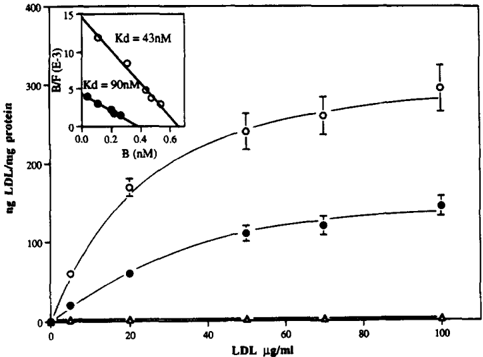
- LDL binding to brain capillary endothelial cells was enhanced threefold when cocultured with astrocytes; maximal binding was 358 ng LDL/mg protein with astrocytes versus 215 ng LDL/mg protein without, with apparent Kd values of 43 nM versus 90 nM, respectively.
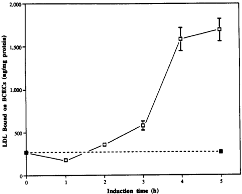
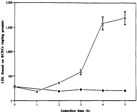
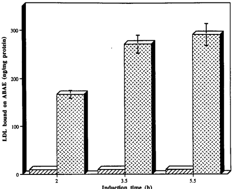
- Cholesterol depletion of astrocytes by 36 h in lipoprotein-deficient serum decreased astrocyte cholesterol content by 32% (27.00 ± 3.37 vs 39.25 ± 4.03 µg cholesterol/mg protein, n = 6) and led to upregulation of endothelial LDL receptors, reaching a maximum of 600% of control binding at 4–5 h.
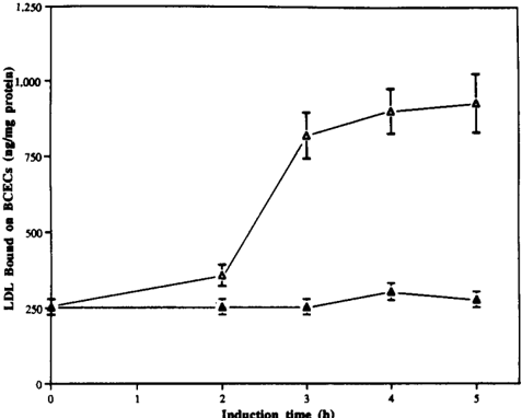
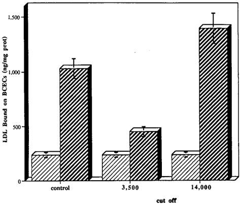
- Astrocyte-conditioned medium increased LDL binding by 300% at 4–5 h; upregulation occurred with a 14,000 mol wt cutoff dialysis membrane but not with a 3,500 cutoff, indicating a soluble factor(s) between 3,500 and 14,000 mol wt.
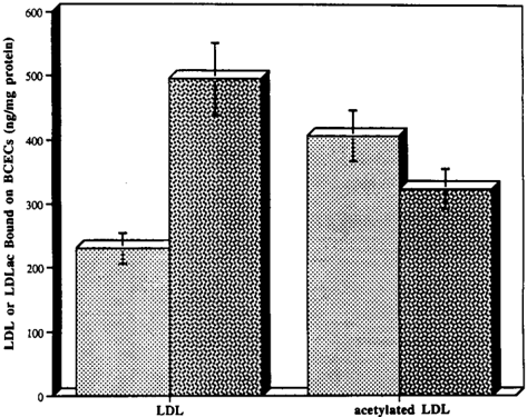
- The effect was specific: substituting astrocytes with smooth muscle cells or brain endothelium with aortic or adrenal cortex endothelium did not enhance luminal LDL receptor expression; acetylated LDL/scavenger receptor binding was not upregulated, and cycloheximide blocked the increase, indicating new LDL receptor synthesis.
Methods
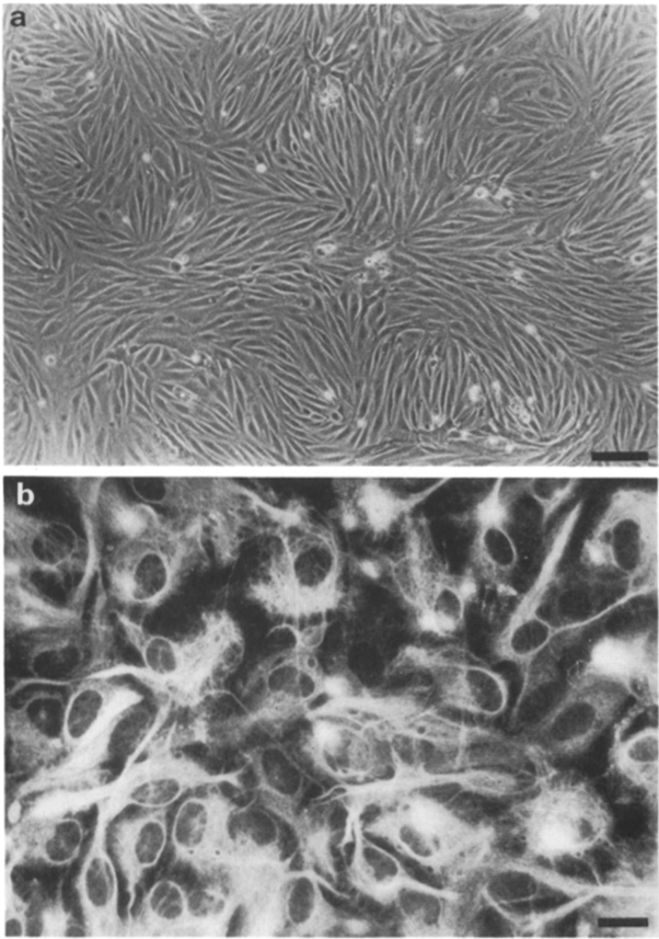
- Bovine brain capillary endothelial cells were cocultured with rat astrocytes on opposite sides of collagen-coated filter inserts; astrocytes were >95% GFAP positive. Bovine aortic endothelial cells, adrenal cortex endothelial cells, and smooth muscle cells were used for specificity comparisons.
- LDL binding was performed at 4°C with [125I]LDL, with nonspecific binding determined by 20-fold excess unlabeled LDL; Scatchard analysis, C7 monoclonal antibody blocking, acetylated LDL binding, and HPLC cholesterol measurement were used.
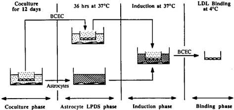
- Regulation experiments followed four phases: 12 d coculture in 15% calf serum, 36 h astrocyte incubation in 10% lipoprotein-deficient serum, induction with cholesterol-depleted astrocytes or conditioned medium at 37°C, then LDL binding at 4°C; dialysis membranes with 3,500 or 14,000 mol wt cutoffs and cycloheximide (10 µg/ml) were used in modified induction experiments.
Limitations
- The paper states that the chemical nature of the astrocyte-regulating LDL receptor factor(s) is under investigation, that additional studies are underway to determine whether bound lipoproteins are degraded or transcytosed, and that 36 h LPDS incubation was used because cell lysis could be detected after 48 h.
- The paper also notes that upregulation was lower with astrocyte-conditioned medium, possibly because the secreted molecule(s) is unstable.
- Evident from the text: the findings rely on an in vitro bovine/rat coculture model, the soluble factor is not purified or chemically identified, and in vivo confirmation is not provided.
Model-written summary. Check it against the paper before relying on it.
Integrity signals
Registry
Cites
Statistics
Figures
Verification
Claims the paper makes
30 Own result · 27 Own interpretation · 10 No full text of the sources · 9 Sources not found · 4 Not checked yet · 2 Own method · 2 No basis given · 1 Partly supported · accuracy not yet measured
The paper states this without a source or data behind it here, so there is nothing to check it against. The quote shows it is in the paper.
In the paper
“Since aortic ECs and adrenal cortex ECs cultured in the same medium do not express this LDL receptor at confluence”
plain- Asserted: passes — The authors put it forward as true.
- Checkable: passes — It can be checked against evidence.
- Load-bearing: passes — The paper’s conclusions depend on it.
- Based: passes — The paper gives a reason why it holds.
A result the paper reports from its own data — it cites no other source for it, so there is none to check. The quote shows it is in the paper.
In the paper
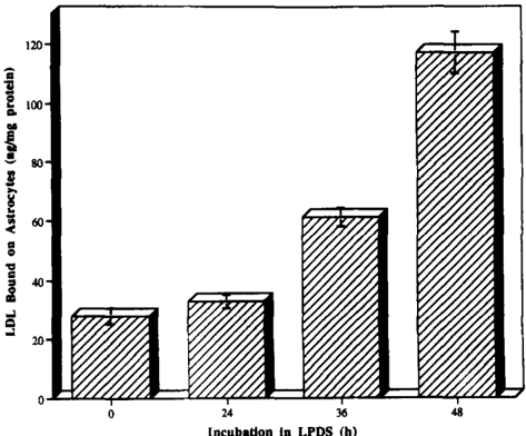
“The pretreat- ment of astrocytes with LPDS led to a time-dependent in- crease in the binding of [t2SI]LDL to the cells (Fig. 4), indi- cating that the cells have modified their metabolism to obtain more exogenous cholesterol.”
plaindirect- Asserted: passes — The authors put it forward as true.
- Checkable: passes — It can be checked against evidence.
- Load-bearing: passes — The paper’s conclusions depend on it.
- Based: passes — The paper gives a reason why it holds.
The paper states this without a source or data behind it here, so there is nothing to check it against. The quote shows it is in the paper.
In the paper
“Furthermore, the AUF has no effect if ECs are incubated with cycloheximide.”
plain- Asserted: passes — The authors put it forward as true.
- Checkable: passes — It can be checked against evidence.
- Load-bearing: passes — The paper’s conclusions depend on it.
- Based: passes — The paper gives a reason why it holds.
A result the paper reports from its own data — it cites no other source for it, so there is none to check. The quote shows it is in the paper.
In the paper
“when aortic ECs, originating from peripheral large vessels, or adrenal cortex ECs, origi- nating from peripheral microvessels, were cocultivated with astrocytes, no effect on the upregulation of the LDL recep- tors was observed when astrocytes were fed with LPDS.”
plain- Asserted: passes — The authors put it forward as true.
- Checkable: passes — It can be checked against evidence.
- Load-bearing: passes — The paper’s conclusions depend on it.
- Based: passes — The paper gives a reason why it holds.
A result the paper reports from its own data — it cites no other source for it, so there is none to check. The quote shows it is in the paper.
In the paper
“labeled LDL bound specifically to brain capillary ECs that were 5 d postconfluence (as visualized by phase contrast mi- croscopy) and in the presence of a medium containing 15 % calf serum.”
plaincorrelational- Asserted: passes — The authors put it forward as true.
- Checkable: passes — It can be checked against evidence.
- Load-bearing: passes — The paper’s conclusions depend on it.
- Based: passes — The paper gives a reason why it holds.
A result the paper reports from its own data — it cites no other source for it, so there is none to check. The quote shows it is in the paper.
In the paper
“Similarly, SMC did not induce an upregulation of LDL receptors on brain capillary ECs (data not shown).”
plain- Asserted: passes — The authors put it forward as true.
- Checkable: passes — It can be checked against evidence.
- Load-bearing: passes — The paper’s conclusions depend on it.
- Based: passes — The paper gives a reason why it holds.
A result the paper reports from its own data — it cites no other source for it, so there is none to check. The quote shows it is in the paper.
In the paper
“When astrocytes were pre-incubated in lipoprotein-deficient medium before the induction phase, binding of LDL to en- dothelial cells was increased a further sixfold.”
plaindirect- Asserted: passes — The authors put it forward as true.
- Checkable: passes — It can be checked against evidence.
- Load-bearing: passes — The paper’s conclusions depend on it.
- Based: passes — The paper gives a reason why it holds.
A result the paper reports from its own data — it cites no other source for it, so there is none to check. The quote shows it is in the paper.
In the paper
“This factor(s) is exclusively secreted by astrocytes cocultured with brain capillary endothelial cells, but it also upregulates the LDL receptor on other cell types.”
plaindirect- Asserted: passes — The authors put it forward as true.
- Checkable: passes — It can be checked against evidence.
- Load-bearing: unclear — It is unclear whether the paper’s conclusions depend on it.
- Based: passes — The paper gives a reason why it holds.
A result the paper reports from its own data — it cites no other source for it, so there is none to check. The quote shows it is in the paper.
In the paper
“Our findings also showed that the capacity of ECs to bind LDL is greater when cocultured with astrocytes than in their absence.”
plaincorrelational- Asserted: passes — The authors put it forward as true.
- Checkable: passes — It can be checked against evidence.
- Load-bearing: passes — The paper’s conclusions depend on it.
- Based: passes — The paper gives a reason why it holds.
A result the paper reports from its own data — it cites no other source for it, so there is none to check. The quote shows it is in the paper.
In the paper
“Furthermore, when binding of acetylated LDL is per- formed instead of LDL to brain capillary ECs, no upregula- tion of the scavenger receptor occurs (Fig. 9).”
plain- Asserted: passes — The authors put it forward as true.
- Checkable: passes — It can be checked against evidence.
- Load-bearing: unclear — It is unclear whether the paper’s conclusions depend on it.
- Based: passes — The paper gives a reason why it holds.
A result the paper reports from its own data — it cites no other source for it, so there is none to check. The quote shows it is in the paper.
In the paper
“Using a cell culture model of the blood-brain barrier consisting of a coculture of brain capillary endothelial cells and astrocytes, we observed that the capacity of endothelial cells to bind LDL is enhanced threefold when cocultured with astrocytes.”
plaindirect- Asserted: passes — The authors put it forward as true.
- Checkable: passes — It can be checked against evidence.
- Load-bearing: passes — The paper’s conclusions depend on it.
- Based: passes — The paper gives a reason why it holds.
A result the paper reports from its own data — it cites no other source for it, so there is none to check. The quote shows it is in the paper.
In the paper
“However, the maximal binding was higher (358 ng LDL/mg protein) when the ECs were cocultured with astrocytes than when they were cultured alone (215 ng LDL/mg protein).”
plaincorrelational- Asserted: passes — The authors put it forward as true.
- Checkable: passes — It can be checked against evidence.
- Load-bearing: passes — The paper’s conclusions depend on it.
- Based: passes — The paper gives a reason why it holds.
A result the paper reports from its own data — it cites no other source for it, so there is none to check. The quote shows it is in the paper.
In the paper
“When brain capillary ECs were incubated with cycloheximide during the”
plain- Asserted: passes — The authors put it forward as true.
- Checkable: passes — It can be checked against evidence.
- Load-bearing: passes — The paper’s conclusions depend on it.
- Based: passes — The paper gives a reason why it holds.
A result the paper reports from its own data — it cites no other source for it, so there is none to check. The quote shows it is in the paper.
In the paper
“Labeled LDL bound with saturation kinetic on brain capri- lary ECs cultured alone or cocultured with astrocytes in 15 % CS, in contrast to what was observed on aortic endothelial cells (Fig. 3).”
plaincorrelational- Asserted: passes — The authors put it forward as true.
- Checkable: passes — It can be checked against evidence.
- Load-bearing: passes — The paper’s conclusions depend on it.
- Based: passes — The paper gives a reason why it holds.
A result the paper reports from its own data — it cites no other source for it, so there is none to check. The quote shows it is in the paper.
In the paper
“The results showed that the AUF(s) secreted by astrocytes triggered the appearance of specific binding on aortic ECs and smooth muscle cells, even when they were cultured at confluence and in the presence of a high concen- tration of lipoproteins.”
plaindirect- Asserted: passes — The authors put it forward as true.
- Checkable: passes — It can be checked against evidence.
- Load-bearing: passes — The paper’s conclusions depend on it.
- Based: passes — The paper gives a reason why it holds.
A result the paper reports from its own data — it cites no other source for it, so there is none to check. The quote shows it is in the paper.
In the paper
“We have shown that the lipid requirement of astrocytes increases the expression of endothelial cell LDL receptors.”
boosteddirect- Asserted: passes — The authors put it forward as true.
- Checkable: passes — It can be checked against evidence.
- Load-bearing: passes — The paper’s conclusions depend on it.
- Based: passes — The paper gives a reason why it holds.
A result the paper reports from its own data — it cites no other source for it, so there is none to check. The quote shows it is in the paper.
In the paper
“Furthermore, HPLC analysis shows a 32 % decrease of the cholesterol content of astro- cytes (27.00 + 3.37 #g cholesterol/rag protein vs 39.25 + 4.03 #g cholesterol/mg protein, n = 6), when they were in- cubated at 37C for 36 h in LPDS.”
plaindirectn = 6- Asserted: passes — The authors put it forward as true.
- Checkable: passes — It can be checked against evidence.
- Load-bearing: passes — The paper’s conclusions depend on it.
- Based: passes — The paper gives a reason why it holds.
A result the paper reports from its own data — it cites no other source for it, so there is none to check. The quote shows it is in the paper.
In the paper
“These data revealed, for the culture with or without astrocytes, the presence on brain cap- illary ECs of a single class of high affinity LDL binding sites with a Kd of the order of riM.”
plain- Asserted: passes — The authors put it forward as true.
- Checkable: passes — It can be checked against evidence.
- Load-bearing: passes — The paper’s conclusions depend on it.
- Based: passes — The paper gives a reason why it holds.
A result the paper reports from its own data — it cites no other source for it, so there is none to check. The quote shows it is in the paper.
In the paper
“No specific binding was ob- served on aortic ECs cultured alone”
plain- Asserted: passes — The authors put it forward as true.
- Checkable: passes — It can be checked against evidence.
- Load-bearing: passes — The paper’s conclusions depend on it.
- Based: passes — The paper gives a reason why it holds.
A result the paper reports from its own data — it cites no other source for it, so there is none to check. The quote shows it is in the paper.
In the paper
“we could see a large increase in the specific binding of the LDL to the aortic ECs when they were in the presence of cholesterol-depleted astro- cytes”
hedgeddirect- Asserted: passes — The authors put it forward as true.
- Checkable: passes — It can be checked against evidence.
- Load-bearing: passes — The paper’s conclusions depend on it.
- Based: passes — The paper gives a reason why it holds.
A result the paper reports from its own data — it cites no other source for it, so there is none to check. The quote shows it is in the paper.
In the paper
“Furthermore, there was an increase in the num- ber of the LDL receptors on brain capillary ECs in the pres- ence of astrocytes.”
plaindirect- Asserted: passes — The authors put it forward as true.
- Checkable: passes — It can be checked against evidence.
- Load-bearing: passes — The paper’s conclusions depend on it.
- Based: passes — The paper gives a reason why it holds.
A result the paper reports from its own data — it cites no other source for it, so there is none to check. The quote shows it is in the paper.
In the paper
“Under these conditions, no upregulation on aortic ECs and adrenal cortex ECs was observed when the astrocytes were cultured in LPDS for 36 h (data not shown).”
plain- Asserted: passes — The authors put it forward as true.
- Checkable: passes — It can be checked against evidence.
- Load-bearing: passes — The paper’s conclusions depend on it.
- Based: passes — The paper gives a reason why it holds.
A result the paper reports from its own data — it cites no other source for it, so there is none to check. The quote shows it is in the paper.
In the paper
“we have also shown this kind of specific interaction because substituting astrocytes with smooth muscle cells in coculture did not enhance the expression of the LDL receptors on brain capillary ECs.”
boosted- Asserted: passes — The authors put it forward as true.
- Checkable: passes — It can be checked against evidence.
- Load-bearing: passes — The paper’s conclusions depend on it.
- Based: passes — The paper gives a reason why it holds.
A result the paper reports from its own data — it cites no other source for it, so there is none to check. The quote shows it is in the paper.
In the paper
“Furthermore, astrocytes cultured alone do not up- regulate LDL receptors on brain capillary ECs during the induction phase.”
plain- Asserted: passes — The authors put it forward as true.
- Checkable: passes — It can be checked against evidence.
- Load-bearing: passes — The paper’s conclusions depend on it.
- Based: passes — The paper gives a reason why it holds.
None of the sources cited for this claim have been checked yet.
In the paper
“In vivo, normal circulating concentration of lipoproteins downregulates the LDL receptor levels on vas- cular endothelial cells (ECs).”
What the sources say
- Not checked yetcites Vasile et al., 1983 source
Checked before — its earlier verdict is held back until a new check runs.
plaindirect- Asserted: passes — The authors put it forward as true.
- Checkable: passes — It can be checked against evidence.
- Load-bearing: passes — The paper’s conclusions depend on it.
- Based: passes — The paper gives a reason why it holds.
- Not checked yet
A conclusion the paper draws from its own results — it cites no other source for it, so there is none to check. The quote shows it is in the paper.
In the paper
“Experiments with dialysis membranes of different pore size showed that this effect is mediated by a soluble factor(s) with relative molecular mass somewhere between 3,500 and 14,000.”
plaindirect- Asserted: passes — The authors put it forward as true.
- Checkable: passes — It can be checked against evidence.
- Load-bearing: passes — The paper’s conclusions depend on it.
- Based: passes — The paper gives a reason why it holds.
A conclusion the paper draws from its own results — it cites no other source for it, so there is none to check. The quote shows it is in the paper.
In the paper
“As already stated for the upregulation of the LDL receptor, the mole- cule(s) could not be a product of the cell lysis that could oc- cur during the incubation with LPDS.”
hedged- Asserted: passes — The authors put it forward as true.
- Checkable: passes — It can be checked against evidence.
- Load-bearing: passes — The paper’s conclusions depend on it.
- Based: unclear — It is unclear why the paper says it holds.
A conclusion the paper draws from its own results — it cites no other source for it, so there is none to check. The quote shows it is in the paper.
In the paper
“We docu- ment specific EC-astrocyte interactions, and we propose that a soluble factor(s) derived from astrocytes modulates LDL receptor expression at the luminal surface of the brain capillary endothelium.”
hedgeddirect- Asserted: passes — The authors put it forward as true.
- Checkable: passes — It can be checked against evidence.
- Load-bearing: passes — The paper’s conclusions depend on it.
- Based: passes — The paper gives a reason why it holds.
A conclusion the paper draws from its own results — it cites no other source for it, so there is none to check. The quote shows it is in the paper.
In the paper
“Reciprocal communications between ECs and as- trocytes are required to activate the mechanism of the up- regulation during the coculture phase.”
plaindirect- Asserted: passes — The authors put it forward as true.
- Checkable: passes — It can be checked against evidence.
- Load-bearing: passes — The paper’s conclusions depend on it.
- Based: passes — The paper gives a reason why it holds.
A conclusion the paper draws from its own results — it cites no other source for it, so there is none to check. The quote shows it is in the paper.
In the paper
“This LDL recep- tor was present at the luminal side of ECs.”
plain- Asserted: passes — The authors put it forward as true.
- Checkable: passes — It can be checked against evidence.
- Load-bearing: passes — The paper’s conclusions depend on it.
- Based: passes — The paper gives a reason why it holds.
A conclusion the paper draws from its own results — it cites no other source for it, so there is none to check. The quote shows it is in the paper.
In the paper
“the two close neighbors in vivo, brain endotheliumand astrocytes, interact specifically in vitro to induce the expres- sion of the LDL receptor at the luminal side of brain capil- lary ECs.”
plaindirect- Asserted: passes — The authors put it forward as true.
- Checkable: passes — It can be checked against evidence.
- Load-bearing: passes — The paper’s conclusions depend on it.
- Based: passes — The paper gives a reason why it holds.
A conclusion the paper draws from its own results — it cites no other source for it, so there is none to check. The quote shows it is in the paper.
In the paper
“The significant conclusion from the cholesterol-depleted astro- cyte conditioned medium experiments is that a soluble fac- tor(s) is involved in the upregulation mechanism.”
boosteddirect- Asserted: passes — The authors put it forward as true.
- Checkable: passes — It can be checked against evidence.
- Load-bearing: passes — The paper’s conclusions depend on it.
- Based: passes — The paper gives a reason why it holds.
In the paper
“This estimate of affinity is similar to that reported by Vlodasky et al. (1978) in bovine aortic ECs cultured in lipoprotein deficient serum (Kd = 27 nM).”
What the sources say
- Not matchedcites Vlodasky et al. (1978)
Couldn’t be matched to a paper in Crossref, so it can’t be checked.
plain- Asserted: passes — The authors put it forward as true.
- Checkable: passes — It can be checked against evidence.
- Load-bearing: unclear — It is unclear whether the paper’s conclusions depend on it.
- Based: passes — The paper gives a reason why it holds.
- Not matched
In the paper
“In attempts to investigate such interactions, we have developed an in vitro model of the blood-brain barrier that imitates an in vivo situation by culturing brain capillary ECs and astrocytes on opposite sides of a filter (Dehouck et al., 1990a, 1992).”
What the sources say
- Not matchedcites Dehouck et al., 1990a, 1992
Couldn’t be matched to a paper in Crossref, so it can’t be checked.
plain- Asserted: passes — The authors put it forward as true.
- Checkable: passes — It can be checked against evidence.
- Load-bearing: passes — The paper’s conclusions depend on it.
- Based: passes — The paper gives a reason why it holds.
- Not matched
A conclusion the paper draws from its own results — it cites no other source for it, so there is none to check. The quote shows it is in the paper.
In the paper
“This discrepancy may be related to the origin of the ECs (brain capillaries)”
hedgedconditional- Asserted: passes — The authors put it forward as true.
- Checkable: passes — It can be checked against evidence.
- Load-bearing: passes — The paper’s conclusions depend on it.
- Based: passes — The paper gives a reason why it holds.
In the paper
“In addition, we have shown that cultured brain capillary ECs have LDL receptors with the same apparent molecular weight as in vivo (Meresse et al., 1991).”
What the sources say
- Not checked — no full text of the sourcecites Meresse et al., 1991 source
plain- Asserted: passes — The authors put it forward as true.
- Checkable: passes — It can be checked against evidence.
- Load-bearing: unclear — It is unclear whether the paper’s conclusions depend on it.
- Based: passes — The paper gives a reason why it holds.
In the paper
“We have determined (M6resse et al., 1991) by ligand blotting that ECs in culture express an LDL receptor with the same apparent molecular weight as that in vivo (132,000 D).”
What the sources say
- Not matchedcites M6resse et al., 1991
Couldn’t be matched to a paper in Crossref, so it can’t be checked.
boosted- Asserted: passes — The authors put it forward as true.
- Checkable: passes — It can be checked against evidence.
- Load-bearing: unclear — It is unclear whether the paper’s conclusions depend on it.
- Based: passes — The paper gives a reason why it holds.
- Not matched
In the paper
“This receptor exhibits the same characteristic properties as the LDL receptor on human fibroblasts (Goldstein and Brown, 1977).”
What the sources say
- Not checked — no full text of the sourcecites Goldstein and Brown, 1977 source
plain- Asserted: passes — The authors put it forward as true.
- Checkable: passes — It can be checked against evidence.
- Load-bearing: passes — The paper’s conclusions depend on it.
- Based: passes — The paper gives a reason why it holds.
A conclusion the paper draws from its own results — it cites no other source for it, so there is none to check. The quote shows it is in the paper.
In the paper
“This study confirms the notion that the final fine tuning of cell differentiation is under local control.”
boosteddirect- Asserted: passes — The authors put it forward as true.
- Checkable: passes — It can be checked against evidence.
- Load-bearing: passes — The paper’s conclusions depend on it.
- Based: passes — The paper gives a reason why it holds.
In the paper
“Astroeytes, the nearest neighbor of brain capillaries, have been shown to in- duce some of the specialized properties of ECs (Janzer and Raft, 1987).”
What the sources say
- Not matchedcites Janzer and Raft, 1987
Couldn’t be matched to a paper in Crossref, so it can’t be checked.
plaindirect- Asserted: passes — The authors put it forward as true.
- Checkable: passes — It can be checked against evidence.
- Load-bearing: passes — The paper’s conclusions depend on it.
- Based: passes — The paper gives a reason why it holds.
- Not matched
A conclusion the paper draws from its own results — it cites no other source for it, so there is none to check. The quote shows it is in the paper.
In the paper
“Our results integrated into this model, and they strongly sug- gest that the cerebral endothelial receptor plays a role in cho- lesterol transport across the blood-brain barrier.”
hedgedcorrelational- Asserted: passes — The authors put it forward as true.
- Checkable: passes — It can be checked against evidence.
- Load-bearing: passes — The paper’s conclusions depend on it.
- Based: passes — The paper gives a reason why it holds.
A conclusion the paper draws from its own results — it cites no other source for it, so there is none to check. The quote shows it is in the paper.
In the paper
“This result indicates that the lipid requirement of astrocytes modulates the expression of endothelial cell LDL receptors.”
hedgeddirect- Asserted: passes — The authors put it forward as true.
- Checkable: passes — It can be checked against evidence.
- Load-bearing: passes — The paper’s conclusions depend on it.
- Based: passes — The paper gives a reason why it holds.
A conclusion the paper draws from its own results — it cites no other source for it, so there is none to check. The quote shows it is in the paper.
In the paper
“the culture medium used could not have been responsible for the absence of downregulationobserved with brain capillary ECs.”
hedged- Asserted: passes — The authors put it forward as true.
- Checkable: passes — It can be checked against evidence.
- Load-bearing: passes — The paper’s conclusions depend on it.
- Based: passes — The paper gives a reason why it holds.
In the paper
“This receptor exhibits the same characteristic properties as the LDL recep- tor on human fibroblasts (Goldstein and Brown, 1977).”
What the sources say
- Not checked — no full text of the sourcecites Goldstein and Brown, 1977 source
plain- Asserted: passes — The authors put it forward as true.
- Checkable: passes — It can be checked against evidence.
- Load-bearing: unclear — It is unclear whether the paper’s conclusions depend on it.
- Based: passes — The paper gives a reason why it holds.
A conclusion the paper draws from its own results — it cites no other source for it, so there is none to check. The quote shows it is in the paper.
In the paper
“This factor(s) is exclusively secreted by astrocytes cocultured with brain capillary endothelial cells, but it also upregulates the LDL receptor on other cell types.”
plaindirect- Asserted: passes — The authors put it forward as true.
- Checkable: passes — It can be checked against evidence.
- Load-bearing: passes — The paper’s conclusions depend on it.
- Based: passes — The paper gives a reason why it holds.
In the paper
“several authors have shown that capillary ECs differ from the ECs of other vessels in their biochemical and functional characteristics (Zetter, 1988).”
What the sources say
- Not matchedcites Zetter, 1988
Couldn’t be matched to a paper in Crossref, so it can’t be checked.
boosted- Asserted: passes — The authors put it forward as true.
- Checkable: passes — It can be checked against evidence.
- Load-bearing: passes — The paper’s conclusions depend on it.
- Based: passes — The paper gives a reason why it holds.
- Not matched
A conclusion the paper draws from its own results — it cites no other source for it, so there is none to check. The quote shows it is in the paper.
In the paper
“So, the AUF seems to regulate LDL receptor expres- sion specifically.”
hedgedconditional- Asserted: passes — The authors put it forward as true.
- Checkable: passes — It can be checked against evidence.
- Load-bearing: passes — The paper’s conclusions depend on it.
- Based: passes — The paper gives a reason why it holds.
A conclusion the paper draws from its own results — it cites no other source for it, so there is none to check. The quote shows it is in the paper.
In the paper
“These results show that the upregula- tion is caused by new synthesis of the LDL receptor.”
plaindirect- Asserted: passes — The authors put it forward as true.
- Checkable: passes — It can be checked against evidence.
- Load-bearing: passes — The paper’s conclusions depend on it.
- Based: passes — The paper gives a reason why it holds.
- Rests on an assumption: Aortic endothelial cells and smooth muscle cells have no intracellular pool of LDL receptor.
- Rests on an assumption: Cycloheximide inhibits peptidyl transferase on the eukaryotic large ribosomal subunit and therefore protein synthesis in the cell.
A conclusion the paper draws from its own results — it cites no other source for it, so there is none to check. The quote shows it is in the paper.
In the paper
“This indicated that a cross-talk takes place between brain capillary ECs and astrocytes during the cocul- ture.”
hedgeddirect- Asserted: passes — The authors put it forward as true.
- Checkable: passes — It can be checked against evidence.
- Load-bearing: passes — The paper’s conclusions depend on it.
- Based: passes — The paper gives a reason why it holds.
What the paper did — it cites no source for this, so there is none to check. The quote shows it is in the paper.
In the paper
“In our experimental conditions, only the albuminal face was in contact with LPDS”
plain- Asserted: passes — The authors put it forward as true.
- Checkable: passes — It can be checked against evidence.
- Load-bearing: passes — The paper’s conclusions depend on it.
- Based: passes — The paper gives a reason why it holds.
In the paper
“tight junctions, very well developed in our coculture (Dehouck et al., 1990a), prevent [~ff]LDL leak into the abluminal com- partment when the experiment is performed at 4C (data not shown).”
What the sources say
- Not matchedcites Dehouck et al., 1990a
Couldn’t be matched to a paper in Crossref, so it can’t be checked.
boosteddirect- Asserted: passes — The authors put it forward as true.
- Checkable: passes — It can be checked against evidence.
- Load-bearing: passes — The paper’s conclusions depend on it.
- Based: passes — The paper gives a reason why it holds.
- Not matched
A conclusion the paper draws from its own results — it cites no other source for it, so there is none to check. The quote shows it is in the paper.
In the paper
“This study confirms the notion that endothelial ceils gener- ally not express their final destination-specific differentiated features until these features are induced by local, environ- ment-produced conditions.”
boosteddirect- Asserted: passes — The authors put it forward as true.
- Checkable: passes — It can be checked against evidence.
- Load-bearing: unclear — It is unclear whether the paper’s conclusions depend on it.
- Based: passes — The paper gives a reason why it holds.
A conclusion the paper draws from its own results — it cites no other source for it, so there is none to check. The quote shows it is in the paper.
In the paper
“suggesting that binding on endothelial ceils is mediated by the receptor.”
hedgeddirect- Asserted: passes — The authors put it forward as true.
- Checkable: passes — It can be checked against evidence.
- Load-bearing: passes — The paper’s conclusions depend on it.
- Based: passes — The paper gives a reason why it holds.
In the paper
“The observations that brain ECs can increase the inci- dence of intramembraneous particle arrays in astrocytes (Tao-Cheng et al., 1990) are in agreement with our results showing that endothelial cells can in turn induce changes in associated glia.”
What the sources say
- Not checked — no full text of the sourcecites Tao-Cheng et al., 1990 source
plaindirect- Asserted: passes — The authors put it forward as true.
- Checkable: passes — It can be checked against evidence.
- Load-bearing: unclear — It is unclear whether the paper’s conclusions depend on it.
- Based: passes — The paper gives a reason why it holds.
A conclusion the paper draws from its own results — it cites no other source for it, so there is none to check. The quote shows it is in the paper.
In the paper
“Since these EC-astrocyte effects are a cross- species (bovine-rat), it is likely to be a fundamental property of the two cell types.”
hedgedcorrelational- Asserted: passes — The authors put it forward as true.
- Checkable: passes — It can be checked against evidence.
- Load-bearing: unclear — It is unclear whether the paper’s conclusions depend on it.
- Based: passes — The paper gives a reason why it holds.
A conclusion the paper draws from its own results — it cites no other source for it, so there is none to check. The quote shows it is in the paper.
In the paper
“Considered together, these results indicate that the 12 d of the coculture are necessary for enhanced LDL binding.”
hedgeddirect- Asserted: passes — The authors put it forward as true.
- Checkable: passes — It can be checked against evidence.
- Load-bearing: passes — The paper’s conclusions depend on it.
- Based: passes — The paper gives a reason why it holds.
A conclusion the paper draws from its own results — it cites no other source for it, so there is none to check. The quote shows it is in the paper.
In the paper
“These results indicate that we are dealing with an LDL receptor.”
hedged- Asserted: passes — The authors put it forward as true.
- Checkable: passes — It can be checked against evidence.
- Load-bearing: passes — The paper’s conclusions depend on it.
- Based: passes — The paper gives a reason why it holds.
None of the sources cited for this claim have been checked yet.
In the paper
“These modificationsresult from specific inter- actions between brain capillary ECs and brain parenchyma (Risau et al., 1986; Stewart and Wiley, 1981).”
What the sources say
plaindirect- Asserted: passes — The authors put it forward as true.
- Checkable: passes — It can be checked against evidence.
- Load-bearing: passes — The paper’s conclusions depend on it.
- Based: passes — The paper gives a reason why it holds.
A conclusion the paper draws from its own results — it cites no other source for it, so there is none to check. The quote shows it is in the paper.
In the paper
“The experiments using conditioned medium clearly dem- onstrate that coculmre increases the probability of detecting the effects of a short-lived agent produced by astrocytes.”
boosteddirect- Asserted: passes — The authors put it forward as true.
- Checkable: passes — It can be checked against evidence.
- Load-bearing: passes — The paper’s conclusions depend on it.
- Based: passes — The paper gives a reason why it holds.
- Probably not a substantial claim. 14
- It may state two separate claims as one. 15
The cited sources support only part of this claim — read each source’s verdict below.
In the paper
“Brain capillary ECs, which constitute the blood-brain barrier, are sealed together by high electrical resistance tight junctions, and they display a low rate of transcellular vesicular trans- pert (Reese and Karnovsky, 1967).”
What the sources say
- Partly supportedcites Reese and Karnovsky, 1967 sourceExperimental — accuracy not yet measured
The Discussion (pp. 211–213) identifies cerebral endothelium as the barrier, describes apparently continuous tight junctions, and reports few transport-associated vesicles and no observed peroxidase discharge beyond the endothelium. It assesses junction tightness structurally, not by electrical-resistance measurement; that specific qualifier is unsupported. (LLM, conf=0.88)
plain- Asserted: passes — The authors put it forward as true.
- Checkable: passes — It can be checked against evidence.
- Load-bearing: passes — The paper’s conclusions depend on it.
- Based: passes — The paper gives a reason why it holds.
Probably not a substantial claim. It may state two separate claims as one.
- Partly supported
A result the paper reports from its own data — it cites no other source for it, so there is none to check. The quote shows it is in the paper.
In the paper
“although brain capillary ECs bind specif- ically acetylated LDL, astrocytes cocultured with brain cap- illary ECs for 12 d and fed with LPDS cannot upregulate scavenger receptor expression on brain capillary ECs sur- face.”
plain- Asserted: passes — The authors put it forward as true.
- Checkable: passes — It can be checked against evidence.
- Load-bearing: passes — The paper’s conclusions depend on it.
- Based: passes — The paper gives a reason why it holds.
It may state two separate claims as one.
A result the paper reports from its own data — it cites no other source for it, so there is none to check. The quote shows it is in the paper.
In the paper
“when a complete serum-containing medium was changed to a medium containing 10% LPDS 36 h before the binding ex- periments, the cholesterol content of the astrocytes was depleted, and the expression of the apolipoprotein (B, E) receptors was upregulated.”
plaindirect- Asserted: passes — The authors put it forward as true.
- Checkable: passes — It can be checked against evidence.
- Load-bearing: passes — The paper’s conclusions depend on it.
- Based: passes — The paper gives a reason why it holds.
It may state two separate claims as one.
A result the paper reports from its own data — it cites no other source for it, so there is none to check. The quote shows it is in the paper.
In the paper
“No increase of LDL receptor expression was observed in the coculture with a cut-off membrane of 3,500. In contrast, the upregulation was observed in the coculture with a cut-off membrane of 14,000 (Fig. 7).”
plain- Asserted: passes — The authors put it forward as true.
- Checkable: passes — It can be checked against evidence.
- Load-bearing: passes — The paper’s conclusions depend on it.
- Based: passes — The paper gives a reason why it holds.
It may state two separate claims as one.
A result the paper reports from its own data — it cites no other source for it, so there is none to check. The quote shows it is in the paper.
In the paper
“The C7-monoclonai antibody, known to interact with the rl~ceptor binding domain, totally blocked the binding of LDL to bovine brain capillary ECs. Control irrelevant IgG had no effecton LDL binding (results not shown)”
plaindirect- Asserted: passes — The authors put it forward as true.
- Checkable: passes — It can be checked against evidence.
- Load-bearing: passes — The paper’s conclusions depend on it.
- Based: passes — The paper gives a reason why it holds.
It may state two separate claims as one.
A result the paper reports from its own data — it cites no other source for it, so there is none to check. The quote shows it is in the paper.
In the paper
“ECs form a monolayer of small, tightly packed, nonoverlap- ping and contact-inhibited cells.”
plain- Asserted: passes — The authors put it forward as true.
- Checkable: passes — It can be checked against evidence.
- Load-bearing: fails — The paper’s conclusions may not depend on it.
- Based: passes — The paper gives a reason why it holds.
Probably not a substantial claim. The paper’s conclusions may not depend on it.
A result the paper reports from its own data — it cites no other source for it, so there is none to check. The quote shows it is in the paper.
In the paper
“upregulation experi- ments performed with SMC plated instead of astrocytes, or with astrocytes not cocultured with brain capillary ECs, did not enhance LDL receptor expression, even if astrocytes were fed with LPDS during the induction phase.”
plain- Asserted: passes — The authors put it forward as true.
- Checkable: passes — It can be checked against evidence.
- Load-bearing: passes — The paper’s conclusions depend on it.
- Based: passes — The paper gives a reason why it holds.
It may state two separate claims as one.
A result the paper reports from its own data — it cites no other source for it, so there is none to check. The quote shows it is in the paper.
In the paper
“These results indicate that brain capillary ECs at con- fluence, cultured in a medium containing lipoproteins, ex- press LDL receptors in contrast to what was observed on aortic ECs.”
hedged- Asserted: passes — The authors put it forward as true.
- Checkable: passes — It can be checked against evidence.
- Load-bearing: passes — The paper’s conclusions depend on it.
- Based: passes — The paper gives a reason why it holds.
It may state two separate claims as one.
A result the paper reports from its own data — it cites no other source for it, so there is none to check. The quote shows it is in the paper.
In the paper
“As shown in Fig. 5, increased binding of lipoproteins was al- ready manifested 3 h after coculture of the two cell types at 37C, and the maximum effect(600% of the control binding) was observed at 4 and 5 h.”
plaindirect- Asserted: passes — The authors put it forward as true.
- Checkable: passes — It can be checked against evidence.
- Load-bearing: passes — The paper’s conclusions depend on it.
- Based: passes — The paper gives a reason why it holds.
It may state two separate claims as one.
A claim is checked only against the cited paper’s full text, never its abstract, and none is available for its sources.
In the paper
“Like other cells in the body, astrocytes possess an apo (B, E) receptor, and at the cellular level, this receptor provides a regulated mechanism for supplying cells with cholesterol (Volpe et al., 1978; Pitas et al., 1987).”
What the sources say
boosteddirect- Asserted: passes — The authors put it forward as true.
- Checkable: passes — It can be checked against evidence.
- Load-bearing: fails — The paper’s conclusions may not depend on it.
- Based: passes — The paper gives a reason why it holds.
Probably not a substantial claim. The paper’s conclusions may not depend on it.
A conclusion the paper draws from its own results — it cites no other source for it, so there is none to check. The quote shows it is in the paper.
In the paper
“The secretion of this factor re- quires cocultured cells, indicating that the reciprocal differ- entiation of both cell types might happen.”
hedgedconditional- Asserted: passes — The authors put it forward as true.
- Checkable: passes — It can be checked against evidence.
- Load-bearing: unclear — It is unclear whether the paper’s conclusions depend on it.
- Based: passes — The paper gives a reason why it holds.
It may state two separate claims as one.
A conclusion the paper draws from its own results — it cites no other source for it, so there is none to check. The quote shows it is in the paper.
In the paper
“The fact that the up- regulation is lower when the astrocyte conditioned medium was used could be explained by the secretion of an unstable molecule(s) by astrocytes.”
hedgedconditional- Asserted: passes — The authors put it forward as true.
- Checkable: passes — It can be checked against evidence.
- Load-bearing: fails — The paper’s conclusions may not depend on it.
- Based: passes — The paper gives a reason why it holds.
Probably not a substantial claim. The paper’s conclusions may not depend on it.
None of the sources cited for this claim have been checked yet.
In the paper
“A number of different studies have confirmed that growing ECs in culture also bind LDL (Vlodavsky et al., 1978).”
What the sources say
- Not checked yetcites Vlodavsky et al., 1978 source
Checked before — its earlier verdict is held back until a new check runs.
boostedcorrelational- Asserted: passes — The authors put it forward as true.
- Checkable: passes — It can be checked against evidence.
- Load-bearing: fails — The paper’s conclusions may not depend on it.
- Based: passes — The paper gives a reason why it holds.
Probably not a substantial claim. The paper’s conclusions may not depend on it.
- Not checked yet
In the paper
“It is well established that coculturing ECs with primary glial cultures results in marked changes of several endothelial parameters, which suggests that several structural and functional charac- teristics of cerebral capillaries are determined by the sur- rounding astrocytes 0o6, 1992).”
What the sources say
- Not matchedcites 0o6, 1992
Couldn’t be matched to a paper in Crossref, so it can’t be checked.
boostedconditional- Asserted: passes — The authors put it forward as true.
- Checkable: passes — It can be checked against evidence.
- Load-bearing: fails — The paper’s conclusions may not depend on it.
- Based: passes — The paper gives a reason why it holds.
Probably not a substantial claim. The paper’s conclusions may not depend on it. It may state two separate claims as one.
- Not matched
A conclusion the paper draws from its own results — it cites no other source for it, so there is none to check. The quote shows it is in the paper.
In the paper
“the occurrence of the LDL receptor on brain capillary endothelial cells at confluence and in the presence of a high concentration of lipoproteins could be explained not only by the originality of the cell type used (brain capillary en- dothelial cells), but also by the local control of astrocytes.”
hedgedconditional- Asserted: passes — The authors put it forward as true.
- Checkable: passes — It can be checked against evidence.
- Load-bearing: passes — The paper’s conclusions depend on it.
- Based: passes — The paper gives a reason why it holds.
It may state two separate claims as one.
A conclusion the paper draws from its own results — it cites no other source for it, so there is none to check. The quote shows it is in the paper.
In the paper
“AUF liberation is specific and occurs only with coculmred brain capillary endothelial cells and astrocytes.”
plain- Asserted: passes — The authors put it forward as true.
- Checkable: passes — It can be checked against evidence.
- Load-bearing: passes — The paper’s conclusions depend on it.
- Based: passes — The paper gives a reason why it holds.
It may state two separate claims as one.
In the paper
“Estrada et al. (1990) have shown that a peptide with molecu- lar weight >50000 derived from cerebral capillary ECs could be involved in the local signaling between cell types that control new vessel formation in development (i.e., brain capillary ECs and astrocytes).”
What the sources say
- Not checked — no full text of the sourcecites Estrada et al. (1990) source
hedgedconditional- Asserted: passes — The authors put it forward as true.
- Checkable: passes — It can be checked against evidence.
- Load-bearing: fails — The paper’s conclusions may not depend on it.
- Based: passes — The paper gives a reason why it holds.
Probably not a substantial claim. The paper’s conclusions may not depend on it.
In the paper
“The astrocytes were characterized with glial flbtiUary acidic protein (GFAP), and >95 % of the population was GFAP positive (Dehouck et al., 1990/,).”
What the sources say
- Not matchedcites Dehouck et al., 1990b
Couldn’t be matched to a paper in Crossref, so it can’t be checked.
plain- Asserted: passes — The authors put it forward as true.
- Checkable: passes — It can be checked against evidence.
- Load-bearing: fails — The paper’s conclusions may not depend on it.
- Based: passes — The paper gives a reason why it holds.
Probably not a substantial claim. The paper’s conclusions may not depend on it. It may state two separate claims as one.
- Not matched
A conclusion the paper draws from its own results — it cites no other source for it, so there is none to check. The quote shows it is in the paper.
In the paper
“Substituting astrocytes with smooth muscle cells or brain endothelium with endothelium from the aorta or the adrenal cortex did not enhance the luminal LDL receptor expression on endothelial cells, demonstrating the specificity of the interactions.”
boosted- Asserted: passes — The authors put it forward as true.
- Checkable: passes — It can be checked against evidence.
- Load-bearing: passes — The paper’s conclusions depend on it.
- Based: passes — The paper gives a reason why it holds.
It may state two separate claims as one.
What the paper did — it cites no source for this, so there is none to check. The quote shows it is in the paper.
In the paper
“This culture is a pure en- dothelial cell population without contaminationby pericytes.”
plain- Asserted: passes — The authors put it forward as true.
- Checkable: passes — It can be checked against evidence.
- Load-bearing: passes — The paper’s conclusions depend on it.
- Based: fails — The paper may give no reason why it holds.
Probably not a substantial claim. The paper may give no reason why it holds.
None of the sources cited for this claim have been checked yet.
In the paper
“In contrast to the endothelial cells in large vessels where LDL receptors are downregulated, brain capillary endothelial cells in vivo express an LDL receptor.”
What the sources say
plaincorrelational- Asserted: passes — The authors put it forward as true.
- Checkable: passes — It can be checked against evidence.
- Load-bearing: passes — The paper’s conclusions depend on it.
- Based: passes — The paper gives a reason why it holds.
It may state two separate claims as one.
In the paper
“Pitas et al. (1987) have proposed a model for cholesterol transport and homeostasis within the central nervous sys- tem.”
What the sources say
- Not checked — no full text of the sourcecites Pitas et al. (1987) source
plain- Asserted: passes — The authors put it forward as true.
- Checkable: passes — It can be checked against evidence.
- Load-bearing: fails — The paper’s conclusions may not depend on it.
- Based: passes — The paper gives a reason why it holds.
Probably not a substantial claim. The paper’s conclusions may not depend on it.
In the paper
“These receptors appear to be regulated by a feedback mechanism responsive to the avail- ability of LDL in the surrounding medium. These authors have suggested that LDL suppresses the synthesis of new receptor sites.”
What the sources say
- Not checked — no full text of the sourcecites Brown et al., 1981 source
hedgeddirect- Asserted: passes — The authors put it forward as true.
- Checkable: passes — It can be checked against evidence.
- Load-bearing: fails — The paper’s conclusions may not depend on it.
- Based: passes — The paper gives a reason why it holds.
Probably not a substantial claim. The paper’s conclusions may not depend on it.
In the paper
“we showed that an- giogenin was mitogenic only for brain capillary ECs (Cha- moux et al., 1991).”
What the sources say
- Not checked — no full text of the sourcecites Chamoux et al., 1991 source
plaindirect- Asserted: passes — The authors put it forward as true.
- Checkable: passes — It can be checked against evidence.
- Load-bearing: fails — The paper’s conclusions may not depend on it.
- Based: passes — The paper gives a reason why it holds.
Probably not a substantial claim. The paper’s conclusions may not depend on it.
In the paper
“Cancilla and DeBault (1983) have already shown that the proximity in vitro of astrocytes enhances the uptake of neutral amino acids by ECs.”
What the sources say
- Not checked — no full text of the sourcecites Cancilla and DeBault (1983) source
boosteddirect- Asserted: passes — The authors put it forward as true.
- Checkable: passes — It can be checked against evidence.
- Load-bearing: fails — The paper’s conclusions may not depend on it.
- Based: passes — The paper gives a reason why it holds.
Probably not a substantial claim. The paper’s conclusions may not depend on it.
In the paper
“Apolipoprotein E, secreted by astrocytes within the brain, transports and redistributes cholesterol via brain in- terstitial fluid to cells that require cholesterol and express apolipoprotein B,E (LDL) receptors (Hofman et al., 1987).”
What the sources say
- Not matchedcites Hofman et al., 1987
Couldn’t be matched to a paper in Crossref, so it can’t be checked.
plaindirect- Asserted: passes — The authors put it forward as true.
- Checkable: passes — It can be checked against evidence.
- Load-bearing: fails — The paper’s conclusions may not depend on it.
- Based: passes — The paper gives a reason why it holds.
Probably not a substantial claim. The paper’s conclusions may not depend on it.
- Not matched
The paper relies on this without support.
Aortic endothelial cells and smooth muscle cells have no intracellular pool of LDL receptor.
“aortic ECs and smooth muscle cells have no intracellular pool of LDL receptor”
The paper relies on this without support.
Astrocytes are capable of influencing the differentiation of brain endothelial cells in culture, as they are in vivo.
“It is well known that astrocytes are capable of influencingthe differentiationofbrain ECs in culture, as they are in vivo.”
The paper relies on this without support.
The direct action of LPDS on endothelial cells is well known, and lipoprotein receptor activity of endothelial cells is upregulated by exposure to LPDS after a minimum of 24 h.
“the direct action of LPDS on endothelial cells is very well known, and lipoprotein receptor activity of EC is up- regulated by exposure to LPDS after a minimum of 24 h.”
The paper relies on this without support.
Cycloheximide inhibits peptidyl transferase on the eukaryotic large ribosomal subunit and therefore protein synthesis in the cell.
“Cycloheximide, known to inhibit peptidyl transferase on eu- karyiotic large ribosomal subunit, and therefore protein syn- thesis in the cell”
Claims other papers make about this one
Each is checked against this paper’s own text: does it say what the citing paper says it does?
The check wasn’t confident either way, so it gives no verdict — never disputed.
In the citing paper
“Moreover, incubation with LDL has been shown to alter astrocyte morphology, although the specific consequences and mechanisms by which LDL cholesterol affects astrocytes remain incompletely understood (Dehouck et al. 1994; Engel et al. 2019; Pitas et al. 1987).”
From LDL Cholesterol Modulates Astrocyte Metabolism, Lipid Handling, and Morphology: Evidence From In Vitro and In Vivo Models (2026) · cites this paper as Dehouck et al. 1994
Experimental — accuracy not yet measuredIn the citing paper
“Moreover, incubation with LDL has been shown to alter astrocyte morphology, although the specific consequences and mechanisms by which LDL cholesterol affects astrocytes remain incompletely understood (Dehouck et al. 1994; Engel et al. 2019; Pitas et al. 1987).”
From LDL Cholesterol Modulates Astrocyte Metabolism, Lipid Handling, and Morphology: Evidence From In Vitro and In Vivo Models (2026) · cites this paper as Dehouck et al. 1994
Citations
Cites
FINE STRUCTURAL LOCALIZATION OF A BLOOD-BRAIN BARRIER TO EXOGENOUS PEROXIDASE
Reese, T. S., & Karnovsky, M. J. (1967) · The Journal of Cell Biology · doi:10.1083/jcb.34.1.207 · Cited by 2,629
Fat Transport in Lipoproteins — An Integrated Approach to Mechanisms and Disorders
Fredrickson, D. S., Levy, R. I., & Lees, R. S. (1967) · New England Journal of Medicine · doi:10.1056/nejm196701052760107 · Cited by 1,911
Prescribing Personalized Nutrition for Cardiovascular Health: Are We Ready?
Juma, S., Imrhan, V., Vijayagopal, P., & Prasad, C. (2014) · Lifestyle Genomics · doi:10.1159/000370213 · Cited by 1,908
The Low-Density Lipoprotein Pathway and its Relation to Atherosclerosis
Goldstein, L. J., & Brown, S. M. (1977) · Annual Review of Biochemistry · doi:10.1146/annurev.bi.46.070177.004341 · Cited by 1,906
Bilheimer, D. W., Eisenberg, S., & Levy, R. I. (1972) · Biochimica et Biophysica Acta (BBA) - Lipids and Lipid Metabolism · doi:10.1016/0005-2760(72)90034-3 · Cited by 1,815
Astrocytes induce blood–brain barrier properties in endothelial cells
Janzer, R. C., & RAFF, M. C. (1987) · Nature · doi:10.1038/325253a0 · Cited by 1,613
Basu, S. K., Goldstein, J. L., Anderson, G. W., & Brown, M. S. (1976) · Proceedings of the National Academy of Sciences · doi:10.1073/pnas.73.9.3178 · Cited by 1,019
Booher, J., & Sensenbrenner, M. (1972) · PubMed · Cited by 760
Regulation of Plasma Cholesterol by Lipoprotein Receptors
Brown, M. S., Kovanen, P. T., & Goldstein, J. L. (1981) · Science · doi:10.1126/science.6261329 · Cited by 702
Astrocytes synthesize apolipoprotein E and metabolize apolipoprotein E-containing lipoproteins
Pitas, R. E., Boyles, J. K., Lee, S. H., Foss, D., & Mahley, R. W. (1987) · Biochimica et Biophysica Acta (BBA) - Lipids and Lipid Metabolism · doi:10.1016/0005-2760(87)90295-5 · Cited by 693
Cited by
Sorted by citations
Tight junctions of the blood-brain barrier: development, composition and regulation.
Wolburg, H., & Lippoldt, A. (2002) · PubMed · doi:10.1016/s1537-1891(02)00200-8 · Cited by 1,236
Astrocyte-endothelial Interactions and Blood-Brain Barrier Permeability*
Abbott, N. J. (2002) · Journal of Anatomy · doi:10.1046/j.1469-7580.2002.00064.x · Cited by 1,168
Nanoparticulate systems for brain delivery of drugs.
Kreuter, J. (2001) · Advanced Drug Delivery Reviews · doi:10.1016/s0169-409x(00)00122-8 · Cited by 1,107
Brain Cholesterol: Long Secret Life Behind a Barrier
Björkhem, I., & Meaney, S. (2004) · Arteriosclerosis, Thrombosis, and Vascular Biology · doi:10.1161/01.atv.0000120374.59826.1b · Cited by 1,044
Nanoparticulate systems for brain delivery of drugs
Kreuter, J. (2012) · Advanced Drug Delivery Reviews · doi:10.1016/j.addr.2012.09.015 · Cited by 965
Metadata
- Authors
- Dehouck, B., Dehouck, M. P., Fruchart, J. C., & Cecchelli, R.
- Affiliations
- Inserm (FR)
- ORCID
- Dehouck, M P
- Venue
- The Journal of cell biology
- Publisher
- Rockefeller University Press
- Published
- 1994-07-15
- Volume
- 126
- Issue
- 2
- Pages
- 465-473
- ISSN
- 0021-9525 (ISSN-L)
- 1540-8140
- PMC
- PMC2200038
- OpenAlex
- W2159464926
- Semantic Scholar
- cf88aa0075b09115170892dbefe55b2f59dfa9db
- Cited by
- 240
- References
- 34
- Citations by year
- 2026: 3 · 2025: 4 · 2024: 3 · 2023: 9 · 2022: 7 · 2021: 6 · 2020: 8 · 2019: 3 · 2018: 5 · 2017: 10 · 2016: 7 · 2015: 7 · 2014: 11 · 2013: 13 · 2012: 7
- FWCI
- 7.04
- Type
- journal article
- Language
- en
- Open access
- green
- www.ncbi.nlm.nih.gov
- Licence
- cc-by-nc-sa
- Locations
- The Journal of Cell Biology (publishedVersion)
- Europe PMC (PubMed Central) (publishedVersion, cc-by-nc-sa)
- PubMed Central (publishedVersion, cc-by-nc-sa)
- PubMed (publishedVersion)
- Topics
- Life Sciences › Neuroscience › Neurology › Barrier Structure and Function Studies
- Life Sciences › Biochemistry, Genetics and Molecular Biology › Biochemistry › Cholesterol and Lipid Metabolism
- Health Sciences › Medicine › Oncology › Drug Transport and Resistance Mechanisms
- Keywords
- LDL receptor, blood-brain barrier, brain microvascular endothelial cells, astrocytes, LDL binding, soluble factors, receptor up-regulation, cell differentiation
- MeSH terms
- Adrenal Cortex, Animals, Aorta, Astrocytes, Binding Sites, Biological Factors, Blood-Brain Barrier, Brain, Capillaries, Cattle, Cell Communication, Cholesterol, Endothelium, Vascular, Kinetics, Lipoproteins, LDL, Muscle, Smooth, Receptors, LDL, Up-Regulation, Rats
About these checks
Registry, retraction, Cites and statistics results are computed, not judged, with one exception: a printed reference that no index lists is read once by a model before it counts as a citation error. Claims are extracted by a model and kept only when their quote appears verbatim in the text; each cited source is then checked, with a model’s help, for whether it says what the claim relies on. Those verdicts can be wrong — read the claim’s quote and the cited source before relying on one.
A language model drew the claims from the paper on 2026-11-10, under version 3 of the claim definition (model deepseek-v4.1-flash, prompt extract-v3).
A second model labelled each claim on 2026-11-10 (model jev-1.13-20260917, definition revision gold-guideline-v0.2).
Cited sources are checked with a language model’s help (gpt-6-sol).
Statistics and arithmetic are recomputed, not judged.
Confidence bands are shown only for fields calibrated on our annotated test set. None is calibrated yet, so no band is shown.
What counts as a claim ›How we measure confidence ›How we check this ›
Are you an author, or do you see a problem with this page? Request a review
