Cited by 63

The DOI matches its Crossref record; 3 of the 3 references it cites were found in a registry or an index.

Reticular dysgenesis–associated AK2 protects hematopoietic stem and progenitor cell development from oxidative stress

Rissone, Alberto, Weinacht, Katja Gabriele, la Marca, Giancarlo, Bishop, Kevin, Giocaliere, Elisa, Jagadeesh, Jayashree, Felgentreff, Kerstin, Dobbs, Kerry, Al-Herz, Waleed, Jones, Marypat, Chandrasekharappa, Settara, Kirby, Martha, Wincovitch, Stephen, Simon, Karen Lyn, Itan, Yuval, DeVine, Alex, Schlaeger, Thorsten, Schambach, Axel, Sood, Raman, Notarangelo, Luigi D., Candotti, Fabio·2015·Journal of Experimental Medicine

Abstract

Adenylate kinases (AKs) are phosphotransferases that regulate the cellular adenine nucleotide composition and play a critical role in the energy homeostasis of all tissues. The AK2 isoenzyme is expressed in the mitochondrial intermembrane space and is mutated in reticular dysgenesis (RD), a rare form of severe combined immunodeficiency (SCID) in humans. RD is characterized by a maturation arrest in the myeloid and lymphoid lineages, leading to early onset, recurrent, and overwhelming infections. To gain insight into the pathophysiology of RD, we studied the effects of AK2 deficiency using the zebrafish model and induced pluripotent stem cells (iPSCs) derived from fibroblasts of an RD patient. In zebrafish, Ak2 deficiency affected hematopoietic stem and progenitor cell (HSPC) development with increased oxidative stress and apoptosis. AK2-deficient iPSCs recapitulated the characteristic myeloid maturation arrest at the promyelocyte stage and demonstrated an increased AMP/ADP ratio, indicative of an energy-depleted adenine nucleotide profile. Antioxidant treatment rescued the hematopoietic phenotypes in vivo in ak2 mutant zebrafish and restored differentiation of AK2-deficient iPSCs into mature granulocytes. Our results link hematopoietic cell fate in AK2 deficiency to cellular energy depletion and increased oxidative stress. This points to the potential use of antioxidants as a supportive therapeutic modality for patients with RD.

Overview

The claims and statistics of this paper haven’t been checked yet.

Run the check — free, no account needed. It reads the open-access text and checks the sources it cites.

A few free checks a day · papers already verified open instantly · results are public

Integrity signals

Registry

Crossref · 10.1084/jem.20141286

Cites

3 of 3 references verified0 citation errors · 0 not checkable

Statistics

Figures

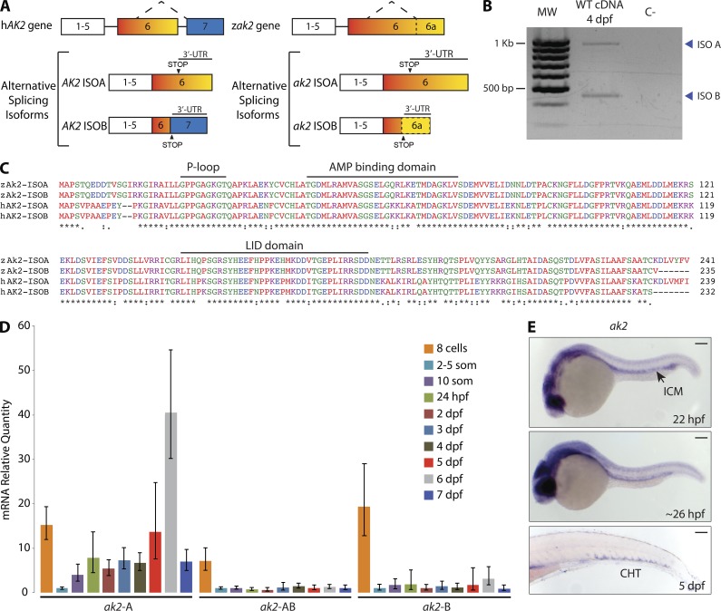
Figure 1. Zebrafish AK2 alternative splicing isoforms are expressed in hematopoietic regions during embryo development. (A) Schematic representation of human (hAK2) and zebrafish (zak2) conserved gene structure and alternative splicing mechanism. STOP indicates the position of the STOP codons. Neither exons nor introns are drawn to scale. (B) RT-PCR analysis on cDNA of 4 dpf WT embryos. The reverse primer AK2-ISO B R2 (Table S1) was used in the PCR reaction to bind to isoforms A and B. MW, molecular weight size marker; C−, negative control. Blue arrowheads point to the two different alternative splicing isoforms. The results are representative of two independent experiments. (C) Multiprotein sequence alignment of human (hAK2) and zebrafish (zAk2) AK2 splicing isoforms (indicated as ISOA and ISOB). Features of human AK2 structure are depicted above the amino acid sequences. Different colors indicate different physicochemical properties. (D) mRNA quantitative real-time PCR analysis showing the relative expression of AK2 splicing isoforms during embryo development using specific probes for isoforms A or B (ak2-A and ak2-B) or a common probe for A and B isoforms (ak2-AB). Expression levels were normalized to β-actin 2 (β-act 2) and elongation factor 1 α (ef1α), and mRNA from 2–5-somite stage embryos was used as reference. Error bars indicate the calculated maximum (RQmax) and minimum (RQmin) expression levels that represent the standard error of the mean expression level (RQ value). Data are pooled from at least three independent experiments. (E) Spatiotemporal analysis by stereomicroscopy of AK2 expression by WISH using a common probe for A and B isoforms at the indicated times. For each stage, at least 25 embryos have been analyzed. Data show one representative experiment out of two independently performed. ICM, intermediate cell mass; CHT, caudal hematopoietic tissue. Bars, 100 µm.
Figure 1. Zebrafish AK2 alternative splicing isoforms are expressed in hematopoietic regions during embryo development. (A) Schematic representation of human (hAK2) and zebrafish (zak2) conserved gene structure and alternative splicing mechanism. STOP indicates the position of the STOP codons. Neither exons nor introns are drawn to scale. (B) RT-PCR analysis on cDNA of 4 dpf WT embryos. The reverse primer AK2-ISO B R2 (Table S1) was used in the PCR reaction to bind to isoforms A and B. MW, molecular weight size marker; C−, negative control. Blue arrowheads point to the two different alternative splicing isoforms. The results are representative of two independent experiments. (C) Multiprotein sequence alignment of human (hAK2) and zebrafish (zAk2) AK2 splicing isoforms (indicated as ISOA and ISOB). Features of human AK2 structure are depicted above the amino acid sequences. Different colors indicate different physicochemical properties. (D) mRNA quantitative real-time PCR analysis showing the relative expression of AK2 splicing isoforms during embryo development using specific probes for isoforms A or B (ak2-A and ak2-B) or a common probe for A and B isoforms (ak2-AB). Expression levels were normalized to β-actin 2 (β-act 2) and elongation factor 1 α (ef1α), and mRNA from 2–5-somite stage embryos was used as reference. Error bars indicate the calculated maximum (RQmax) and minimum (RQmin) expression levels that represent the standard error of the mean expression level (RQ value). Data are pooled from at least three independent experiments. (E) Spatiotemporal analysis by stereomicroscopy of AK2 expression by WISH using a common probe for A and B isoforms at the indicated times. For each stage, at least 25 embryos have been analyzed. Data show one representative experiment out of two independently performed. ICM, intermediate cell mass; CHT, caudal hematopoietic tissue. Bars, 100 µm.CC BY-NC-SA · PubMed Central
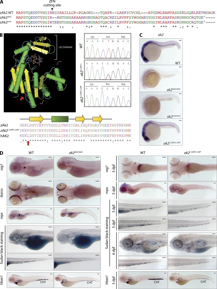
Figure 2. Zebrafish AK2 mutants present a wide array of hematopoietic defects. (A) Alignment of protein sequences encoded by the WT and ZFN-induced mutant alleles (del2 and ins4) shows the predicted frameshift after the Arg 15 codon targeted by the ZFN cutting site. Premature stop codons are also indicated (^). (B) 3D structure of human AK2 (left). (bottom) Partial sequence multi-alignment of WT (zAk2) and missense mutant zebrafish Ak2 (zAk2L124P/L124P) and human AK2 (hAK2). The mutated amino acid position is highlighted in both panels (red arrows). (right) Sequence chromatograms showing the nucleotide change for L124P missense mutation in heterozygous (ak2L124P/+) and homozygous (ak2L124P/L124P) embryos compared with the WT sequence. (C) Stereomicroscope images of WISH analysis on 24 hpf WT and mutant embryos using an antisense probe against ak2 mRNA (A and B transcripts). For each sample, at least 25 embryos have been analyzed. (D) Analysis of definitive hematopoietic phenotypes in ak2del2/del2 and ak2L124P/L124P mutants by WISH and stereomicroscopy. Red and black dashed circles indicate thymus and kidney regions, respectively. (left) Lateral views of embryos hybridized with rag1, mpx, and hbae1 and ventral views of ikaros hybridization at 5 dpf. Lateral views of embryos stained using Sudan black solution to mark granulocytes at 5 dpf. (right) Ventral views of rag1 and lateral views of mpx and hbae1 hybridized embryos at 5 dpf and lateral views of embryos stained using Sudan black solution at different stages of development. For each sample, at least 25 embryos have been analyzed. Data in C and D show one representative experiment out of two independently performed. Bars, 100 µm.
Figure 2. Zebrafish AK2 mutants present a wide array of hematopoietic defects. (A) Alignment of protein sequences encoded by the WT and ZFN-induced mutant alleles (del2 and ins4) shows the predicted frameshift after the Arg 15 codon targeted by the ZFN cutting site. Premature stop codons are also indicated (^). (B) 3D structure of human AK2 (left). (bottom) Partial sequence multi-alignment of WT (zAk2) and missense mutant zebrafish Ak2 (zAk2L124P/L124P) and human AK2 (hAK2). The mutated amino acid position is highlighted in both panels (red arrows). (right) Sequence chromatograms showing the nucleotide change for L124P missense mutation in heterozygous (ak2L124P/+) and homozygous (ak2L124P/L124P) embryos compared with the WT sequence. (C) Stereomicroscope images of WISH analysis on 24 hpf WT and mutant embryos using an antisense probe against ak2 mRNA (A and B transcripts). For each sample, at least 25 embryos have been analyzed. (D) Analysis of definitive hematopoietic phenotypes in ak2del2/del2 and ak2L124P/L124P mutants by WISH and stereomicroscopy. Red and black dashed circles indicate thymus and kidney regions, respectively. (left) Lateral views of embryos hybridized with rag1, mpx, and hbae1 and ventral views of ikaros hybridization at 5 dpf. Lateral views of embryos stained using Sudan black solution to mark granulocytes at 5 dpf. (right) Ventral views of rag1 and lateral views of mpx and hbae1 hybridized embryos at 5 dpf and lateral views of embryos stained using Sudan black solution at different stages of development. For each sample, at least 25 embryos have been analyzed. Data in C and D show one representative experiment out of two independently performed. Bars, 100 µm.CC BY-NC-SA · PubMed Central
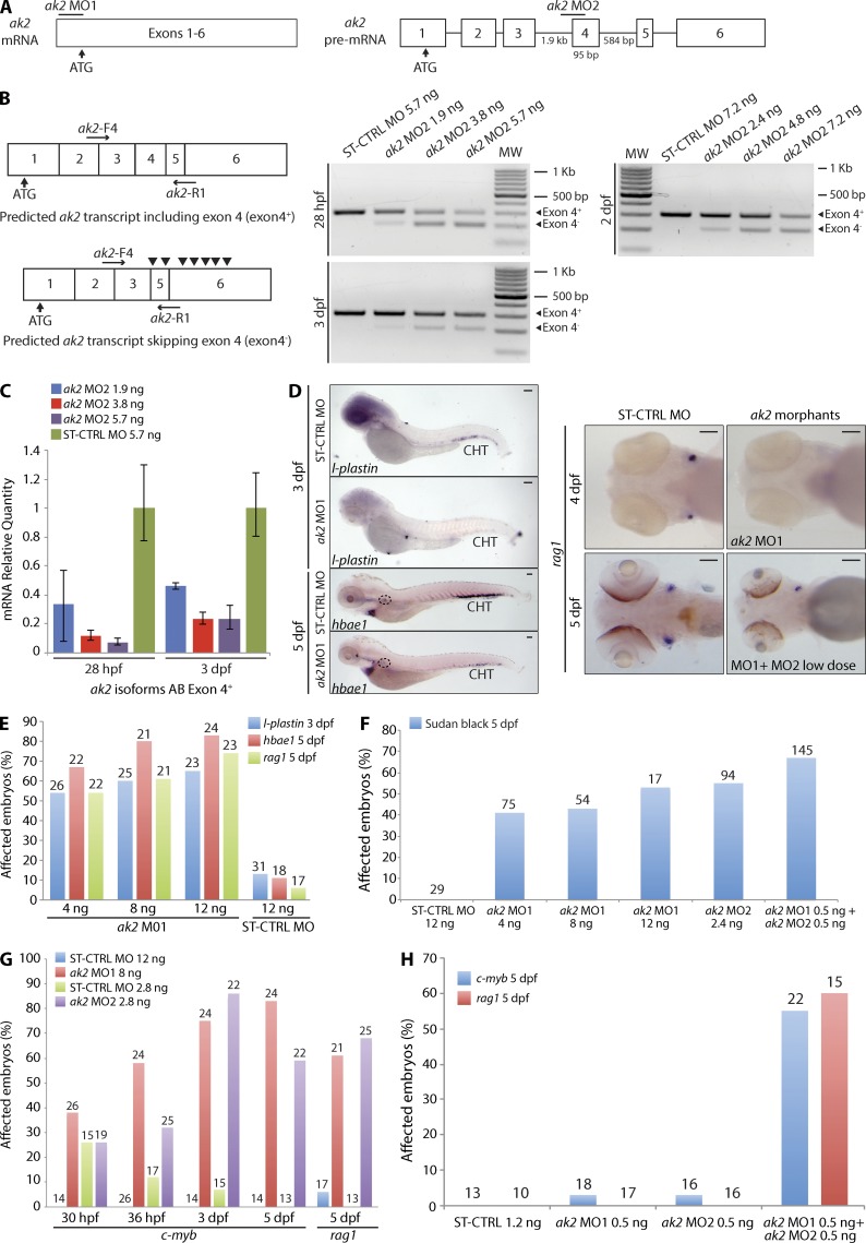
Figure 3. ak2 knockdown by MO injection phenocopies hematopoietic defects observed in ak2 zebrafish mutants. (A) Schematic representation of the two different MOs used in the study: ak2 MO1 blocking the mRNA translation and ak2 MO2 targeting the intron 3–exon 4 splice junction of the ak2 gene. Neither exons nor introns are drawn to scale. Numbers below the introns (straight lines) and exons (white boxes) indicate their size in bases. (B) Schematic representation of the predicted AK2 transcripts in MO-treated embryos. Black arrowheads in the bottom left panel indicate noncanonical STOP codons introduced by ak2 MO2–induced skipping of exon 4. Horizontal black arrows indicate the pair of primers used in RT-PCR (right panels) to test the effects of different doses of ak2 MO2 during development as indicated. MW, molecular weight size marker. (C) mRNA quantitative real-time PCR analysis of splicing MO activity (three different doses) at 28 hpf and 3 dpf. In both cases, ST-CTRL MO–injected embryos serve as reference. The error bars indicate the calculated maximum (RQmax) and minimum (RQmin) expression levels that represent the standard error of the mean expression levels (RQ value). (D–H) Qualitative and quantitative analysis of hematopoietic defects induced by ak2 MOs injection during definitive hematopoiesis assessed by WISH and stereomicroscopy. WT Ekkwill (EK) embryos were injected with different doses (4, 8, and 12 ng) of ak2 MO1 or ak2 MO2 (2.4 ng) and analyzed by WISH at different developmental stages using specific hematopoietic probes. ST-CTRL MO at the highest dose used for ak2 MO1 was used as a control. Numbers above bars indicate the total count of embryos analyzed. (D, left) Stereomicroscope lateral views of l-plastin (marker of macrophages and monocytes) and hbae1 WISH signals in ST-CTRL and ak2 MO1 morphants at 3 and 5 dpf. Black dashed circles indicate the position of the kidney. (right) Stereomicroscope ventral views of ST-CTRL MO, ak2 MO1 morphants, or ak2 MO1- and MO2-coinjected embryos hybridized with rag1 ISH probe to visualize differentiating lymphocytes in the thymus. For each sample, at least 25 embryos have been analyzed. Bars, 100 µm. (E) Effect of ak2 MO1 and ST-CTRL MO injection on the expression of l-plastin, hbae1, and rag1. (F) Effect of single ak2 MO injection and low dose coinjection on granulopoiesis (Sudan black stain). (G and H) Effect of single ak2 MO injection or low dose coinjection on the expression of c-myb and rag1 from 30 hpf to 5 dpf.. Data are presented as percentage of affected embryos. All data (B–H) show one representative experiment out of at least two independently performed.
Figure 3. ak2 knockdown by MO injection phenocopies hematopoietic defects observed in ak2 zebrafish mutants. (A) Schematic representation of the two different MOs used in the study: ak2 MO1 blocking the mRNA translation and ak2 MO2 targeting the intron 3–exon 4 splice junction of the ak2 gene. Neither exons nor introns are drawn to scale. Numbers below the introns (straight lines) and exons (white boxes) indicate their size in bases. (B) Schematic representation of the predicted AK2 transcripts in MO-treated embryos. Black arrowheads in the bottom left panel indicate noncanonical STOP codons introduced by ak2 MO2–induced skipping of exon 4. Horizontal black arrows indicate the pair of primers used in RT-PCR (right panels) to test the effects of different doses of ak2 MO2 during development as indicated. MW, molecular weight size marker. (C) mRNA quantitative real-time PCR analysis of splicing MO activity (three different doses) at 28 hpf and 3 dpf. In both cases, ST-CTRL MO–injected embryos serve as reference. The error bars indicate the calculated maximum (RQmax) and minimum (RQmin) expression levels that represent the standard error of the mean expression levels (RQ value). (D–H) Qualitative and quantitative analysis of hematopoietic defects induced by ak2 MOs injection during definitive hematopoiesis assessed by WISH and stereomicroscopy. WT Ekkwill (EK) embryos were injected with different doses (4, 8, and 12 ng) of ak2 MO1 or ak2 MO2 (2.4 ng) and analyzed by WISH at different developmental stages using specific hematopoietic probes. ST-CTRL MO at the highest dose used for ak2 MO1 was used as a control. Numbers above bars indicate the total count of embryos analyzed. (D, left) Stereomicroscope lateral views of l-plastin (marker of macrophages and monocytes) and hbae1 WISH signals in ST-CTRL and ak2 MO1 morphants at 3 and 5 dpf. Black dashed circles indicate the position of the kidney. (right) Stereomicroscope ventral views of ST-CTRL MO, ak2 MO1 morphants, or ak2 MO1- and MO2-coinjected embryos hybridized with rag1 ISH probe to visualize differentiating lymphocytes in the thymus. For each sample, at least 25 embryos have been analyzed. Bars, 100 µm. (E) Effect of ak2 MO1 and ST-CTRL MO injection on the expression of l-plastin, hbae1, and rag1. (F) Effect of single ak2 MO injection and low dose coinjection on granulopoiesis (Sudan black stain). (G and H) Effect of single ak2 MO injection or low dose coinjection on the expression of c-myb and rag1 from 30 hpf to 5 dpf.. Data are presented as percentage of affected embryos. All data (B–H) show one representative experiment out of at least two independently performed.CC BY-NC-SA · PubMed Central
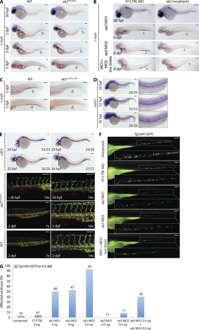
Figure 4. AK2 deficiency impairs HSPC development in zebrafish. (A–C) Representative stereomicroscope images of WISH analyses of c-myb expression in ak2del2/del2 null mutants, ak2 morphants, and ak2L124P/L124P mutants. Red and black dashed circles mark thymus and kidney regions, respectively; open arrowheads indicate CHT regions. (A) Lateral views of right side of mutant embryos at different developmental stages compared with their WT siblings. White arrows mark insulin expression (positive control). (B) Lateral views of ST-CTRL MO and ak2 MO morphants at different developmental stages as indicated. Two different ak2 MOs (MO1 or MO2) were injected separately or in combination at low dose (MO1 + MO2 low doses). (C) Lateral views of the trunk/tail regions of ak2L124P/L124P mutant embryos at 3 and 5 dpf compared with their WT siblings. (D) Stereomicroscope images of WISH analysis of runx1 expression from 29 to 36 hpf in ak2del2 embryos from an intercross of heterozygous (ak2del2/+) adults (35, 32, and 26 embryos analyzed at 29, 32, and 36 hpf, respectively). (E) Analysis of vascular system development from 24 to 48 hpf in ak2del2 mutant embryos. (top) Stereomicroscope lateral views of WISH analysis with a cdh5 antisense probe on embryos from an intercross of heterozygous (ak2del2/+) adults (33, 29, 20, and 27 embryos analyzed at 24, 29, 32, and 36 hpf, respectively). (bottom) Confocal microscopy lateral views of the trunk/tail regions of ak2del2/del2 double transgenic Tg(fli1a:EGFP)y1;Tg(gata1a:dsRed)sd2 embryos and WT controls at different developmental stages; fli1a (EGFP) marks vascular development, and gata-1 (dsRed) marks red blood cells as indicated. (F) Stereomicroscope lateral views of trunk/tail region of 4.5 dpf Tg(cd41:GFP) embryos injected with different ak2 MOs. (G) Quantitative analysis of ak2 MO injections on Tg(cd41:GFP) embryos. Numbers above bars indicate the total number of embryos analyzed. Data are presented as percentage of affected embryos. For each panel (A–G), results of one representative experiment out of at least two independent replicates are shown. (A–F) For each sample at least, 20 embryos have been analyzed. Bars, 100 µm.
Figure 4. AK2 deficiency impairs HSPC development in zebrafish. (A–C) Representative stereomicroscope images of WISH analyses of c-myb expression in ak2del2/del2 null mutants, ak2 morphants, and ak2L124P/L124P mutants. Red and black dashed circles mark thymus and kidney regions, respectively; open arrowheads indicate CHT regions. (A) Lateral views of right side of mutant embryos at different developmental stages compared with their WT siblings. White arrows mark insulin expression (positive control). (B) Lateral views of ST-CTRL MO and ak2 MO morphants at different developmental stages as indicated. Two different ak2 MOs (MO1 or MO2) were injected separately or in combination at low dose (MO1 + MO2 low doses). (C) Lateral views of the trunk/tail regions of ak2L124P/L124P mutant embryos at 3 and 5 dpf compared with their WT siblings. (D) Stereomicroscope images of WISH analysis of runx1 expression from 29 to 36 hpf in ak2del2 embryos from an intercross of heterozygous (ak2del2/+) adults (35, 32, and 26 embryos analyzed at 29, 32, and 36 hpf, respectively). (E) Analysis of vascular system development from 24 to 48 hpf in ak2del2 mutant embryos. (top) Stereomicroscope lateral views of WISH analysis with a cdh5 antisense probe on embryos from an intercross of heterozygous (ak2del2/+) adults (33, 29, 20, and 27 embryos analyzed at 24, 29, 32, and 36 hpf, respectively). (bottom) Confocal microscopy lateral views of the trunk/tail regions of ak2del2/del2 double transgenic Tg(fli1a:EGFP)y1;Tg(gata1a:dsRed)sd2 embryos and WT controls at different developmental stages; fli1a (EGFP) marks vascular development, and gata-1 (dsRed) marks red blood cells as indicated. (F) Stereomicroscope lateral views of trunk/tail region of 4.5 dpf Tg(cd41:GFP) embryos injected with different ak2 MOs. (G) Quantitative analysis of ak2 MO injections on Tg(cd41:GFP) embryos. Numbers above bars indicate the total number of embryos analyzed. Data are presented as percentage of affected embryos. For each panel (A–G), results of one representative experiment out of at least two independent replicates are shown. (A–F) For each sample at least, 20 embryos have been analyzed. Bars, 100 µm.CC BY-NC-SA · PubMed Central
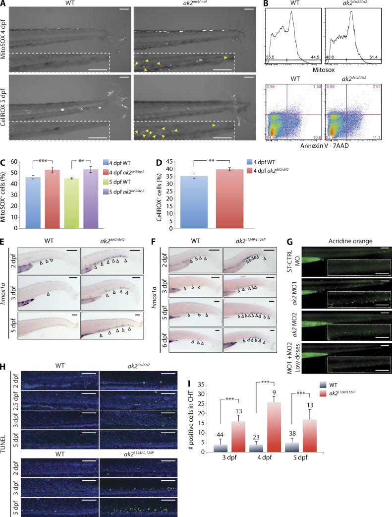
Figure 5. AK2 mutant zebrafish demonstrate increased levels of cellular oxidative stress and apoptosis in hematopoietic tissues. (A) Confocal microscopy assessment of oxidative stress in the CHT region of WT and ak2ins4/ins4 mutant embryos probed with the MitoSOX or CellROX indicators at the indicated stages. Yellow arrowheads in the dashed insets indicate MitoSOX- or CellROX-positive cells in CHT regions. (B) Representative quantitative analysis by flow cytometry of oxidative stress, apoptosis, and cell death on 5 dpf ak2del2/del2 and control embryos (60 embryos each) using MitoSOX Red and Annexin V + 7-AAD, respectively. (C and D) Quantitative analysis of MitoSOX Red (C) and CellROX Green (D) staining at 4 and 5 dpf in ak2del2/del2 mutants and WT siblings (60 embryos each). (E) WISH analysis of hmox1a expression in ak2del2/del2 mutants and their WT siblings at different stages of development. (F) WISH analysis of hmox1a expression in ak2L124P/L124P mutants and their WT siblings at different stages of development. (E and F) Open arrowheads indicate hmox1a-positive cells. (G) Stereomicroscope analysis of acridine orange staining of AK2 morphants at 3 dpf. Dashed insets show CHT regions. (H) Confocal analysis of fluorescent TUNEL staining in CHT regions of WT siblings and ak2del2/del2 and ak2L124P/L124P mutant embryos from 2 to 5 dpf. Each panel represents a crop of the CHT region from a 14× magnification image. The green signal indicates TUNEL-positive cells and the blue signal indicates DAPI staining. (A and E–H) For each sample, at least 25 embryos have been analyzed. Bars, 100 µm. (I) Quantitative analysis of TUNEL AP staining on WT siblings and ak2L124P/L124P missense mutant embryos from 3 to 5 dpf. The number of embryos analyzed is shown above each column. In panels C, D, and I, error bars indicate standard deviation; **, P < 0.01; ***, P < 0.001 for the indicated comparisons using an unpaired Student’s t test. Data in C and D are pooled from at least three independent experiments. Data in A, B, and E–I are representative of at least two independent experiments.
Figure 5. AK2 mutant zebrafish demonstrate increased levels of cellular oxidative stress and apoptosis in hematopoietic tissues. (A) Confocal microscopy assessment of oxidative stress in the CHT region of WT and ak2ins4/ins4 mutant embryos probed with the MitoSOX or CellROX indicators at the indicated stages. Yellow arrowheads in the dashed insets indicate MitoSOX- or CellROX-positive cells in CHT regions. (B) Representative quantitative analysis by flow cytometry of oxidative stress, apoptosis, and cell death on 5 dpf ak2del2/del2 and control embryos (60 embryos each) using MitoSOX Red and Annexin V + 7-AAD, respectively. (C and D) Quantitative analysis of MitoSOX Red (C) and CellROX Green (D) staining at 4 and 5 dpf in ak2del2/del2 mutants and WT siblings (60 embryos each). (E) WISH analysis of hmox1a expression in ak2del2/del2 mutants and their WT siblings at different stages of development. (F) WISH analysis of hmox1a expression in ak2L124P/L124P mutants and their WT siblings at different stages of development. (E and F) Open arrowheads indicate hmox1a-positive cells. (G) Stereomicroscope analysis of acridine orange staining of AK2 morphants at 3 dpf. Dashed insets show CHT regions. (H) Confocal analysis of fluorescent TUNEL staining in CHT regions of WT siblings and ak2del2/del2 and ak2L124P/L124P mutant embryos from 2 to 5 dpf. Each panel represents a crop of the CHT region from a 14× magnification image. The green signal indicates TUNEL-positive cells and the blue signal indicates DAPI staining. (A and E–H) For each sample, at least 25 embryos have been analyzed. Bars, 100 µm. (I) Quantitative analysis of TUNEL AP staining on WT siblings and ak2L124P/L124P missense mutant embryos from 3 to 5 dpf. The number of embryos analyzed is shown above each column. In panels C, D, and I, error bars indicate standard deviation; **, P < 0.01; ***, P < 0.001 for the indicated comparisons using an unpaired Student’s t test. Data in C and D are pooled from at least three independent experiments. Data in A, B, and E–I are representative of at least two independent experiments.CC BY-NC-SA · PubMed Central
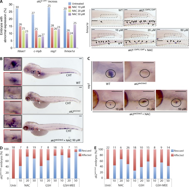
Figure 6. Antioxidant treatment induces rescue of hematopoietic phenotypes in AK2 zebrafish mutants. (A) Embryos from an incross of heterozygous ak2L124P mutants were treated with different doses of NAC (10, 20, or 50 µM) until 5 dpf when the expression of different markers was assessed by WISH. (left) Quantification of rescue induced by NAC treatment of ak2L124P missense mutants. Data are presented as the percentage of abnormal expression of each marker by WISH analysis at the indicated concentrations. The number of embryos analyzed is shown above each column. All observed differences compared with the untreated embryos are significant (P < 0.005, Z-test). (right) Representative stereomicroscopy images of tail regions of WT siblings and ak2L124P/L124P mutants untreated or treated with different doses of NAC. Black arrowheads indicate hmox1a-positive cells in CHT regions (boxed insets). (B) Lateral views of WT siblings and ak2del2/del2 mutants showing c-myb expression (WISH) in untreated and treated with 50 µM NAC embryos at 5 dpf. Red and black insets show thymus and kidney regions, respectively; white arrows mark insulin expression (positive control). (C) Lateral views of rag 1 expression (WISH) in thymic region of WT siblings, ak2del2/del2 mutants untreated and treated with increasing concentrations of NAC at 5 dpf. Black circles show thymic region in each sample. (A [right], B, and C) For each sample, at least 25 embryos have been analyzed. Bars, 100 µm. (D and E) Quantitative analysis of the effect of antioxidant treatment on c-myb (D) and rag1 (E) expression at 5 dpf in embryos from an incross of heterozygous ak2del2 mutants. Data are presented as the percentage of ak2del2/del2 null embryos. Numbers above columns indicate the total number of ak2del2/del2-null mutants found in each group of genotyped embryos. Data (A–E) are representative of at least two independent experiments.
Figure 6. Antioxidant treatment induces rescue of hematopoietic phenotypes in AK2 zebrafish mutants. (A) Embryos from an incross of heterozygous ak2L124P mutants were treated with different doses of NAC (10, 20, or 50 µM) until 5 dpf when the expression of different markers was assessed by WISH. (left) Quantification of rescue induced by NAC treatment of ak2L124P missense mutants. Data are presented as the percentage of abnormal expression of each marker by WISH analysis at the indicated concentrations. The number of embryos analyzed is shown above each column. All observed differences compared with the untreated embryos are significant (P < 0.005, Z-test). (right) Representative stereomicroscopy images of tail regions of WT siblings and ak2L124P/L124P mutants untreated or treated with different doses of NAC. Black arrowheads indicate hmox1a-positive cells in CHT regions (boxed insets). (B) Lateral views of WT siblings and ak2del2/del2 mutants showing c-myb expression (WISH) in untreated and treated with 50 µM NAC embryos at 5 dpf. Red and black insets show thymus and kidney regions, respectively; white arrows mark insulin expression (positive control). (C) Lateral views of rag 1 expression (WISH) in thymic region of WT siblings, ak2del2/del2 mutants untreated and treated with increasing concentrations of NAC at 5 dpf. Black circles show thymic region in each sample. (A [right], B, and C) For each sample, at least 25 embryos have been analyzed. Bars, 100 µm. (D and E) Quantitative analysis of the effect of antioxidant treatment on c-myb (D) and rag1 (E) expression at 5 dpf in embryos from an incross of heterozygous ak2del2 mutants. Data are presented as the percentage of ak2del2/del2 null embryos. Numbers above columns indicate the total number of ak2del2/del2-null mutants found in each group of genotyped embryos. Data (A–E) are representative of at least two independent experiments.CC BY-NC-SA · PubMed Central
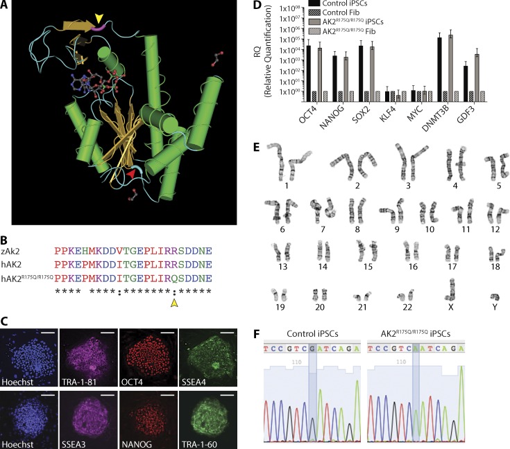
Figure 7. Generation and characterization of human AK2R175Q/R175Q iPSCs. (A) Schematic representation of the annotated 3D structure of the human AK2 protein. The amino acid position mutated in patient-derived AK2R175Q/R175Q mutant fibroblasts and iPSC lines is highlighted in violet (yellow arrowhead). The amino acid position mutated in ak2L124P/L124P mutant zebrafish is marked in light blue (red arrowhead). (B) Partial sequence multi-alignment of the LID domain of zebrafish (zAk2) and human (hAK2) proteins and the human mutated form (hAK2R175Q/R175Q). Yellow arrowhead marks the amino acid position mutated in patient-derived cell lines. (C–F) iPSCs were generated from AK2-deficient dermal and control foreskin fibroblasts. (C) Representative confocal microscopy images showing the expression of human pluripotency markers (TRA-1-81 and SSEA3 [magenta], Oct4 and NANOG [red], and SSEA4 and TRA-1-60 [green]) using immunofluorescently labeled antibodies in AK2R175Q/R175Q iPSCs; cellular content is highlighted by nuclear staining with Hoechst 33342 (blue). Bars, 100 µm. (D) mRNA analysis using quantitative real-time PCR of the indicated pluripotency-associated genes in AK2R175Q/R175Q and control iPSCs. AK2R175Q/R175Q and control iPSCs gene expression was compared with the respective parental fibroblasts (Fib), and human β-actin gene expression (hACTB) was used as housekeeping gene. Error bars indicate standard error. (E) Karyotype and G-banding analysis of AK2R175Q/R175Q iPSCs. (F) Sequencing of the genomic region surrounding the mutation in the control and patient-derived AK2-mutated iPSC line. Data (C–F) are representative of at least two independent experiments.
Figure 7. Generation and characterization of human AK2R175Q/R175Q iPSCs. (A) Schematic representation of the annotated 3D structure of the human AK2 protein. The amino acid position mutated in patient-derived AK2R175Q/R175Q mutant fibroblasts and iPSC lines is highlighted in violet (yellow arrowhead). The amino acid position mutated in ak2L124P/L124P mutant zebrafish is marked in light blue (red arrowhead). (B) Partial sequence multi-alignment of the LID domain of zebrafish (zAk2) and human (hAK2) proteins and the human mutated form (hAK2R175Q/R175Q). Yellow arrowhead marks the amino acid position mutated in patient-derived cell lines. (C–F) iPSCs were generated from AK2-deficient dermal and control foreskin fibroblasts. (C) Representative confocal microscopy images showing the expression of human pluripotency markers (TRA-1-81 and SSEA3 [magenta], Oct4 and NANOG [red], and SSEA4 and TRA-1-60 [green]) using immunofluorescently labeled antibodies in AK2R175Q/R175Q iPSCs; cellular content is highlighted by nuclear staining with Hoechst 33342 (blue). Bars, 100 µm. (D) mRNA analysis using quantitative real-time PCR of the indicated pluripotency-associated genes in AK2R175Q/R175Q and control iPSCs. AK2R175Q/R175Q and control iPSCs gene expression was compared with the respective parental fibroblasts (Fib), and human β-actin gene expression (hACTB) was used as housekeeping gene. Error bars indicate standard error. (E) Karyotype and G-banding analysis of AK2R175Q/R175Q iPSCs. (F) Sequencing of the genomic region surrounding the mutation in the control and patient-derived AK2-mutated iPSC line. Data (C–F) are representative of at least two independent experiments.CC BY-NC-SA · PubMed Central
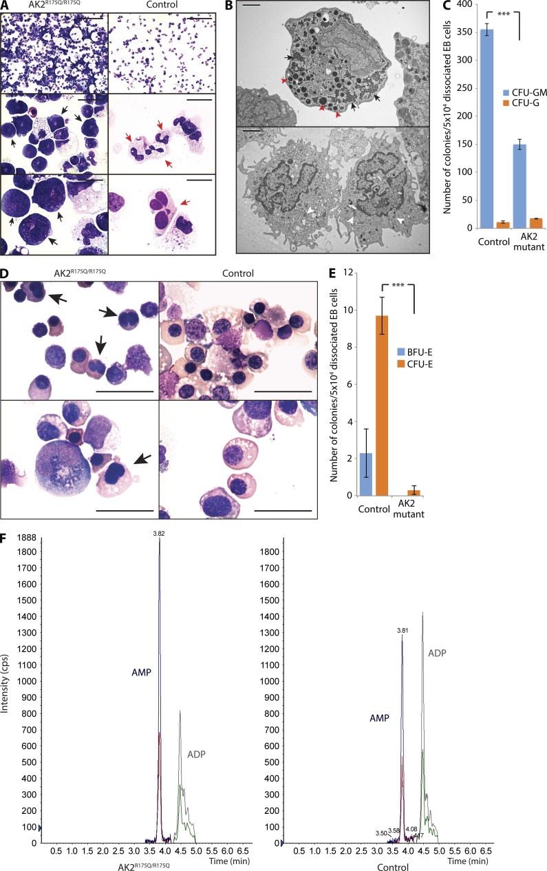
Figure 8. AK2 deficiency affects in vitro granulopoiesis, erythropoiesis, and the adenine nucleotide profile of human myeloid cells. (A) Microscopic analysis of in vitro myeloid differentiation of AK2R175Q/R175Q and control iPSCs; black arrows indicate promyelocytes; red arrows indicate mature neutrophils. (B) Electron microscopy assessment of AK2R175Q/R175Q iPSC-derived myeloid precursors and control cells. Red arrowheads mark large electron-dense primary granules, and black arrows indicate glycogen storage in the cytoplasm (top). White arrowheads highlight pale secondary granules (bottom). (C) The number of myeloid colonies grown from 50k dissociated EB cells after 14 d of culture on methylcellulose was assessed (***, P < 0.001, χ2 test). Data represent the mean of three experiments, and error bars depict standard error. (D) In vitro erythroid differentiation of AK2R175Q/R175Q and control iPSCs was assessed by microscopy. Black arrows indicate incomplete nuclear separation. (A, B, and D) Data are representative of at least three independent experiments. Bars: (A, top) 100 µm; (A, middle) 20 µm; (A and D, bottom) 10 µm; (B) 2 µm; (D, top) 25 µm. (E) Number of red blood cell–forming colonies (BFU-E and CFU-E) grown from 50k dissociated EB cells after 14 d of culture on methylcellulose (***, P < 0.003, χ2 test). Data represent the mean of three independent experiments, and error bars depict standard error. (F) Quantification of cellular AMP and ADP content in AK2R175Q/R175Q iPSC-derived myeloid cells and control myeloid cells by tandem mass spectrometry. Data are representative of three independent experiments. iPSCs generated from a human foreskin fibroblast line were used as WT control in all experiments (A–F).
Figure 8. AK2 deficiency affects in vitro granulopoiesis, erythropoiesis, and the adenine nucleotide profile of human myeloid cells. (A) Microscopic analysis of in vitro myeloid differentiation of AK2R175Q/R175Q and control iPSCs; black arrows indicate promyelocytes; red arrows indicate mature neutrophils. (B) Electron microscopy assessment of AK2R175Q/R175Q iPSC-derived myeloid precursors and control cells. Red arrowheads mark large electron-dense primary granules, and black arrows indicate glycogen storage in the cytoplasm (top). White arrowheads highlight pale secondary granules (bottom). (C) The number of myeloid colonies grown from 50k dissociated EB cells after 14 d of culture on methylcellulose was assessed (***, P < 0.001, χ2 test). Data represent the mean of three experiments, and error bars depict standard error. (D) In vitro erythroid differentiation of AK2R175Q/R175Q and control iPSCs was assessed by microscopy. Black arrows indicate incomplete nuclear separation. (A, B, and D) Data are representative of at least three independent experiments. Bars: (A, top) 100 µm; (A, middle) 20 µm; (A and D, bottom) 10 µm; (B) 2 µm; (D, top) 25 µm. (E) Number of red blood cell–forming colonies (BFU-E and CFU-E) grown from 50k dissociated EB cells after 14 d of culture on methylcellulose (***, P < 0.003, χ2 test). Data represent the mean of three independent experiments, and error bars depict standard error. (F) Quantification of cellular AMP and ADP content in AK2R175Q/R175Q iPSC-derived myeloid cells and control myeloid cells by tandem mass spectrometry. Data are representative of three independent experiments. iPSCs generated from a human foreskin fibroblast line were used as WT control in all experiments (A–F).CC BY-NC-SA · PubMed Central
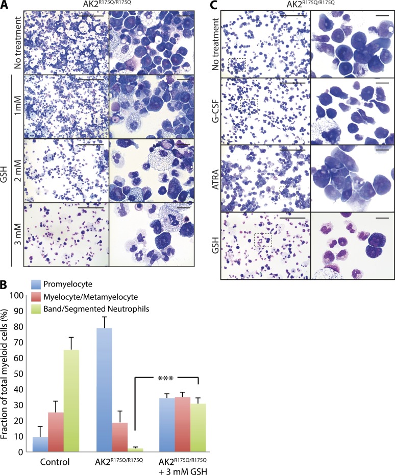
Figure 9. Antioxidant treatment rescues myeloid differentiation of human AK2R175Q/R175Q iPSCs. (A) Microscopy analysis of in vitro myeloid maturation of AK2R175Q/R175Q iPSCs in the presence of different concentrations of the antioxidant agent GSH in the culture medium as indicated. Data are representative of at least three independent experiments. (B) Maturation stage–specific comparison of GSH-treated and untreated AK2R175Q/R175Q myeloid lineage cells versus control. Data are presented as the percent fraction of total myeloid cells and represent the mean of three experiments. Error bars depict standard error (***, P < 0.0001, χ2 test). iPSCs generated from a human foreskin fibroblast line were used as WT control. (C) Microscopy analysis of in vitro myeloid maturation of AK2R175Q/R175Q iPSCs in the presence of 25 ng/ml G-CSF, 1 µM ATRA, or 3 mM GSH. Magnified images of boxed areas are presented in the right panels. Data are representative of at least two independent experiments. Bars: (A and C, left) 100 µm; (A and C, right) 10 µm.
Figure 9. Antioxidant treatment rescues myeloid differentiation of human AK2R175Q/R175Q iPSCs. (A) Microscopy analysis of in vitro myeloid maturation of AK2R175Q/R175Q iPSCs in the presence of different concentrations of the antioxidant agent GSH in the culture medium as indicated. Data are representative of at least three independent experiments. (B) Maturation stage–specific comparison of GSH-treated and untreated AK2R175Q/R175Q myeloid lineage cells versus control. Data are presented as the percent fraction of total myeloid cells and represent the mean of three experiments. Error bars depict standard error (***, P < 0.0001, χ2 test). iPSCs generated from a human foreskin fibroblast line were used as WT control. (C) Microscopy analysis of in vitro myeloid maturation of AK2R175Q/R175Q iPSCs in the presence of 25 ng/ml G-CSF, 1 µM ATRA, or 3 mM GSH. Magnified images of boxed areas are presented in the right panels. Data are representative of at least two independent experiments. Bars: (A and C, left) 100 µm; (A and C, right) 10 µm.CC BY-NC-SA · PubMed Central

Verification

Claims the paper makes

The claims and statistics of this paper haven’t been checked yet.

Run the check — free, no account needed. It reads the open-access text and checks the sources it cites.

A few free checks a day · papers already verified open instantly · results are public

Claims other papers make about this one

Each is checked against this paper’s own text: does it say what the citing paper says it does?

Self-check

Percentages, sums and reported differences re-computed from the paper’s own numbers.

The self-check needs the paper’s full text.

Citations

Cites

Show all 3 references

Research works printed but not linked: 56

  1. Al-Zahrani, 2013, Skeletal abnormalities and successful hematopoietic stem cell transplantation in patients with reticular dysgenesis, J. Allergy Clin. Immunol., 132, 993, 10.1016/j.jaci.2013.04.055 · DOI
  2. Aloj, 2012, Severe combined immunodeficiences: new and old scenarios, Int. Rev. Immunol., 31, 43, 10.3109/08830185.2011.644607 · DOI
  3. Antoine, 2003, Long-term survival and transplantation of haemopoietic stem cells for immunodeficiencies: report of the European experience 1968-99, Lancet., 361, 553, 10.1016/S0140-6736(03)12513-5 · DOI
  4. Argenton, 2004, Ectopic expression and knockdown of a zebrafish sox21 reveal its role as a transcriptional repressor in early development, Mech. Dev., 121, 131, 10.1016/j.mod.2004.01.001 · DOI · Paper page
  5. Bandlow, 1988, Yeast adenylate kinase is active simultaneously in mitochondria and cytoplasm and is required for non-fermentative growth, Eur. J. Biochem., 178, 451, 10.1111/j.1432-1033.1988.tb14469.x · DOI
  6. Bertrand, 2008, CD41+ cmyb+ precursors colonize the zebrafish pronephros by a novel migration route to initiate adult hematopoiesis, Development., 135, 1853, 10.1242/dev.015297 · DOI
  7. Bertrand, 2002, Reticular dysgenesis: HLA non-identical bone marrow transplants in a series of 10 patients, Bone Marrow Transplant., 29, 759, 10.1038/sj.bmt.1703531 · DOI
  8. Bujan, 1993, Effect of recombinant human granulocyte colony-stimulating factor in reticular dysgenesis, Blood., 82, 1684, 10.1182/blood.V82.5.1684.bloodjournal8251684a · DOI
  9. Burkart, 2011, Adenylate kinase 2 links mitochondrial energy metabolism to the induction of the unfolded protein response, J. Biol. Chem., 286, 4081, 10.1074/jbc.M110.134106 · DOI
  10. Castaigne, 1990, All-trans retinoic acid as a differentiation therapy for acute promyelocytic leukemia. I. Clinical results, Blood., 76, 1704, 10.1182/blood.V76.9.1704.1704 · DOI
  11. Chen, 2012, Adenylate kinase 2 (AK2) promotes cell proliferation in insect development, BMC Mol. Biol., 13, 31, 10.1186/1471-2199-13-31 · DOI
  12. Choi, 2012, Predicting the functional effect of amino acid substitutions and indels, PLoS ONE., 7, e46688, 10.1371/journal.pone.0046688 · DOI · Paper page
  13. Craven, 2005, Loss of Hspa9b in zebrafish recapitulates the ineffective hematopoiesis of the myelodysplastic syndrome, Blood., 105, 3528, 10.1182/blood-2004-03-1089 · DOI
  14. Daily, 2010, Many local motions cooperate to produce the adenylate kinase conformational transition, J. Mol. Biol., 400, 618, 10.1016/j.jmb.2010.05.015 · DOI
  15. De Vaal, 1959, Reticular dysgenesia, Lancet., 274, 1123, 10.1016/S0140-6736(59)90105-9 · DOI
  16. Dzeja, 2003, Phosphotransfer networks and cellular energetics, J. Exp. Biol., 206, 2039, 10.1242/jeb.00426 · DOI
  17. Dzeja, 2009, Adenylate kinase and AMP signaling networks: metabolic monitoring, signal communication and body energy sensing, Int. J. Mol. Sci., 10, 1729, 10.3390/ijms10041729 · DOI
  18. Finckbeiner, 2011, Transient knockdown and overexpression reveal a developmental role for the zebrafish enosf1b gene, Cell Biosci., 1, 32, 10.1186/2045-3701-1-32 · DOI
  19. Fischer, 2005, Severe combined immunodeficiency. A model disease for molecular immunology and therapy, Immunol. Rev., 203, 98, 10.1111/j.0105-2896.2005.00223.x · DOI
  20. Flicek, 2013, Ensembl 2013, Nucleic Acids Res., 41, D48, 10.1093/nar/gks1236 · DOI
  21. Fujisawa, 2009, Adenylate kinase isozyme 2 is essential for growth and development of Drosophila melanogaster, Comp. Biochem. Physiol. B Biochem. Mol. Biol., 153, 29, 10.1016/j.cbpb.2009.01.006 · DOI
  22. Goujon, 2010, A new bioinformatics analysis tools framework at EMBL-EBI, Nucleic Acids Res., 38, W695, 10.1093/nar/gkq313 · DOI · Paper page
  23. Hatefi, 1985, The mitochondrial electron transport and oxidative phosphorylation system, Annu. Rev. Biochem., 54, 1015, 10.1146/annurev.bi.54.070185.005055 · DOI
  24. Henderson, 2013, First reported case of Omenn syndrome in a patient with reticular dysgenesis, J. Allergy Clin. Immunol., 131, 1227, 10.1016/j.jaci.2012.07.045 · DOI
  25. Jang, 2007, A low level of reactive oxygen species selects for primitive hematopoietic stem cells that may reside in the low-oxygenic niche, Blood., 110, 3056, 10.1182/blood-2007-05-087759 · DOI · Paper page
  26. Janssen, 2000, Adenylate kinase 1 gene deletion disrupts muscle energetic economy despite metabolic rearrangement, EMBO J., 19, 6371, 10.1093/emboj/19.23.6371 · DOI
  27. Kimmel, 1995, Stages of embryonic development of the zebrafish, Dev. Dyn., 203, 253, 10.1002/aja.1002030302 · DOI · Paper page
  28. Kissa, 2008, Live imaging of emerging hematopoietic stem cells and early thymus colonization, Blood., 111, 1147, 10.1182/blood-2007-07-099499 · DOI
  29. Köhler, 1999, Release of adenylate kinase 2 from the mitochondrial intermembrane space during apoptosis, FEBS Lett., 447, 10, 10.1016/S0014-5793(99)00251-3 · DOI
  30. Lagresle-Peyrou, 2009, Human adenylate kinase 2 deficiency causes a profound hematopoietic defect associated with sensorineural deafness, Nat. Genet., 41, 106, 10.1038/ng.278 · DOI
  31. Lagresle-Peyrou, 2011, Occurrence of myelodysplastic syndrome in 2 patients with reticular dysgenesis, J. Allergy Clin. Immunol., 128, 230, 10.1016/j.jaci.2011.02.008 · DOI
  32. Lawson, 2002, In vivo imaging of embryonic vascular development using transgenic zebrafish, Dev. Biol., 248, 307, 10.1006/dbio.2002.0711 · DOI
  33. Le Guyader, 2008, Origins and unconventional behavior of neutrophils in developing zebrafish, Blood., 111, 132, 10.1182/blood-2007-06-095398 · DOI
  34. Lee, 2007, AK2 activates a novel apoptotic pathway through formation of a complex with FADD and caspase-10, Nat. Cell Biol., 9, 1303, 10.1038/ncb1650 · DOI
  35. Lee, 1998, Cloning and expression of human adenylate kinase 2 isozymes: differential expression of adenylate kinase 1 and 2 in human muscle tissues, J. Biochem., 123, 47, 10.1093/oxfordjournals.jbchem.a021915 · DOI
  36. Liesa, 2013, Mitochondrial dynamics in the regulation of nutrient utilization and energy expenditure, Cell Metab., 17, 491, 10.1016/j.cmet.2013.03.002 · DOI · Paper page
  37. Lin, 2005, Analysis of thrombocyte development in CD41-GFP transgenic zebrafish, Blood., 106, 3803, 10.1182/blood-2005-01-0179 · DOI
  38. Liu, 2009, Enzymatically inactive adenylate kinase 4 interacts with mitochondrial ADP/ATP translocase, Int. J. Biochem. Cell Biol., 41, 1371, 10.1016/j.biocel.2008.12.002 · DOI
  39. Mugoni, 2014, Analysis of oxidative stress in zebrafish embryos, J. Vis. Exp., e51328, 10.3791/51328 · DOI
  40. Park, 2008, Reprogramming of human somatic cells to pluripotency with defined factors, Nature., 451, 141, 10.1038/nature06534 · DOI · Paper page
  41. Poliani, 2009, Early defects in human T-cell development severely affect distribution and maturation of thymic stromal cells: possible implications for the pathophysiology of Omenn syndrome, Blood., 114, 105, 10.1182/blood-2009-03-211029 · DOI
  42. Pucar, 2000, Compromised energetics in the adenylate kinase AK1 gene knockout heart under metabolic stress, J. Biol. Chem., 275, 41424, 10.1074/jbc.M007903200 · DOI
  43. Pucar, 2002, Adenylate kinase AK1 knockout heart: energetics and functional performance under ischemia-reperfusion, Am. J. Physiol. Heart Circ. Physiol., 283, H776, 10.1152/ajpheart.00116.2002 · DOI
  44. Renaud, 2011, Studying cell behavior in whole zebrafish embryos by confocal live imaging: application to hematopoietic stem cells, Nat. Protoc., 6, 1897, 10.1038/nprot.2011.408 · DOI
  45. Rissone, 2012, The synaptic proteins β-neurexin and neuroligin synergize with extracellular matrix-binding vascular endothelial growth factor a during zebrafish vascular development, Arterioscler. Thromb. Vasc. Biol., 32, 1563, 10.1161/ATVBAHA.111.243006 · DOI
  46. Ryter, 2002, Heme oxygenase-1: molecular mechanisms of gene expression in oxygen-related stress, Antioxid. Redox Signal., 4, 625, 10.1089/15230860260220120 · DOI
  47. Sauer, 2001, Reactive oxygen species as intracellular messengers during cell growth and differentiation, Cell. Physiol. Biochem., 11, 173, 10.1159/000047804 · DOI
  48. Shapiro, 2000, Backbone dynamics of Escherichia coli adenylate kinase at the extreme stages of the catalytic cycle studied by 15N NMR relaxation, Biochemistry., 39, 6634, 10.1021/bi992076h · DOI
  49. Sheehan, 1947, An improved method of staining leucocyte granules with Sudan black B, J. Pathol. Bacteriol., 59, 336, 10.1002/path.1700590142 · DOI
  50. Single, 1998, Simultaneous release of adenylate kinase and cytochrome c in cell death, Cell Death Differ., 5, 1001, 10.1038/sj.cdd.4400462 · DOI
  51. Sood, 2006, Methods for reverse genetic screening in zebrafish by resequencing and TILLING, Methods., 39, 220, 10.1016/j.ymeth.2006.04.012 · DOI
  52. Sood, 2013, Efficient methods for targeted mutagenesis in zebrafish using zinc-finger nucleases: data from targeting of nine genes using CompoZr or CoDA ZFNs, PLoS ONE., 8, e57239, 10.1371/journal.pone.0057239 · DOI
  53. Takubo, 2010, Regulation of the HIF-1alpha level is essential for hematopoietic stem cells, Cell Stem Cell., 7, 391, 10.1016/j.stem.2010.06.020 · DOI · Paper page
  54. Thisse, 2008, High-resolution in situ hybridization to whole-mount zebrafish embryos, Nat. Protoc., 3, 59, 10.1038/nprot.2007.514 · DOI
  55. Warlich, 2011, Lentiviral vector design and imaging approaches to visualize the early stages of cellular reprogramming, Mol. Ther., 19, 782, 10.1038/mt.2010.314 · DOI
  56. Whitford, 2007, Conformational transitions of adenylate kinase: switching by cracking, J. Mol. Biol., 366, 1661, 10.1016/j.jmb.2006.11.085 · DOI

Cited by

Sorted by citations

Metadata

Authors
Rissone, A., Weinacht, K. G., la Marca, G., Bishop, K., Giocaliere, E., Jagadeesh, J., Felgentreff, K., Dobbs, K., Al-Herz, W., Jones, M., Chandrasekharappa, S., Kirby, M., Wincovitch, S., Simon, K. L., Itan, Y., DeVine, A., Schlaeger, T., Schambach, A., Sood, R., Notarangelo, L. D., & Candotti, F.
Venue
Journal of Experimental Medicine
Published
2015-07-06
Volume
212
Issue
8
Pages
1185-1202
ISSN
  • 1540-9538
  • 0022-1007
Cited by
63
References
3
Language
en

About these checks

Registry, retraction, Cites and statistics results are computed, not judged, with one exception: a printed reference that no index lists is read once by a model before it counts as a citation error. Claims are extracted by a model and kept only when their quote appears verbatim in the text; each cited source is then checked, with a model’s help, for whether it says what the claim relies on. Those verdicts can be wrong — read the claim’s quote and the cited source before relying on one.

Statistics and arithmetic are recomputed, not judged.

Confidence bands are shown only for fields calibrated on our annotated test set. None is calibrated yet, so no band is shown.

What counts as a claim ›How we measure confidence ›How we check this ›

Are you an author, or do you see a problem with this page? Request a review