

The DOI matches its Crossref record; 62 of the 63 references it cites were found in a registry or an index; 13 of the 70 sources its claims cite were checked for support.
Directed stimulation with interfascicular interfaces for peripheral nerve stimulation
Open accessAbstract
Abstract Objective. Computational models have shown that directional electrical contacts placed within the epineurium, between the fascicles, and not penetrating the perineurium, can achieve selectivity levels similar to point source contacts placed within the fascicle. The objective of this study is to test, in a murine model, the hypothesis that directed interfascicular contacts are selective. Approach. Multiple interfascicular electrodes with directional contacts, exposed on a single face, were implanted in the sciatic nerves of 32 rabbits. Fine-wire intramuscular wire electrodes were implanted to measure electromyographic (EMG) activity from medial and lateral gastrocnemius, soleus, and tibialis anterior muscles. Main results. The recruitment data demonstrated that directed interfascicular interfaces, which do not penetrate the perineurium, selectively activate different axon populations. Significance. Interfascicular interfaces that are inside the nerve, but do not penetrate the perineurium are an alternative to intrafascicular interfaces and may offer additional selectivity compared to extraneural approaches.
Overview
CoChat Summary
## TL;DR
This feasibility study tested directed interfascicular peripheral nerve electrodes in rabbit sciatic nerves and found they could selectively recruit different axon populations and muscles without penetrating the perineurium. Selectivity was observed for both multiple-contact designs and a single directional contact rotated to different orientations.
## Key findings
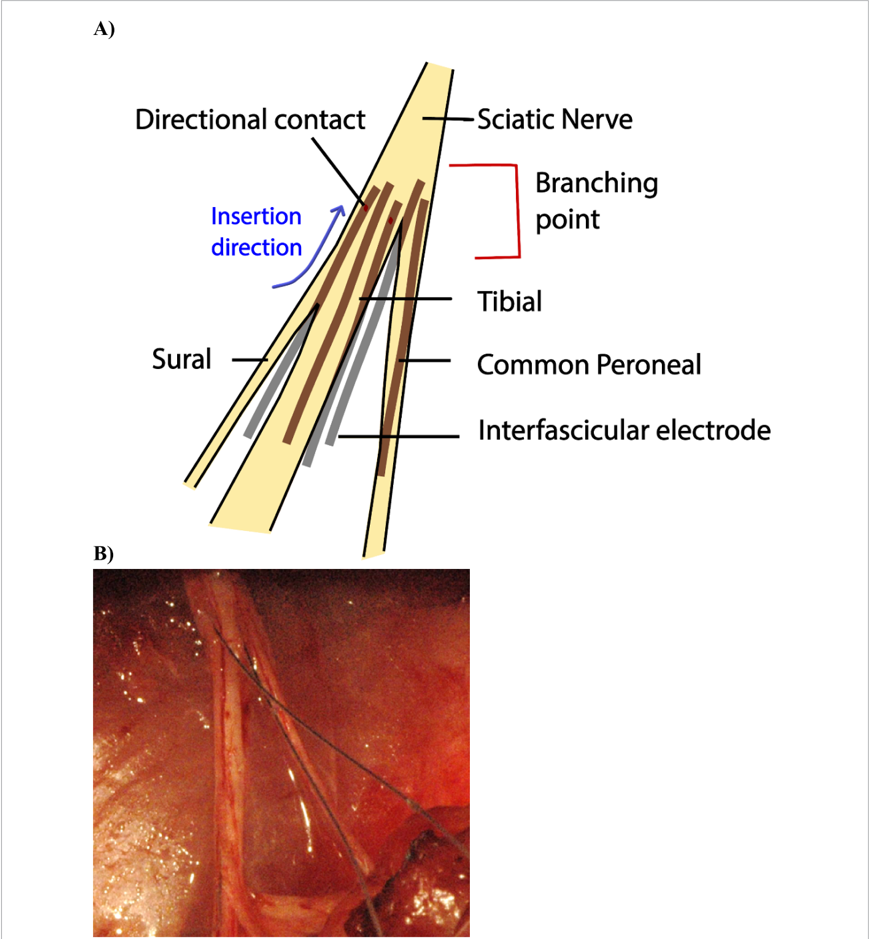
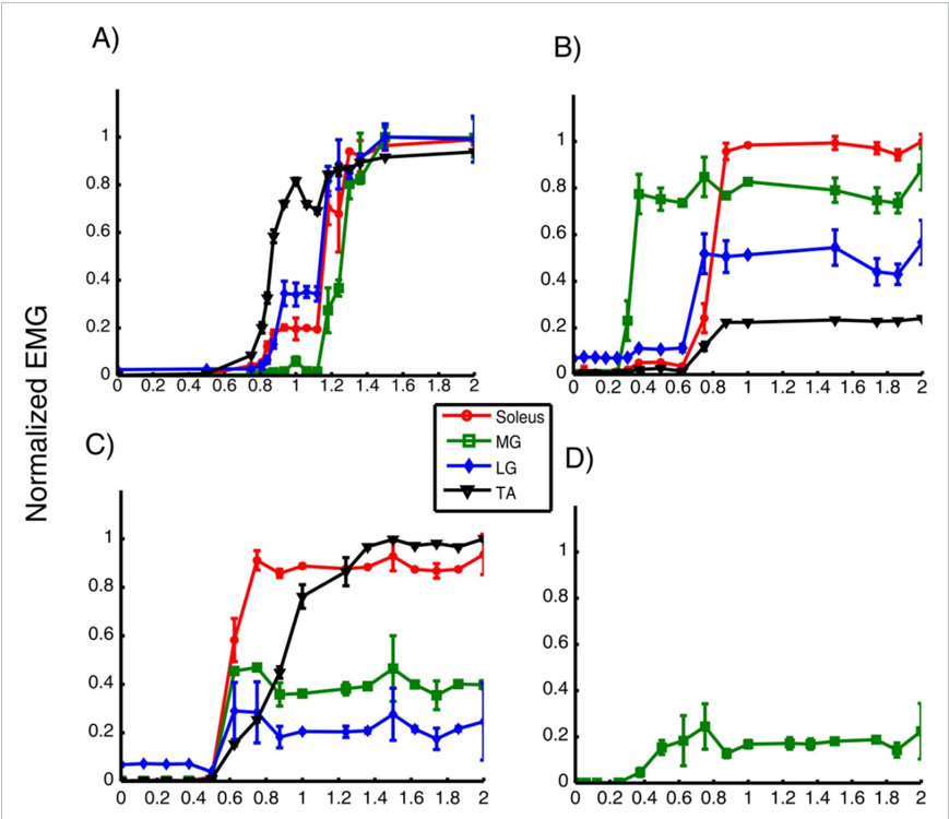
- In a trial with six longitudinally inserted interfascicular contacts, the overall selectivity was Λ = 15/15 = 1.
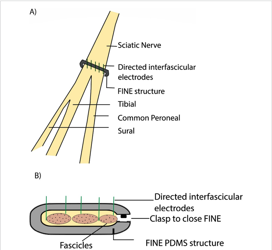
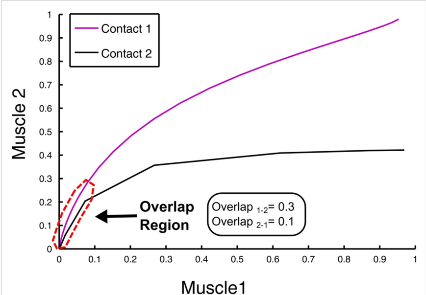
- In a trial with five transverse contacts, 4 of 5 contacts selectively recruited different axon groups; overall selectivity was Λ = 6/10 = 0.6, and after removing redundant Channel 5 it became Λ = 6/6 = 1.0.
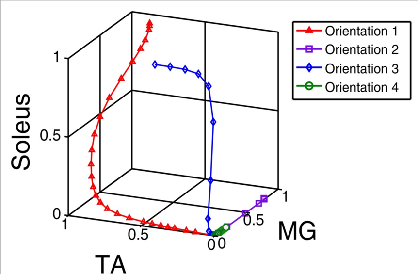
- For a single interfascicular electrode tested at four orientations, all orientations had less than 50% overlap and overall selectivity was Λ = 6/6 = 1.0.
- Across 32 trials of single wire electrodes, the average axonal selectivity was Λ = 0.8 ± 0.3, with 4.75 ± 0.8 selective orientations per wire; for 34 longitudinal trials the average was 0.85 ± 0.2 with 6.53 ± 1.03 contacts, and for 34 transverse trials it was 0.73 ± 0.3 with 5 contacts.
## Methods
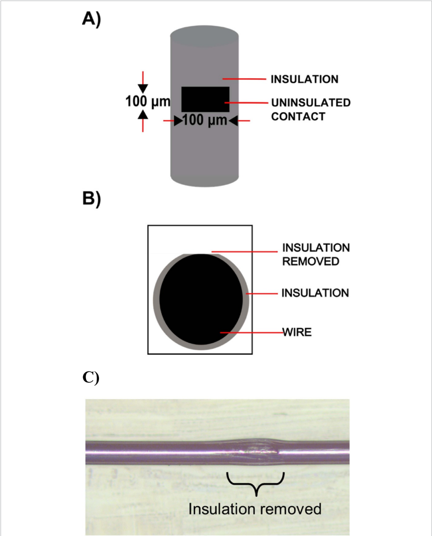
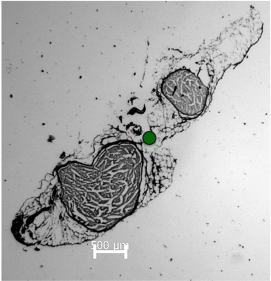
- Design: Feasibility experiment in rabbit sciatic nerves comparing three interfascicular interface configurations: single directed contact, multiple longitudinal contacts, and multiple transverse contacts mounted on a FINE structure.
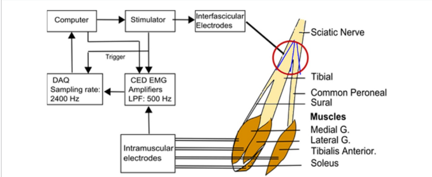
- Sample/data: 32 New Zealand rabbits; tabled trials included 32 single-wire trials from 14 animals, 34 longitudinal trials from 14 animals, and 34 transverse trials from 12 animals. EMG was recorded from lateral gastrocnemius, medial gastrocnemius, soleus, and tibialis anterior using intramuscular wire electrodes.
- Analysis: Stimulation used pulse-amplitude-modulated biphasic pulses; recruitment curves were summarized as EMG activation and compared using overlap of EMG recruitment trajectories. Overall selectivity was defined as the percentage of contact pairs with <50% overlap.
## Limitations
- The paper states that histology did not confirm that the transverse configuration did not penetrate the perineurium, though the authors say this is a reasonable assumption.
- The study was done in a rabbit sciatic nerve with 2–3 fascicles in the popliteal fossa region, so the text itself indicates limited immediate generalizability to larger, more fasciculated human nerves.
- The paper notes that chronic studies are necessary; the reported work is an initial feasibility study, not a long-term implant evaluation.
Model-written summary. Check it against the paper before relying on it.
Integrity signals
Registry
Cites
Statistics
Figures
Verification
Claims the paper makes
8 Valid · 1 Not valid · 28 Not checked · 63 Own results and interpretations
Every source the paper cites for this claim was checked and supports it.
In the paper
“These interfaces have been used clinically for fascicleselective stimulation [13, 14].”
What the sources say
- Supportedcites [13] Freeberg, 2017, The design of and chronic tissue response to a composite nerve electrode with patterned stiffness, J. Neural Eng. source
The Introduction (page not provided) describes non-penetrating nerve cuff electrodes placed around nerves and says they can selectively stimulate target fascicles. It also reports that the selectivity of the flat interface nerve electrode was demonstrated through motor responses to stimulation in human subjects, supporting the claim of clinical use. (LLM, conf=0.88)
- Supportedcites [14] Christie, 2017, Long-term stability of stimulating spiral nerve cuff electrodes on human peripheral nerves, J. Neuroeng. Rehabil. source
The Background (page unavailable) describes spiral nerve cuffs as non-penetrating electrodes implanted in humans and says their contacts can activate different fascicles. The Results, under “Percent overlap of recruited motor unit populations” (page unavailable), report selective activation of different motor populations by implanted cuffs on human femoral nerves. (LLM, conf=0.88)
plain- Asserted: passes — The authors put it forward as true.
- Checkable: passes — It can be checked against evidence.
- Load-bearing: unclear — It is unclear whether the paper’s conclusions depend on it.
- Based: passes — The paper gives a reason why it holds.
- Supported
The cited sources support only part of this claim — read each source’s verdict below.
In the paper
“The human nerve, however, has many more fascicles than the pre-clinical animal models [26].”
What the sources say
- Partly supportedcites [26] Brill, 2017, Quantification of human upper extremity nerves and fascicular anatomy, Muscle Nerve sourceExperimental — accuracy not yet measured
The Results (Fascicular Characterization; page unavailable) quantify fascicle counts in human upper-extremity nerves. The Discussion (Implications for Neural Interface Design; page unavailable) says preclinical animal nerve structures differ significantly from human ones, but does not say that animal models have fewer fascicles. (LLM, conf=0.83)
plain- Asserted: passes — The authors put it forward as true.
- Checkable: passes — It can be checked against evidence.
- Load-bearing: passes — The paper’s conclusions depend on it.
- Based: passes — The paper gives a reason why it holds.
- Partly supported
Every source the paper cites for this claim was checked and supports it.
In the paper
“The threshold for activation of a smaller fascicle is generally lower than a larger fascicle [51].”
What the sources say
- Supportedcites [51] Grinberg, 2008, Fascicular perineurium thickness, size, and position affect model predictions of neural excitation, IEEE Trans. Neural Syst. Rehabil. Eng. source
The Discussion, section IV.B (page not supplied), states that small fascicles had lower activation thresholds than large fascicles when modeled with physiologic perineurium thickness. It also describes large fascicles as more difficult to stimulate, supporting the claim’s general wording. (LLM, conf=0.93)
plainstates an association- Asserted: passes — The authors put it forward as true.
- Checkable: passes — It can be checked against evidence.
- Load-bearing: passes — The paper’s conclusions depend on it.
- Based: passes — The paper gives a reason why it holds.
- Supported
Every source the paper cites for this claim was checked and supports it.
In the paper
“This shunting can cause the current from an extraneural source to be directed away from a larger, more resistive fascicle toward a smaller, less resistive fascicle [51].”
What the sources say
- Supportedcites [51] Grinberg, 2008, Fascicular perineurium thickness, size, and position affect model predictions of neural excitation, IEEE Trans. Neural Syst. Rehabil. Eng. source
The Discussion, section IV.C, proposes that current may be shunted away from a large, more resistive fascicle toward a smaller, less resistive neighbor. The claim’s “can” preserves the paper’s framing of shunting as a possible mechanism, rather than an established one. (LLM, conf=0.91)
plainstates a cause- Asserted: passes — The authors put it forward as true.
- Checkable: passes — It can be checked against evidence.
- Load-bearing: passes — The paper’s conclusions depend on it.
- Based: passes — The paper gives a reason why it holds.
- Supported
Every source the paper cites for this claim was checked and supports it.
In the paper
“This is because the size of the perineurium is directly proportional to the size of the fascicle.”
What the sources say
- Supportedcites [51] Grinberg, 2008, Fascicular perineurium thickness, size, and position affect model predictions of neural excitation, IEEE Trans. Neural Syst. Rehabil. Eng. source
The Results (section III.A; page unavailable) report that perineurial thickness averaged 3.0% of fascicle diameter and that a linear fit through zero was 2.6%. This supports the stated proportional relationship, though the measured ratio varied among fascicles. (LLM, conf=0.90)
plainstates an association- Asserted: passes — The authors put it forward as true.
- Checkable: passes — It can be checked against evidence.
- Load-bearing: passes — The paper’s conclusions depend on it.
- Based: passes — The paper gives a reason why it holds.
- Supported
56 more claims
Create a free account to see all 100 claims and every source verdict.
- It may state two separate claims as one. 20
- Probably not a substantial claim. 26
- The quote only partly states this claim. 1
Citations
Cites
Functional Electrical Stimulation for Neuromuscular Applications
Peckham, P. H., & Knutson, J. S. (2005) · Annual Review of Biomedical Engineering · doi:10.1146/annurev.bioeng.6.040803.140103 · Cited by 784 · cited 1×
“Electrical currents can elicit a response in nerves that are faithfully transmitted to their respective end organs, which respond as if the signal was generated naturally by the body, enabling artificial control of paralyzed musculature [1].”
BackgroundMotor Units: Anatomy, Physiology, and Functional Organization
Burke, R. E. (1981) · Comprehensive Physiology · doi:10.1002/cphy.cp010210 · Cited by 782 · cited 1×
“The gastrocnemii and TA muscles consist of ‘fast-twitch’ fibers primarily [49], that are usually innervated by axons with higher conduction velocities [50].”
BackgroundA neural interface provides long-term stable natural touch perception
Tan, D. W., Schiefer, M. A., Keith, M. W., Anderson, J. R., Tyler, J., & Tyler, D. J. (2014) · Science Translational Medicine · doi:10.1126/scitranslmed.3008669 · Cited by 751
Self-citationLundborg, G., & Rydevik, B. (1973) · The Journal of Bone and Joint Surgery. British volume · doi:10.1302/0301-620x.55b2.390 · Cited by 408 · cited 1×
“Complications due to changes in the endoneurial environment include increased endoneurial pressure, nerve fiber compression, variable shifts in thresholds and loss of axons [29, 30].”
BackgroundLundborg, G., & Rydevik, B. L. (1973) · PubMed · Cited by 403
Functionally selective peripheral nerve stimulation with a flat interface nerve electrode
Tyler, D. J., & Durand, D. M. (2002) · IEEE Transactions on Neural Systems and Rehabilitation Engineering · doi:10.1109/tnsre.2002.806840 · Cited by 398
Self-citationTopp, K. S., & Boyd, B. S. (2006) · Physical Therapy · doi:10.1093/ptj/86.1.92 · Cited by 361 · cited 1×
“In contrast to the highly structured and tight perineurium, the epineurium, the outermost tissue, is a loosely organized collagenous tissue whose main function is generally thought to maintain the structural integrity of the nerve [9, 10].”
Background(1963) · PubMed · Cited by 310
Long-term stimulation and recording with a penetrating microelectrode array in cat sciatic nerve
Branner, A., Stein, R. B., Fernandez, E., Aoyagi, Y., & Normann, R. A. (2004) · IEEE Transactions on Biomedical Engineering · doi:10.1109/tbme.2003.820321 · Cited by 301
Stimulus waveforms for selective neural stimulation
Grill, W. M., & Mortimer, J. T. (1995) · IEEE Engineering in Medicine and Biology Magazine · doi:10.1109/51.395310 · Cited by 280
Cited by
Koppaka, S., Hess‐Dunning, A., & Tyler, D. J. (2022) · Journal of Biomechanics · doi:10.1016/j.jbiomech.2022.111058 · Cited by 21
Self-citationPellot-Cestero, J. E., Herring, E. Z., Graczyk, E. L., Memberg, W. D., Kirsch, R. F., Ajiboye, A. B., & Miller, J. P. (2023) · Neurosurgery · doi:10.1227/neu.0000000000002561 · Cited by 11
“46”
Akouissi, O., Lacour, S. P., Micera, S., & DeSimone, A. (2022) · Journal of Neural Engineering · doi:10.1088/1741-2552/ac7d0e · Cited by 7
Multi-Channel Microscale Nerve Cuffs for Spatially Selective Neuromodulation
Riley, M., Tala, F., Johnson, K. J., & Johnson, B. C. (2024) · Micromachines · doi:10.3390/mi15081036 · Cited by 6
“…intensity varies with distance, where fascicles deeper in the nerve receive less stimulation than fascicles closer to the electrode interface [9,10]; and (3) the stimulation threshold varies with fiber diameter, where larger-diameter fibers have a lower threshold than smaller-diameter fibers…”
BackgroundPetriv, T. I., QR Health Solutions, K., Almhairat, R. M. D., Vorodi, M. V., Luzan, B., Tsymbaliuk, Y., Vaslovych, V. V., Малышева, Т. А., & Tsymbaliuk, V. (2023) · Cell and Organ Transplantology · doi:10.22494/cot.v11i1.151
Metadata
- Authors
- Koppaka, S., Hess-Dunning, A., & Tyler, D. J.
- Affiliations
- Louis Stokes Cleveland VA Medical Center (US)
- Corresponding author
- Tyler, Dustin J
- Venue
- Journal of Neural Engineering
- Publisher
- IOP Publishing
- Published
- 2021-11-12
- Volume
- 18
- Issue
- 6
- Pages
- 066006
- ISSN
- 1741-2552 (ISSN-L)
- 1741-2560
- OpenAlex
- W3208515159
- Cited by
- 7
- References
- 63
- Citations by year
- 2025: 2 · 2024: 1 · 2023: 2 · 2022: 2
- FWCI
- 0.58
- Type
- journal article
- Language
- en
- Open access
- hybrid
- doi.org
- Licence
- cc-by
- creativecommons.org/licenses/by/4.0 (vor)
- iopscience.iop.org/info/page/text-and-data-mining (tdm)
- Locations
- Journal of Neural Engineering (publishedVersion, cc-by)
- PubMed (publishedVersion)
- Funders
- Defense Advanced Research Projects Agency (HR0011-10-1-0074)
- Topics
- Life Sciences › Neuroscience › Cellular and Molecular Neuroscience › Neuroscience and Neural Engineering
- Life Sciences › Neuroscience › Neurology › Vagus Nerve Stimulation Research
- Physical Sciences › Engineering › Biomedical Engineering › Muscle activation and electromyography studies
- Keywords
- peripheral nerve stimulation, sciatic nerve, perineurium, muscle electrical activity, neuromodulation, rabbit model
- MeSH terms
- Animals, Axons, Electric Stimulation, Transcutaneous Electric Nerve Stimulation, Electrodes, Implanted, Peripheral Nerves, Rabbits, Sciatic Nerve, Mice
About these checks
Registry, retraction, Cites and statistics results are computed, not judged, with one exception: a printed reference that no index lists is read once by a model before it counts as a citation error. Claims are extracted by a model and kept only when their quote appears verbatim in the text; each cited source is then checked, with a model’s help, for whether it says what the claim relies on. Those verdicts can be wrong — read the claim’s quote and the cited source before relying on one.
The claim verdicts this report counts are experimental: their accuracy is not yet measured.
A language model drew the claims from the paper on 2026-10-11, under version 3 of the claim definition (model deepseek-v4.1-flash, prompt extract-v3).
A second model labelled each claim on 2026-10-11 (model jev-1.13-20260917, definition revision gold-guideline-v0.2).
Cited sources are checked with a language model’s help (gpt-6-sol).
Statistics and arithmetic are recomputed, not judged.
Confidence bands are shown only for fields calibrated on our annotated test set. None is calibrated yet, so no band is shown.
What counts as a claim ›How we measure confidence ›How we check this ›
Are you an author, or do you see a problem with this page? Request a review
How to cite
Cite CoChat for the checks on this page, and the paper for its own findings.
- This page
- CoChat. (2026). Citation check: Directed stimulation with interfascicular interfaces for peripheral nerve stimulation. https://app.cochat.ai/papers/10.1088/1741-2552/ac33e8
- The paper
- Koppaka, S., Hess-Dunning, A., & Tyler, D. J. (2021). Directed stimulation with interfascicular interfaces for peripheral nerve stimulation. Journal of Neural Engineering. https://doi.org/10.1088/1741-2552/ac33e8
Data sources
Bibliographic data: Semantic Scholar, OpenAlex, Crossref.
Contains information from the Semantic Scholar Academic Graph Datasets, made available under the ODC Attribution License (ODC-BY 1.0).
Semantic Scholar’s data is described in: Kinney, R., et al. (2023). The Semantic Scholar Open Data Platform. arXiv:2301.10140.
