

Mitochondrial defects in acute multiple sclerosis lesions
Open accessThe DOI matches its Crossref record; 50 of the 50 references it cites were found in a registry or an index.
Abstract
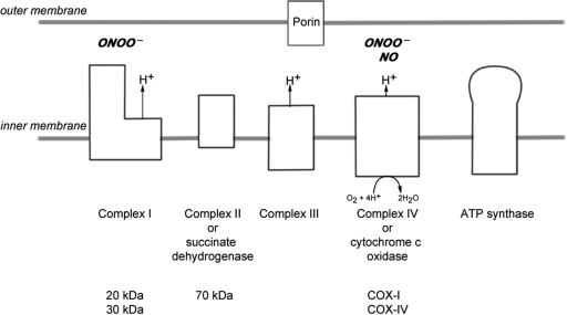
Multiple sclerosis is a chronic inflammatory disease, which leads to focal plaques of demyelination and tissue injury in the CNS. The structural and immunopathological patterns of demyelination suggest that different immune mechanisms may be involved in tissue damage. In a subtype of lesions, which are mainly found in patients with acute fulminant multiple sclerosis with Balo's type concentric sclerosis and in a subset of early relapsing remitting multiple sclerosis, the initial myelin changes closely resemble those seen in white matter stroke (WMS), suggesting a hypoxia-like tissue injury. Since mitochondrial injury may be involved in the pathogenesis of such lesions, we analysed a number of mitochondrial respiratory chain proteins in active lesions from acute multiple sclerosis and from WMS using immunohistochemistry. Functionally important defects of mitochondrial respiratory chain complex IV [cytochrome c oxidase (COX)] including its catalytic component (COX-I) are present in Pattern III but not in Pattern II multiple sclerosis lesions. The lack of immunohistochemically detected COX-I is apparent in oligodendrocytes, hypertrophied astrocytes and axons, but not in microglia. The profile of immunohistochemically detected mitochondrial respiratory chain complex subunits differs between multiple sclerosis and WMS. The findings suggest that hypoxia-like tissue injury in Pattern III multiple sclerosis lesions may be due to mitochondrial impairment.
Overview
The claims and statistics of this paper haven’t been checked yet.
Free: it reads the open-access text and checks the first 5 claims that cite a source we can check. Create a free account to check every claim.
Integrity signals
Registry
Cites
Statistics
Figures


Verification
Claims the paper makes
The claims and statistics of this paper haven’t been checked yet.
Free: it reads the open-access text and checks the first 5 claims that cite a source we can check. Create a free account to check every claim.
Claims other papers make about this one
Each is checked against this paper’s own text: does it say what the citing paper says it does?
The source backs part of the claim, not all of it — or backs it for a different population, condition or comparison.
In the citing paper
“Oxidative damage and mitochondrial dysfunction are key features of MS pathology [277,278,279,280].”
From Ageing, Cellular Senescence and Neurodegenerative Disease (2018) · cites this paper as [279]
Experimental — accuracy not yet measuredThe source backs part of the claim, not all of it — or backs it for a different population, condition or comparison.
In the citing paper
“Other features common to both cuprizone-induced and type 3 MS lesions are mitochondrial stress, selective loss of myelin-associated glycoprotein (MAG), lesions with indistinct borders that are not perivascular, pronounced glial activation, and lymphocyte infiltration [43, 46, 65, 69–71].”
From Oligodendrocyte death and myelin loss in the cuprizone model: an updated overview of the intrinsic and extrinsic causes of cuprizone demyelination (2022) · cites this paper as [69]
Experimental — accuracy not yet measured
Self-check
Percentages, sums and reported differences re-computed from the paper’s own numbers.
The self-check needs the paper’s full text.
Citations
Cites
Mitochondrial DNA mutations in human disease
Taylor, R. W., & Turnbull, D. M. (2005) · Nature Reviews Genetics · doi:10.1038/nrg1606 · Cited by 1,852 · cited 5×
“…c oxidase Abbreviations: CNPase=cyclic nucleotide phosphodiesterase; COX=cytochrome c oxidase; LFB=Luxol fast blue; MAG=myelin associated glycoprotein; MBP=myelin basic protein; mtDNA=mitochondrial DNA; NO=nitric oxide; NWM=normal white matter; PLP=proteolipid protein; WMS=white matter stroke”
BackgroundMethodInfluentialSelf-citationMultiple Sclerosis — The Plaque and Its Pathogenesis
Frohman, E. M., Racke, M. K., & Raine, C. S. (2006) · New England Journal of Medicine · doi:10.1056/nejmra052130 · Cited by 1,740 · cited 1×
“Multiple sclerosis is an inflammatory demyelinating disease of the central nervous system with axonal degeneration and astrogliosis (Frohman et al., 2006).”
BackgroundMitochondrial Respiratory-Chain Diseases
DiMauro, S., & Schon, E. A. (2003) · New England Journal of Medicine · doi:10.1056/nejmra022567 · Cited by 1,661 · cited 5×
“Keywords: multiple sclerosis; Pattern III lesion; mitochondria; cytochrome c oxidase Abbreviations: CNPase=cyclic nucleotide phosphodiesterase; COX=cytochrome c oxidase; LFB=Luxol fast blue; MAG=myelin associated glycoprotein; MBP=myelin basic protein; mtDNA=mitochondrial DNA; NO=nitric oxide;…”
BackgroundInfluentialvon Büdingen, H.-C., Palanichamy, A., Lehmann-Horn, K., Michel, B. A., & Zamvil, S. S. (2015) · European Neurology · doi:10.1159/000377675 · Cited by 1,463
Cleeter, M. W. J., Cooper, J. M., Darley-Usmar, V. M., Moncada, S., & Schapira, A. H. V. (1994) · FEBS Letters · doi:10.1016/0014-5793(94)00424-2 · Cited by 1,241 · cited 1×
“NO competitively inhibits the binding of oxygen to complex IV (Brown and Cooper, 1994; Cleeter et al., 1994).”
BackgroundHIF-1 Regulates Cytochrome Oxidase Subunits to Optimize Efficiency of Respiration in Hypoxic Cells
Fukuda, R., Zhang, H., Kim, J.-W., Shimoda, L., Dang, C. V., & Semenza, G. L. (2007) · Cell · doi:10.1016/j.cell.2007.01.047 · Cited by 1,225 · cited 5×
“Widespread defects of mitochondrial respiratory chain complexes inWMS lesions The transcription of mtDNA and nuclear DNA encoded subunits of the mitochondrial respiratory chain complexes are modulated by hypoxia (Vijayasarathy et al., 2003; Piruat and Lopez-Barneo, 2005; Fukuda et al., 2007).”
BackgroundInfluentialMicrodomains of Intracellular Ca2+: Molecular Determinants and Functional Consequences
Rizzuto, R., & Pozzan, T. (2006) · Physiological Reviews · doi:10.1152/physrev.00004.2005 · Cited by 1,206 · cited 1×
“Mitochondria also play an important role in apoptosis and calcium homeostasis (Rizzuto and Pozzan, 2006; Kalman et al., 2007).”
BackgroundRelapsing and remitting multiple sclerosis: Pathology of the newly forming lesion
Barnett, M. H., & Prineas, J. W. (2004) · Annals of Neurology · doi:10.1002/ana.20016 · Cited by 1,185 · cited 5×
“Mitochondrial defects in acute multiple sclerosis lesions Don Mahad,1 Iryna Ziabreva,1Hans Lassmann2 and DouglasTurnbull1 1The Mitochondrial Research Group, University of Newcastle uponTyne, UK and 2Centre for Brain Research, Medical University of Vienna, Austria Correspondence to: Prof. Dr Hans Lassmann, Center for B…”
BackgroundInfluentialBrown, G. C., & Cooper, C. E. (1994) · FEBS Letters · doi:10.1016/0014-5793(94)01290-3 · Cited by 1,055 · cited 2×
“…c oxidase Abbreviations: CNPase=cyclic nucleotide phosphodiesterase; COX=cytochrome c oxidase; LFB=Luxol fast blue; MAG=myelin associated glycoprotein; MBP=myelin basic protein; mtDNA=mitochondrial DNA; NO=nitric oxide; NWM=normal white matter; PLP=proteolipid protein; WMS=white matter stroke”
BackgroundSystemic infections and inflammation affect chronic neurodegeneration
Perry, V. H., Cunningham, C., & Holmes, C. (2007) · Nature Reviews Immunology · doi:10.1038/nri2015 · Cited by 1,027 · cited 1×
“These activated microglia, found in close proximity to apoptotic oligodendrocytes, may release a range of mediators (Barnett and Prineas, 2004; Perry et al., 2007).”
Background
Cited by
Items 201–300 of 424
Michaličková, D., Šíma, M., & Slanař, O. (2020) · Physiological Research · doi:10.33549/physiolres.934276 · Cited by 37
Iron Heterogeneity in Early Active Multiple Sclerosis Lesions
Tham, M., Frischer, J. M., Weigand, S. D., Fitz‐Gibbon, P. D., Webb, S. M., Guo, Y., Adiele, R. C., Robinson, C. A., Brück, W., Lassmann, H., et al. (2021) · Annals of Neurology · doi:10.1002/ana.25974 · Cited by 35
Self-citationOmotoso, G. O., Olajide, O. J., Gbadamosi, I., Adebayo, J. O., Enaibe, B. U., Akinola, O. B., & Owoyele, B. V. (2019) · Heliyon · doi:10.1016/j.heliyon.2019.e02102 · Cited by 35
Main Role of Antibodies in Demyelination and Axonal Damage in Multiple Sclerosis
Muñoz, Ú., Sebal, C., Escudero, E., Esiri, M. M., Tzartos, J. S., Sloan, C., & Sádaba, M. C. (2021) · Cellular and Molecular Neurobiology · doi:10.1007/s10571-021-01059-6 · Cited by 34
Iron in multiple sclerosis – Neuropathology, immunology, and real-world considerations
Zierfuss, B., Wang, Z., Jackson, A. N., Moezzi, D., & Yong, V. W. (2023) · Multiple Sclerosis and Related Disorders · doi:10.1016/j.msard.2023.104934 · Cited by 35
Wilms, H., Arnold, P., Mojumder, D. K., DeToledo, J. C., & Lucius, R. (2014) · Clinical Pharmacology Advances and Applications · doi:10.2147/cpaa.s35033 · Cited by 35
Praet, J., Orije, J. E. M. J., Kara, F., Guglielmetti, C., Santermans, E., Daans, J., Hens, N., Verhoye, M., Berneman, Z. N., Ponsaerts, P., et al. (2015) · NMR in Biomedicine · doi:10.1002/nbm.3277 · Cited by 35
Oxysterols and multiple sclerosis: Physiopathology, evolutive biomarkers and therapeutic strategy
Véjux, A., Ghzaiel, I., Nury, T., Schneider, V., Charrière, K., Sghaier, R., Zarrouk, A., Leoni, V., Moreau, T., & Lizard, G. (2021) · The Journal of Steroid Biochemistry and Molecular Biology · doi:10.1016/j.jsbmb.2021.105870 · Cited by 32
Proteomic Analysis of Cerebrospinal Fluid in a Fulminant Case of Multiple Sclerosis
Füvesi, J., Hanrieder, J., Bencsik, K., Rajda, C., Kovács, S. K., Kaizer, L., Beniczky, S., Vécsei, L., & Bergquist, J. (2012) · International Journal of Molecular Sciences · doi:10.3390/ijms13067676 · Cited by 34
Oxidative injury in multiple sclerosis cerebellar grey matter
Kemp, K. C., Redondo, J., Hares, K. M., Rice, C. M., Scolding, N. J., & Wilkins, A. (2016) · Brain Research · doi:10.1016/j.brainres.2016.04.027 · Cited by 32
“The role of GM injury in MS has received less attention than WM injury, yet pathological studies of MS GM have revealed evidence for extensive demyelination, inflammation and neuronal loss (Kutzelnigg et al., 2005; Lucchinetti et al., 2011; Mahad et al., 2008).”
Impact of Exercise on Immunometabolism in Multiple Sclerosis
Afzal, R., Dowling, J. K., & McCoy, C. E. (2020) · Journal of Clinical Medicine · doi:10.3390/jcm9093038 · Cited by 32
Glucose metabolic reprogramming in autoimmune diseases
Jeong, H., Lee, B., Han, S. J., & Sohn, D. H. (2023) · Animal Cells and Systems · doi:10.1080/19768354.2023.2234986 · Cited by 33
“Defects in the mitochondrial respiratory chain complex IV, which are represented by the loss of cytochrome c oxidase-1 and 4, are observed in acute MS lesions (Mahad et al. 2008).”
BackgroundSu, K. G., Savino, C., Marracci, G., Chaudhary, P., Yu, X., Morris, B., Galipeau, D., Giorgio, M., Forte, M., & Bourdette, D. (2012) · European Journal of Neuroscience · doi:10.1111/j.1460-9568.2011.07972.x · Cited by 28
Fourcade, S., Goicoechea, L., Parameswaran, J., Schlüter, A., Launay, N., Ruíz, M., Seyer, A., Colsch, B., Calingasan, N. Y., Ferrer, I., et al. (2020) · Brain Pathology · doi:10.1111/bpa.12869 · Cited by 30
Singh, I., Samuvel, D. J., Choi, S., Saxena, N., Singh, A. K., & Won, J. (2018) · Immunology · doi:10.1111/imm.12893 · Cited by 29
Ineichen, B., Zhu, K.-Y., & Carlström, K. E. (2020) · Journal of Neuroscience Research · doi:10.1002/jnr.24767 · Cited by 21
Further understanding of the immunopathology of multiple sclerosis: impact on future treatments
Bhise, V. V., & Dhib-Jalbut, S. S. (2016) · Expert Review of Clinical Immunology · doi:10.1080/1744666x.2016.1191351 · Cited by 29
“Thus, it is no surprise to see a significant loss of complex IV and cytochrome c oxidase-I in the mitochondrial respiratory chain in inflammatory lesions [42].”
BackgroundRao, V. T. S., Khan, D., Jones, R. G., Nakamura, D. S., Kennedy, T. E., Cui, Q., Rone, M. B., Healy, L. M., Watson, R. L., Ghosh, S., et al. (2016) · Scientific Reports · doi:10.1038/srep30020 · Cited by 28
Biller, A., Pflugmann, I., Badde, S., Diem, R., Wildemann, B., Nagel, A. M., Jordan, J., Benkhedah, N., & Kleesiek, J. (2016) · Scientific Reports · doi:10.1038/srep31269 · Cited by 28
Emamnejad, R., Pagnin, M., & Petratos, S. (2024) · Neuroscience & Biobehavioral Reviews · doi:10.1016/j.neubiorev.2024.105788 · Cited by 30
“MS patients exhibit widespread mitochondrial damage, leading to increased reactive oxygen species production that can worsen iron-induced oxidative damage, causing progressive genomic damage and further impairing mitochondrial function (Yakes et al., 1997; Mahad et al., 2008).”
P2x7 receptors control demyelination and inflammation in the cuprizone model
Bernal‐Chico, A., Manterola, A., Cipriani, R., Katona, I., Matute, C., & Mato, S. (2020) · Brain Behavior & Immunity - Health · doi:10.1016/j.bbih.2020.100062 · Cited by 28
Imaging Axonal Degeneration and Repair in Preclinical Animal Models of Multiple Sclerosis
Yandamuri, S. S., & Lane, T. E. (2016) · Frontiers in Immunology · doi:10.3389/fimmu.2016.00189 · Cited by 27
Kauv, P., Ayache, S. S., Créange, A., Chalah, M. A., Lefaucheur, J., Hodel, J., & Brugières, P. (2017) · European Neurology · doi:10.1159/000475496 · Cited by 27
Anterior visual system imaging to investigate energy failure in multiple sclerosis
Kleerekooper, I., Petzold, A., & Trip, S. A. (2020) · Brain · doi:10.1093/brain/awaa049 · Cited by 27
Metabolic response to glatiramer acetate therapy in multiple sclerosis patients
Riccardis, L. D., Ferramosca, A., Danieli, A., Trianni, G., Zara, V., Robertis, F. D., & Maffia, M. (2016) · BBA Clinical · doi:10.1016/j.bbacli.2016.10.004 · Cited by 27
“under the CC BY-NC dysfunctions contribute to neurological disorders [4,5], supporting the role of mitochondria as a potential therapeutic target in MS [6].”
Autoimmune encephalitis in humans: how closely does it reflect multiple sclerosis ?
Höftberger, R., Leisser, M., Bauer, J., & Lassmann, H. (2015) · Acta Neuropathologica Communications · doi:10.1186/s40478-015-0260-9 · Cited by 27
Self-citationCui, Q., Lin, Y. H., Xu, Y. K. T., Fernandes, M. G. F., Rao, V. T. S., Kennedy, T. E., & Antel, J. P. (2020) · PLoS ONE · doi:10.1371/journal.pone.0233859 · Cited by 25
Increased microglial catalase activity in multiple sclerosis grey matter
Gray, E., Kemp, K. C., Hares, K. M., Redondo, J., Rice, C. M., Scolding, N. J., & Wilkins, A. (2014) · Brain Research · doi:10.1016/j.brainres.2014.02.042 · Cited by 26
Analysis of shared underlying mechanism in neurodegenerative disease
Butler, R., Bradford, D. R., & Rodgers, K. E. (2022) · Frontiers in Aging Neuroscience · doi:10.3389/fnagi.2022.1006089 · Cited by 24
Is there a role for herpes simplex virus type 1 in multiple sclerosis?
Duarte, L. F., Gatica, S., Castillo, A., Kalergis, A. M., Bueno, S. M., Riedel, C. A., & González, P. A. (2022) · Microbes and Infection · doi:10.1016/j.micinf.2022.105084 · Cited by 24
Mechanisms Governing Oligodendrocyte Viability in Multiple Sclerosis and Its Animal Models
Lei, Z., & Lin, W. (2024) · Cells · doi:10.3390/cells13020116 · Cited by 25
“Notably, evidence suggests that inflammation-induced mitochondrial damage contributes to oligodendrocyte apoptosis in MS and EAE [84–86 , 93– 95 ].”
BackgroundRemyelinating strategies in multiple sclerosis
Luessi, F., Kuhlmann, T., & Zipp, F. (2014) · Expert Review of Neurotherapeutics · doi:10.1586/14737175.2014.969241 · Cited by 25
Hnilicová, P., Štrbák, O., Kolísek, M., Kurča, E., Zeleňák, K., Sivák, Š., & Kantorová, E. (2020) · International Journal of Molecular Sciences · doi:10.3390/ijms21176117 · Cited by 24
Kallab, M., Hommer, N., Schlatter, A., Bsteh, G., Altmann, P., Popa‐Cherecheanu, A., Pfister, M., Werkmeister, R. M., Schmidl, D., Schmetterer, L. F., et al. (2021) · Frontiers in Neuroscience · doi:10.3389/fnins.2021.761654 · Cited by 23
Al‐Kafaji, G., Bakheit, H. F., Alali, F., Fattah, M., Alhajeri, S., Alharbi, M. A., Daif, A., Alsabbagh, M. M., Alwehaidah, M. S., & Bakhiet, M. (2022) · PLoS ONE · doi:10.1371/journal.pone.0263606 · Cited by 24
Francesco, M. A. D. (2024) · Viruses · doi:10.3390/v16010133 · Cited by 22
“To support this hypothesis, different studies have shown the presence of T cells and antibodies able to cross-react with the HSV-1 protein UL15, with the antigen myelin basic protein, and with the HSV-1 glycoprotein B and a brain epitope, respectively [150,151].”
BackgroundA Hypoxia-Inflammation Cycle and Multiple Sclerosis: Mechanisms and Therapeutic Implications
Soroush, A., & Dunn, J. F. (2024) · Current Treatment Options in Neurology · doi:10.1007/s11940-024-00816-4 · Cited by 23
Demyelination and neurodegeneration in multiple sclerosis: The role of hypoxia
Lassmann, H. (2016) · Annals of Neurology · doi:10.1002/ana.24632 · Cited by 23
Self-citationThe Na+/Ca2+ exchangers in demyelinating diseases
Boscia, F., de Rosa, V., Cammarota, M., Secondo, A., Pannaccione, A., & Annunziato, L. (2019) · Cell Calcium · doi:10.1016/j.ceca.2019.102130 · Cited by 22
Imaging multiple sclerosis and other neurodegenerative diseases
Inglese, M., & Petracca, M. (2013) · Prion · doi:10.4161/pri.22650 · Cited by 23
“In active MS lesions mitochondrial density is increased in axons and astrocytes while complex IV electron transport chains activity is decreased.(19) In MS chronic lesions intra axonal mitochondrial density and complex IV activity are both increased to supply the major metabolic demand of demyelinated axons.”
BackgroundYao, S., Pandey, P., Ljunggren‐Rose, A., & Sriram, S. (2009) · Nitric Oxide · doi:10.1016/j.niox.2009.12.001 · Cited by 22
The complexities of investigating mitochondria dynamics in multiple sclerosis and mouse models of MS
Atkinson, K. C., Osunde, M., & Tiwari-Woodruff, S. K. (2023) · Frontiers in Neuroscience · doi:10.3389/fnins.2023.1144896 · Cited by 21
“To observe mitochondria expression and function in distinct cell types IHC is still widely utilized.”
BackgroundInfluentialMitochondrial complex IV is lost in neurons in the cuprizone mouse model
Varhaug, K. N., Kråkenes, T., Alme, M. N., Vedeler, C. A., & Bindoff, L. A. (2019) · Mitochondrion · doi:10.1016/j.mito.2019.09.003 · Cited by 23
Current Concepts in the Neuropathology and Pathogenesis of Multiple Sclerosis
Moore, G. R. W. (2010) · Canadian Journal of Neurological Sciences / Journal Canadien des Sciences Neurologiques · doi:10.1017/s0317167100022381 · Cited by 27
Proteome analysis of brain in murine experimental autoimmune encephalomyelitis
Fazeli, S. A. S., Nasrabadi, D., Sanati, M. H., Pouya, A., Ibrahim, S. M., Baharvand, H., & Salekdeh, G. H. (2010) · PROTEOMICS · doi:10.1002/pmic.200900507 · Cited by 21
MS ‐Driven Metabolic Alterations Are Recapitulated in iPSC ‐Derived Astrocytes
Ghirotto, B., Oliveira, D. F., Cipelli, M., Basso, P. J., de Lima, J., Breda, C. N. S., Ribeiro, H. C., Silva, C. C. C., Sertié, A. L., Oliveira, A. E. R., et al. (2022) · Annals of Neurology · doi:10.1002/ana.26336 · Cited by 21
Agarwal, R., Adhikary, S., Bhattacharya, S., Goswami, S., Roy, D., Dutta, S., Ganguly, A., Nanda, S., & Rajak, P. (2024) · Environmental Science Advances · doi:10.1039/d4va00062e · Cited by 26
Subclinical multisystem neurologic disease in “pure” OPA1 autosomal dominant optic atrophy
Baker, M. R., Fisher, K. M., Whittaker, R. G., Griffiths, P. G., Yu‐Wai‐Man, P., & Chinnery, P. F. (2011) · Neurology · doi:10.1212/wnl.0b013e318230a15a · Cited by 20
Hashem, M., Shafqat, Q., Wu, Y., Rho, J. M., & Dunn, J. F. (2022) · NeuroImage · doi:10.1016/j.neuroimage.2022.118935 · Cited by 22
Costantini, E., Masciarelli, E., Casorri, L., Luigi, M. D., & Reale, M. (2022) · Frontiers in Cellular Neuroscience · doi:10.3389/fncel.2022.985943 · Cited by 22
Accelerated Cellular Senescence in Progressive Multiple Sclerosis: A Histopathological Study
Papadopoulos, D., Magliozzi, R., Bandiera, S., Cimignolo, I., Barusolo, E., Probert, L., Gorgoulis, V. G., Reynolds, R., & Nicholas, R. S. (2025) · Annals of Neurology · doi:10.1002/ana.27195 · Cited by 19
“Apart from stochastic and environmental insult-related DNA damage that builds up over time and is also seen in age-matched controls, the excess DNA damage seen in progressive MS may be due to the increased levels of oxidative stress particularly affecting neurons and oligodendrocytes in association with active demyeli…”
InfluentialTryptophan Catabolites and Their Impact on Multiple Sclerosis Progression
Watzlawik, J. O., Wootla, B., & Rodriguez, M. (2015) · Current Pharmaceutical Design · doi:10.2174/1381612822666151215095940 · Cited by 19
Different Fumaric Acid Esters Elicit Distinct Pharmacologic Responses
Wipke, B. T., Hoepner, R., Straßburger-Krogias, K., Thomas, A. M., Gianni, D., Szak, S. T., Brennan, M. S., Pistor, M., Gold, R., Chan, A., et al. (2021) · Neurology Neuroimmunology & Neuroinflammation · doi:10.1212/nxi.0000000000000950 · Cited by 18
Malla, B., Liotta, A., Bros, H., Ulshöfer, R., Paul, F., Hauser, A. E., Niesner, R. A., & Infante‐Duarte, C. (2022) · International Journal of Molecular Sciences · doi:10.3390/ijms23031538 · Cited by 19
Animal Weight Is an Important Variable for Reliable Cuprizone-Induced Demyelination
Leopold, P., Schmitz, C., & Kipp, M. (2019) · Journal of Molecular Neuroscience · doi:10.1007/s12031-019-01312-0 · Cited by 17
Yakimov, V., Schweiger, F., Zhan, J., Behrangi, N., Horn, A., Schmitz, C., Hochstrasser, T., & Kipp, M. (2019) · Histochemistry and Cell Biology · doi:10.1007/s00418-019-01786-4 · Cited by 17
Shim, G., Romero-Morales, A. I., Sripathy, S. R., & Maher, B. J. (2024) · Frontiers in Cellular Neuroscience · doi:10.3389/fncel.2023.1322813 · Cited by 17
Rasool, R., Ullah, I., Shahid, S., Mubeen, B., Imam, S. S., Alshehri, S., Ghoneim, M. M., Alzarea, S. I., Murtaza, B. N., Nadeem, M. S., et al. (2022) · Molecules · doi:10.3390/molecules27051608 · Cited by 17
Filippo, M. D., Tozzi, A., Tantucci, M., Arcangeli, S., Chiasserini, D., Ghiglieri, V., de Iure, A., & Calabresi, P. (2013) · Neurobiology of Disease · doi:10.1016/j.nbd.2013.09.022 · Cited by 17
Samardzic, K., & Rodgers, K. J. (2019) · Amino Acids · doi:10.1007/s00726-019-02763-w · Cited by 17
Teriflunomide preserves peripheral nerve mitochondria from oxidative stress-mediated alterations
Malla, B., Cotten, S., Ulshöfer, R., Paul, F., Hauser, A. E., Niesner, R. A., Bros, H., & Infante‐Duarte, C. (2020) · Therapeutic Advances in Chronic Disease · doi:10.1177/2040622320944773 · Cited by 16
Li, W., Wu, M., Li, Y., & Shen, J. (2023) · Free Radical Biology and Medicine · doi:10.1016/j.freeradbiomed.2023.07.032 · Cited by 17
Identification and investigation of mitochondria lacking cytochrome c oxidase activity in axons
Zambonin, J. L., Engeham, S., Campbell, G. R., Ziabreva, I., Beadle, N. E., Taylor, R. W., & Mahad, D. (2010) · Journal of Neuroscience Methods · doi:10.1016/j.jneumeth.2010.07.022 · Cited by 16
Self-citationPuma, but not noxa is essential for oligodendroglial cell death
Hagemeier, K., Lürbke, A., Hucke, S., Albrecht, S., Preisner, A., Klassen, E., Hoffmann, E., Cui, Q., Antel, J. P., Brück, W., et al. (2013) · Glia · doi:10.1002/glia.22552 · Cited by 16
“In a subset of MS lesions, mitochondrial dysfunction of complex IV of the respiratory chain may contribute to oligodendroglial damage (Mahad et al., 2008).”
Baló's concentric sclerosis – A rare entity within the spectrum of demyelinating diseases
Xie, J. S., Jeeva-Patel, T., & Margolin, E. (2021) · Journal of the Neurological Sciences · doi:10.1016/j.jns.2021.117570 · Cited by 17
Mitochondrial Impairments in Peripheral Blood Mononuclear Cells of Multiple Sclerosis Patients
Domínguez‐Mozo, M. I., García-Frontini, M. C., Gómez-Calcerrada, M. I., Pérez‐Pérez, S., García‐Martínez, M. Á., Villar, L. M., Villarrubia, N., Costa‐Frossard, L., Arroyo, R., & Álvarez‐Lafuente, R. (2022) · Biology · doi:10.3390/biology11111633 · Cited by 15
Alterations in Lymphocytic Metabolism—An Emerging Hallmark of MS Pathophysiology?
Greeck, V. B., Williams, S. K., Haas, J., Wildemann, B., & Fairless, R. (2023) · International Journal of Molecular Sciences · doi:10.3390/ijms24032094 · Cited by 15
Peripheral Nerve Injury Induced by Japanese Encephalitis Virus in C57BL/6 Mouse
Yang, H., Wang, X., Wang, Z., Wang, G., Fu, S., Li, F., Yang, L., Yuan, Y., Shen, K., Wang, H., et al. (2023) · Journal of Virology · doi:10.1128/jvi.01658-22 · Cited by 15
Attig, F., Spitzbarth, I., Kalkuhl, A., Deschl, U., Puff, C., Baumgärtner, W., & Ulrich, R. (2019) · International Journal of Molecular Sciences · doi:10.3390/ijms20133217 · Cited by 16
Millet, V., Marder, M., & Pasquini, L. A. (2011) · Experimental Neurology · doi:10.1016/j.expneurol.2011.11.028 · Cited by 17
Ladakis, D. C., Yao, P. J., Vreones, M. P., Blommer, J. M., Kalaitzidis, G., Sotirchos, E. S., Fitzgerald, K. C., Saidha, S., Calabresi, P. A., Kapogiannis, D., et al. (2022) · Multiple Sclerosis Journal · doi:10.1177/13524585221106290 · Cited by 14
Natarajan, C., Yao, S., Zhang, F., & Sriram, S. (2013) · Journal of Neuroimmunology · doi:10.1016/j.jneuroim.2013.09.009 · Cited by 14
“Increased accumulation of NO products associated with mitochondrial injury is noticed in MS (Mahad et al., 2008a,b).”
Future neuroprotective strategies
Sättler, M. B., & Bähr, M. (2009) · Experimental Neurology · doi:10.1016/j.expneurol.2009.08.016 · Cited by 15
Fazeli, S. A. S., Nasrabadi, D., Pouya, A., Mirshavaladi, S., Sanati, M. H., Baharvand, H., & Salekdeh, G. H. (2013) · Journal of Proteomics · doi:10.1016/j.jprot.2013.06.008 · Cited by 14
Lost in elimination: mechanisms of axonal loss
Misgeld, T. (2011) · e-Neuroforum · doi:10.1007/s13295-011-0017-2 · Cited by 15
Experimental autoimmune encephalomyelitis from a tissue energy perspective
Desai, R. A., & Smith, K. J. (2017) · F1000Research · doi:10.12688/f1000research.11839.1 · Cited by 13
“Mitochondrial defects, which will certainly impair ATP production, have been demonstrated within acute 43 and chronic 28,46,47 lesions and also in the normal appearing white matter 22 of patients with MS.”
BackgroundPerdaens, O., & Pesch, V. V. (2024) · International Journal of Molecular Sciences · doi:10.3390/ijms252312637 · Cited by 13
Wootla, B., Watzlawik, J. O., Đenić, A., & Rodriguez, M. (2013) · Expert Review of Clinical Immunology · doi:10.1586/eci.13.37 · Cited by 12
Ďurfinová, M., Bartová, R., Procházková, Ľ., Balco, M., Líška, B., & Gavurníková, G. (2014) · PubMed · Cited by 13
Age-related changes in multiple sclerosis and experimental autoimmune encephalomyelitis
Zuo, M., Ramaglia, V., & Gommerman, J. L. (2022) · Seminars in Immunology · doi:10.1016/j.smim.2022.101631 · Cited by 12
Health 4.0: The case of multiple sclerosis
Grigoriadis, N., Bakirtzis, C., Politis, C., Danas, K., & Thüemmler, C. (2016) · IEEE International Conference on e-Health Networking, Applications and Services (Healthcom) · doi:10.1109/healthcom.2016.7749437 · Cited by 13
Modulation of OPC Mitochondrial Function by Inhibiting USP30 Promotes Their Differentiation
Soung, A., Kyauk, R. V., Pandey, S., Shen, Y. A., Reichelt, M., Lin, H., Jiang, Z., Kirshnamoorthy, P., Foreman, O., Lauffer, B. E. L., et al. (2024) · Glia · doi:10.1002/glia.24648 · Cited by 11
“Dysfunctional mitochondria lead to increased reactive oxidative species (ROS) in chronic MS lesions resulting in axonal damage (Mahad et al. 2008).”
Mathur, D., Riffo‐Campos, Á. L., Castillo, J., Haines, J. D., Vidaurre, Ó. G., Zhang, F., Ferrer, F. C., Casaccia, P., Casanova, B., & López‐Rodas, G. (2017) · Frontiers in Cellular Neuroscience · doi:10.3389/fncel.2017.00209 · Cited by 11
Curcumin Impact on Multiple Sclerosis
Qureshi, M., Al-Suhaimi, E. A., & Shehzad, A. (2019) · Elsevier eBooks · doi:10.1016/b978-0-12-815461-8.00020-7 · Cited by 13
Elkjaer, M. L., Frisch, T., Reynolds, R., Kacprowski, T., Burton, M., Kruse, T. A., Thomassen, M., Baumbach, J., & Illés, Z. (2019) · Acta Neuropathologica Communications · doi:10.1186/s40478-019-0790-7 · Cited by 11
Lack of association between mitochondrial DNA G15257A and G15812A variations and multiple sclerosis
Andalib, S., Talebi, M., Sakhinia, E., Farhoudi, M., Sadeghi‐Bazargani, H., & GJEDDE, A. H. (2015) · Journal of the Neurological Sciences · doi:10.1016/j.jns.2015.06.022 · Cited by 11
Petry, K. G., Brochet, B., Dousset, V., Vignes, J. R., & Boiziau, C. (2010) · Journal of Neural Transmission · doi:10.1007/s00702-010-0432-5 · Cited by 10
Remyelination in Multiple Sclerosis: What do we know and Where are we Going?
Williams, A. (2015) · Neurodegenerative Disease Management · doi:10.2217/nmt.14.40 · Cited by 13
Mitochondrial Dynamics in Physiology and Pathology of Myelinated Axons
Sui, Y., Nguyen, H. B., Thai, T. Q., Ikenaka, K., & Ohno, N. (2019) · Advances in experimental medicine and biology · doi:10.1007/978-981-32-9636-7_10 · Cited by 10
Direct effects of secretory products of immune cells on neurons and glia
Benjamins, J. A. (2013) · Journal of the Neurological Sciences · doi:10.1016/j.jns.2013.06.001 · Cited by 10
“oligodendroglia and little evidence of infiltration of lymphocyte, showed decreased expression of mitochondrial Complex IV cytochrome oxidase subunit COX I, suggesting that the hypoxia-like damage in this type of lesion may result from mitochondrial dysfunction [35].”
Askari, H., Rabiei, F., Lohrasbi, F., Ghadir, S., Arbastan, A. M., & Ghasemi‐Kasman, M. (2024) · Journal of Cellular Physiology · doi:10.1002/jcp.31230 · Cited by 9
“…yet to be fully understood, emerging research has suggested that mitochondrial malfunction and the presence of oxidative stress in oligodendrocytes and neurons play significant roles in the development of CNS disorders, including MS (Dutta et al., 2006; Mahad et al., 2008; Zambonin et al., 2011).”
BackgroundShen, Y., Zhao, J., Yang, R., Yang, H., Guo, M., Ji, B., Du, G., & Li, L. (2024) · International Journal of Molecular Sciences · doi:10.3390/ijms252212035 · Cited by 9
Elkjaer, M. L., Frisch, T., Reynolds, R., Kacprowski, T., Burton, M., Kruse, T. A., Thomassen, M., Baumbach, J., & Illés, Z. (2019) · Acta Neuropathologica Communications · doi:10.1186/s40478-019-0709-3 · Cited by 9
RetractedDemyelination with preferential MAG loss: A complex message from MS paraffin blocks
Rahmanzadeh, R., Sahraian, M. A., Rahmanzade, R., & Rodriguez, M. (2017) · Journal of the Neurological Sciences · doi:10.1016/j.jns.2017.12.029 · Cited by 9
A comprehensive review of the proline mimic azetidine-2-carboxylic acid (A2C)
Rodgers, K. J., Kabalan, J., & Phillips, C. R. (2024) · Toxicology · doi:10.1016/j.tox.2024.153999 · Cited by 8
Partridge, M. A., Myers, S. J., Gopinath, S., & Coorssen, J. R. (2015) · PROTEOMICS - CLINICAL APPLICATIONS · doi:10.1002/prca.201400141 · Cited by 8
Cortical projection neurons as a therapeutic target in multiple sclerosis
Beutel, T., Dzimiera, J., Kapell, H., Engelhardt, M., Gass, A., & Schirmer, L. (2020) · Expert Opinion on Therapeutic Targets · doi:10.1080/14728222.2020.1842358 · Cited by 9
Hauser, S. L., Oksenberg, J. R., & Baranzini, S. E. (2014) · Elsevier eBooks · doi:10.1016/b978-0-12-410529-4.00085-1 · Cited by 10
A Health 4.0 Based Approach Towards the Management of Multiple Sclerosis
Grigoriadis, N., Bakirtzis, C., Politis, C., Danas, K., Thüemmler, C., & Lim, A. K. (2017) · Springer eBooks · doi:10.1007/978-3-319-47617-9_10 · Cited by 9
The effect of sodium channels on neurological/neuronal disorders: A systematic review
Bagheri, S., Haddadi, R., Saki, S., Kourosh‐Arami, M., & Komaki, A. (2021) · International Journal of Developmental Neuroscience · doi:10.1002/jdn.10153 · Cited by 9
Metadata
- Authors
- Mahad, D., Ziabreva, I., Lassmann, H., & Turnbull, D.
- Affiliations
- Medical University of Vienna (AT)
- Corresponding author
- Lassmann, Hans
- Venue
- Brain
- Publisher
- Oxford University Press (OUP)
- Published
- 2008-05-30
- Volume
- 131
- Issue
- 7
- Pages
- 1722-1735
- ISSN
- 0006-8950 (ISSN-L)
- 1460-2156
- PMC
- PMC2442422
- OpenAlex
- W2149715738
- Cited by
- 406
- References
- 50
- Citations by year
- 2026: 8 · 2025: 8 · 2024: 22 · 2023: 18 · 2022: 33 · 2021: 25 · 2020: 30 · 2019: 26 · 2018: 13 · 2017: 23 · 2016: 25 · 2015: 28 · 2014: 28 · 2013: 30 · 2012: 24
- FWCI
- 15.42
- Type
- journal article
- Language
- en
- Open access
- hybrid
- doi.org
- Licence
- cc-by-nc
- Locations
- Brain (publishedVersion, cc-by-nc)
- Europe PMC (PubMed Central) (publishedVersion, cc-by-nc)
- PubMed Central (publishedVersion, cc-by-nc)
- PubMed (publishedVersion)
- Edinburgh Research Explorer (University of Edinburgh) (submittedVersion, cc-by-nc)
- Newcastle University ePrints (Newcastle Univesity)
- Funders
- Wellcome Trust (078415)
- Austrian Science Fund (P 19854)
- Topics
- Health Sciences › Medicine › Clinical Neurology › Multiple Sclerosis Research Studies
- Life Sciences › Biochemistry, Genetics and Molecular Biology › Molecular Biology › Mitochondrial Function and Pathology
- Health Sciences › Medicine › Clinical Neurology › Parkinson's Disease Mechanisms and Treatments
- Keywords
- multiple sclerosis, mitochondrial dysfunction, cytochrome c oxidase, coxI, mitochondrial respiratory chain, Balo's concentric sclerosis, demyelination, oligodendrocytes, astrocytes, microglia, relapsing-remitting multiple sclerosis
- MeSH terms
- Acute Disease, Adult, Aged, Aged, 80 and over, Astrocytes, Axons, Diffuse Cerebral Sclerosis of Schilder, Electron Transport Complex IV, Electron Transport, Female, Humans, Male, Middle Aged, Mitochondria, Multiple Sclerosis, Oligodendroglia, Mitochondrial Encephalomyopathies, Mitochondrial Proteins, Mitochondrial Diseases
About these checks
Registry, retraction, Cites and statistics results are computed, not judged, with one exception: a printed reference that no index lists is read once by a model before it counts as a citation error. Claims are extracted by a model and kept only when their quote appears verbatim in the text; each cited source is then checked, with a model’s help, for whether it says what the claim relies on. Those verdicts can be wrong — read the claim’s quote and the cited source before relying on one.
Statistics and arithmetic are recomputed, not judged.
Confidence bands are shown only for fields calibrated on our annotated test set. None is calibrated yet, so no band is shown.
What counts as a claim ›How we measure confidence ›How we check this ›
Are you an author, or do you see a problem with this page? Request a review
Data sources
Bibliographic data: Semantic Scholar, OpenAlex, Crossref.
Contains information from the Semantic Scholar Academic Graph Datasets, made available under the ODC Attribution License (ODC-BY 1.0).
Semantic Scholar’s data is described in: Kinney, R., et al. (2023). The Semantic Scholar Open Data Platform. arXiv:2301.10140.
