

Mitochondrial defects in acute multiple sclerosis lesions
Open accessThe DOI matches its Crossref record; 50 of the 50 references it cites were found in a registry or an index.
Abstract
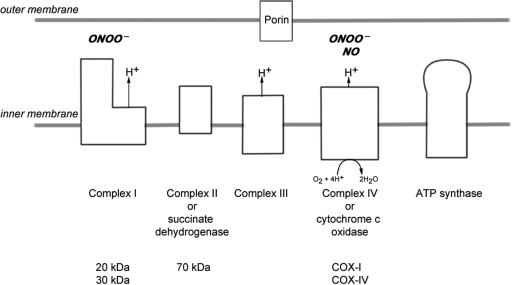
Multiple sclerosis is a chronic inflammatory disease, which leads to focal plaques of demyelination and tissue injury in the CNS. The structural and immunopathological patterns of demyelination suggest that different immune mechanisms may be involved in tissue damage. In a subtype of lesions, which are mainly found in patients with acute fulminant multiple sclerosis with Balo's type concentric sclerosis and in a subset of early relapsing remitting multiple sclerosis, the initial myelin changes closely resemble those seen in white matter stroke (WMS), suggesting a hypoxia-like tissue injury. Since mitochondrial injury may be involved in the pathogenesis of such lesions, we analysed a number of mitochondrial respiratory chain proteins in active lesions from acute multiple sclerosis and from WMS using immunohistochemistry. Functionally important defects of mitochondrial respiratory chain complex IV [cytochrome c oxidase (COX)] including its catalytic component (COX-I) are present in Pattern III but not in Pattern II multiple sclerosis lesions. The lack of immunohistochemically detected COX-I is apparent in oligodendrocytes, hypertrophied astrocytes and axons, but not in microglia. The profile of immunohistochemically detected mitochondrial respiratory chain complex subunits differs between multiple sclerosis and WMS. The findings suggest that hypoxia-like tissue injury in Pattern III multiple sclerosis lesions may be due to mitochondrial impairment.
Overview
The claims and statistics of this paper haven’t been checked yet.
Free: it reads the open-access text and checks the first 5 claims that cite a source we can check. Create a free account to check every claim.
Integrity signals
Registry
Cites
Statistics
Figures


Verification
Claims the paper makes
The claims and statistics of this paper haven’t been checked yet.
Free: it reads the open-access text and checks the first 5 claims that cite a source we can check. Create a free account to check every claim.
Claims other papers make about this one
Each is checked against this paper’s own text: does it say what the citing paper says it does?
The source backs part of the claim, not all of it — or backs it for a different population, condition or comparison.
In the citing paper
“Oxidative damage and mitochondrial dysfunction are key features of MS pathology [277,278,279,280].”
From Ageing, Cellular Senescence and Neurodegenerative Disease (2018) · cites this paper as [279]
Experimental — accuracy not yet measuredThe source backs part of the claim, not all of it — or backs it for a different population, condition or comparison.
In the citing paper
“Other features common to both cuprizone-induced and type 3 MS lesions are mitochondrial stress, selective loss of myelin-associated glycoprotein (MAG), lesions with indistinct borders that are not perivascular, pronounced glial activation, and lymphocyte infiltration [43, 46, 65, 69–71].”
From Oligodendrocyte death and myelin loss in the cuprizone model: an updated overview of the intrinsic and extrinsic causes of cuprizone demyelination (2022) · cites this paper as [69]
Experimental — accuracy not yet measured
Self-check
Percentages, sums and reported differences re-computed from the paper’s own numbers.
The self-check needs the paper’s full text.
Citations
Cites
Mitochondrial DNA mutations in human disease
Taylor, R. W., & Turnbull, D. M. (2005) · Nature Reviews Genetics · doi:10.1038/nrg1606 · Cited by 1,852 · cited 5×
“…c oxidase Abbreviations: CNPase=cyclic nucleotide phosphodiesterase; COX=cytochrome c oxidase; LFB=Luxol fast blue; MAG=myelin associated glycoprotein; MBP=myelin basic protein; mtDNA=mitochondrial DNA; NO=nitric oxide; NWM=normal white matter; PLP=proteolipid protein; WMS=white matter stroke”
BackgroundMethodInfluentialSelf-citationMultiple Sclerosis — The Plaque and Its Pathogenesis
Frohman, E. M., Racke, M. K., & Raine, C. S. (2006) · New England Journal of Medicine · doi:10.1056/nejmra052130 · Cited by 1,740 · cited 1×
“Multiple sclerosis is an inflammatory demyelinating disease of the central nervous system with axonal degeneration and astrogliosis (Frohman et al., 2006).”
BackgroundMitochondrial Respiratory-Chain Diseases
DiMauro, S., & Schon, E. A. (2003) · New England Journal of Medicine · doi:10.1056/nejmra022567 · Cited by 1,661 · cited 5×
“Keywords: multiple sclerosis; Pattern III lesion; mitochondria; cytochrome c oxidase Abbreviations: CNPase=cyclic nucleotide phosphodiesterase; COX=cytochrome c oxidase; LFB=Luxol fast blue; MAG=myelin associated glycoprotein; MBP=myelin basic protein; mtDNA=mitochondrial DNA; NO=nitric oxide;…”
BackgroundInfluentialvon Büdingen, H.-C., Palanichamy, A., Lehmann-Horn, K., Michel, B. A., & Zamvil, S. S. (2015) · European Neurology · doi:10.1159/000377675 · Cited by 1,463
Cleeter, M. W. J., Cooper, J. M., Darley-Usmar, V. M., Moncada, S., & Schapira, A. H. V. (1994) · FEBS Letters · doi:10.1016/0014-5793(94)00424-2 · Cited by 1,241 · cited 1×
“NO competitively inhibits the binding of oxygen to complex IV (Brown and Cooper, 1994; Cleeter et al., 1994).”
BackgroundHIF-1 Regulates Cytochrome Oxidase Subunits to Optimize Efficiency of Respiration in Hypoxic Cells
Fukuda, R., Zhang, H., Kim, J.-W., Shimoda, L., Dang, C. V., & Semenza, G. L. (2007) · Cell · doi:10.1016/j.cell.2007.01.047 · Cited by 1,225 · cited 5×
“Widespread defects of mitochondrial respiratory chain complexes inWMS lesions The transcription of mtDNA and nuclear DNA encoded subunits of the mitochondrial respiratory chain complexes are modulated by hypoxia (Vijayasarathy et al., 2003; Piruat and Lopez-Barneo, 2005; Fukuda et al., 2007).”
BackgroundInfluentialMicrodomains of Intracellular Ca2+: Molecular Determinants and Functional Consequences
Rizzuto, R., & Pozzan, T. (2006) · Physiological Reviews · doi:10.1152/physrev.00004.2005 · Cited by 1,206 · cited 1×
“Mitochondria also play an important role in apoptosis and calcium homeostasis (Rizzuto and Pozzan, 2006; Kalman et al., 2007).”
BackgroundRelapsing and remitting multiple sclerosis: Pathology of the newly forming lesion
Barnett, M. H., & Prineas, J. W. (2004) · Annals of Neurology · doi:10.1002/ana.20016 · Cited by 1,185 · cited 5×
“Mitochondrial defects in acute multiple sclerosis lesions Don Mahad,1 Iryna Ziabreva,1Hans Lassmann2 and DouglasTurnbull1 1The Mitochondrial Research Group, University of Newcastle uponTyne, UK and 2Centre for Brain Research, Medical University of Vienna, Austria Correspondence to: Prof. Dr Hans Lassmann, Center for B…”
BackgroundInfluentialBrown, G. C., & Cooper, C. E. (1994) · FEBS Letters · doi:10.1016/0014-5793(94)01290-3 · Cited by 1,055 · cited 2×
“…c oxidase Abbreviations: CNPase=cyclic nucleotide phosphodiesterase; COX=cytochrome c oxidase; LFB=Luxol fast blue; MAG=myelin associated glycoprotein; MBP=myelin basic protein; mtDNA=mitochondrial DNA; NO=nitric oxide; NWM=normal white matter; PLP=proteolipid protein; WMS=white matter stroke”
BackgroundSystemic infections and inflammation affect chronic neurodegeneration
Perry, V. H., Cunningham, C., & Holmes, C. (2007) · Nature Reviews Immunology · doi:10.1038/nri2015 · Cited by 1,027 · cited 1×
“These activated microglia, found in close proximity to apoptotic oligodendrocytes, may release a range of mediators (Barnett and Prineas, 2004; Perry et al., 2007).”
Background
Cited by
Items 101–200 of 424
Warne, J., Pryce, G., Hill, J. M., Shi, X., Lennerås, F., Puentes, F., Kip, M., Hilditch, L., Walker, P., Simone, M. I., et al. (2016) · Journal of Biological Chemistry · doi:10.1074/jbc.m115.700385 · Cited by 99
“CypD is highly expressed in a subset of astrocytes, microglia, and neurons (19), where it may contribute to excitotoxicity and cell death in MS lesions (12, 16, 17).”
Chamberlain, K. A., Nanescu, S. E., Psachoulia, K., & Huang, J. K. (2016) · Neuropharmacology · doi:10.1016/j.neuropharm.2015.10.010 · Cited by 87
“…(Trapp and Stys, 2009), as abnormalities in mitochondria density, morphology, and activity are consistently found in models of MS (Campbell et al., 2014; Fischer et al., 2012; Joshi et al., 2015; Kiryu-Seo et al., 2010; Mahad et al., 2008, 2009; Qi et al., 2006; Witte et al., 2014, 2010, 2009).”
BackgroundAutophagy and neurodegenerative disorders.
Kesidou, E., Lagoudaki, R., Touloumi, O., Poulatsidou, K.-N., & Simeonidou, C. (2013) · PubMed · doi:10.3969/j.issn.1673-5374.2013.24.007 · Cited by 89
“Defective mitochondria, not subjectable to mitophagy, accumulate within axons rendering them sensitive to energy deficits and prone to degeneration [73] .”
Mitochondrial dysfunction and neurological disorders: A narrative review and treatment overview
Alshial, E. E., Abdulghaney, M. I., Wadan, A. S., Abdellatif, M. A., Ramadan, N. E., Suleiman, A. M., Waheed, N., Abdellatif, M., & Mohammed, H. S. (2023) · Life Sciences · doi:10.1016/j.lfs.2023.122257 · Cited by 98
The disruption of mitochondrial axonal transport is an early event in neuroinflammation
Errea, O., Moreno, B., González-Franquesa, A., García-Rovés, P. M., & Villoslada, P. (2015) · Journal of Neuroinflammation · doi:10.1186/s12974-015-0375-8 · Cited by 92
Contribution of the HNE-immunohistochemistry to modern pathological concepts of major human diseases
Žarković, K., Jakovčević, A., & Žarković, N. (2016) · Free Radical Biology and Medicine · doi:10.1016/j.freeradbiomed.2016.12.009 · Cited by 96
Cell type-specific Nrf2 expression in multiple sclerosis lesions
Licht-Mayer, S., Wimmer, I., Traffehn, S., Metz, I., Brück, W., Bauer, J., Bradl, M., & Lassmann, H. (2015) · Acta Neuropathologica · doi:10.1007/s00401-015-1452-x · Cited by 86
Self-citationAxonal loss in multiple sclerosis
Criste, G. A., Trapp, B. D., & Dutta, R. (2014) · Handbook of clinical neurology · doi:10.1016/b978-0-444-52001-2.00005-4 · Cited by 107
snRNA-seq stratifies multiple sclerosis patients into distinct white matter glial responses
Macnair, W., Calini, D., Agirre, E., Bryois, J., Jäkel, S., Smith, R. S., Kukanja, P., Stokar-Regenscheit, N., Ott, V., Foo, L. C., et al. (2025) · Neuron · doi:10.1016/j.neuron.2024.11.016 · Cited by 88
Neurodegeneration in multiple sclerosis: novel treatment strategies
Luessi, F., Siffrin, V., & Zipp, F. (2012) · Expert Review of Neurotherapeutics · doi:10.1586/ern.12.59 · Cited by 86
Targets of therapy in progressive MS
Lassmann, H. (2017) · Multiple Sclerosis Journal · doi:10.1177/1352458517729455 · Cited by 82
Self-citationThe role of glutamate and its receptors in multiple sclerosis
Stojanović, I. R., Kostić, M. S., & Ljubisavljević, S. (2014) · Journal of Neural Transmission · doi:10.1007/s00702-014-1188-0 · Cited by 93
“In active demyelinating MS lesions, in both axons and neurons, the increased mitochondrial density was associated with the enhanced expression of mtHSP70, the marker of mitochondrial stress (Witte et al. 2009; Mahad et al. 2008).”
BackgroundMitochondria in Multiple Sclerosis: Molecular Mechanisms of Pathogenesis
Patergnani, S., Fossati, V., Bonora, M., Giorgi, C., Marchi, S., Missiroli, S., Rusielewicz, T., Wieckowski, M. R., & Pinton, P. (2016) · International review of cytology · doi:10.1016/bs.ircmb.2016.08.003 · Cited by 88
Oligodendrocyte Progenitor Cell Susceptibility to Injury in Multiple Sclerosis
Cui, Q., Kuhlmann, T., Miron, V. E., Leong, S. Y., Fang, J. Q., Gris, P., Kennedy, T. E., Almazán, G., & Antel, J. P. (2013) · American Journal Of Pathology · doi:10.1016/j.ajpath.2013.04.016 · Cited by 92
“15 e 17 The extent to which OPCs may be susceptible to similar injury as mature OLs, with which they share a common microenvironment, in MS lesions remains to be de fi ned.”
BackgroundZarruk, J. G., Berard, J. L., Passos dos Santos, R., Kroner, A., Lee, J., Arosio, P., & David, S. (2015) · Neurobiology of Disease · doi:10.1016/j.nbd.2015.02.001 · Cited by 82
Multiple sclerosis associated with Leber's Hereditary Optic Neuropathy
Palace, J. A. (2009) · Journal of the Neurological Sciences · doi:10.1016/j.jns.2009.09.009 · Cited by 84
Autophagy of Mitochondria: A Promising Therapeutic Target for Neurodegenerative Disease
Kamat, P. K., Kalani, A., Kyles, P., Tyagi, S. C., & Tyagi, N. (2014) · Cell Biochemistry and Biophysics · doi:10.1007/s12013-014-0006-5 · Cited by 86
Amri, F., Ghouili, I., Amri, M., Carrier, A., & Masmoudi‐Kouki, O. (2016) · Journal of Neurochemistry · doi:10.1111/jnc.13876 · Cited by 87
Potential biological contributers to the sex difference in multiple sclerosis progression
Álvarez‐Sánchez, N., & Dunn, S. E. (2023) · Frontiers in Immunology · doi:10.3389/fimmu.2023.1175874 · Cited by 82
Perturbed Glucose Metabolism: Insights into Multiple Sclerosis Pathogenesis
Mathur, D., pez-Rodas, G. L., Casanova, B., & Marti, M. B. (2014) · Frontiers in Neurology · doi:10.3389/fneur.2014.00250 · Cited by 85
MRI in Leber's hereditary optic neuropathy: the relationship to multiple sclerosis
Matthews, L., Enzinger, C., Fazekas, F., Rovira, À., Ciccarelli, O., Dotti, M. T., Filippi, M., Frederiksen, J. L., Giorgio, A., Küker, W., et al. (2014) · Journal of Neurology Neurosurgery & Psychiatry · doi:10.1136/jnnp-2014-308186 · Cited by 85
Neuromyelitis optica lesions may inform multiple sclerosis heterogeneity debate
Brück, W., Popescu, B., Lucchinetti, C. F., Markovic‐Plese, S., Gold, R., Thal, D. R., & Metz, I. (2012) · Annals of Neurology · doi:10.1002/ana.23621 · Cited by 77
Šišková, Z., Mahad, D., Pudney, C., Campbell, G. R., Cadogan, M., O'Connor, V. M., Asuni, A. A., & Perry, V. H. (2010) · American Journal Of Pathology · doi:10.2353/ajpath.2010.091037 · Cited by 81
“We stained frozen tissue sections using enzyme histochemistry for COX (complex IV) and SDH (complex II) activity, a widely used method for studying mitochondrial function in other pathological and neuropathological conditions, including Alzheimer’s disease.(20,26,27,28) The pattern of the complex IV functional activit…”
Self-citationThe role of mitochondria in axonal degeneration and tissue repair in MS
van Horssen, J., Witte, M. E., & Ciccarelli, O. (2012) · Multiple Sclerosis Journal · doi:10.1177/1352458512452924 · Cited by 82
Oligodendroglia and neurotrophic factors in neurodegeneration
Bankston, A. N., Mandler, M. D., & Feng, Y. (2013) · Neuroscience Bulletin · doi:10.1007/s12264-013-1321-3 · Cited by 70
“the mitochondrial damage seen in MS lesions and mouse models of demyelination([27-29]).”
BackgroundCell type mapping reveals tissue niches and interactions in subcortical multiple sclerosis lesions
Lerma-Martin, C., Badia-i-Mompel, P., Ramirez Flores, R. O., Sekol, P., Schäfer, P. S. L., Riedl, C. J., Hofmann, A., Thäwel, T., Wünnemann, F., Ibarra-Arellano, M. A., et al. (2024) · Nature Neuroscience · doi:10.1038/s41593-024-01796-z · Cited by 76
“It is known that mitochondrial dysfunction and metabolic exhaustion are known features of chronic MS lesions and have also been reported for reactive AS in MS 57,58 .”
Autoimmune and inflammatory mechanisms of CNS damage
Kapadia, M., & Šakić, B. (2011) · Progress in Neurobiology · doi:10.1016/j.pneurobio.2011.08.008 · Cited by 81
“and axons in MS patients (Dutta et al., 2006; Mahad et al., 2008).”
Upadhayay, S., & Mehan, S. (2021) · Brain Disorders · doi:10.1016/j.dscb.2021.100019 · Cited by 77
Stork, L., Ellenberger, D., Beißbarth, T., Friede, T., Lucchinetti, C. F., Brück, W., & Metz, I. (2018) · JAMA Neurology · doi:10.1001/jamaneurol.2017.4842 · Cited by 77
Faizi, M., Salimi, A., Seydi, E., Naserzadeh, P., Kouhnavard, M., Rahimi, A., & Pourahmad, J. (2016) · Toxicology Mechanisms and Methods · doi:10.3109/15376516.2016.1172284 · Cited by 71
Deijns, S. J., Broen, J. C. A., Kruyt, N. D., Schubart, C. D., Andréoli, L., Tincani, A., & Limper, M. (2020) · Autoimmunity Reviews · doi:10.1016/j.autrev.2020.102592 · Cited by 70
Energy failure in multiple sclerosis and its investigation using MR techniques
Paling, D., Golay, X., Wheeler‐Kingshott, C. A. M. G., Kapoor, R., & Miller, D. H. (2011) · Journal of Neurology · doi:10.1007/s00415-011-6117-7 · Cited by 71
“seen in mitochondrial complex I in chronic active MS plaques [15], complex I and III in the normal appearing white matter (NAWM) [16], and complex IV on the peripheries of Balo’s type and pattern III lesions [17], which are characterised by preferential loss of myelin associated glycoprotein and apoptosis of oligodend…”
Kynurenines in the Pathogenesis of Multiple Sclerosis: Therapeutic Perspectives
Biernacki, T., Sandi, D., Bencsik, K., & Vécsei, L. (2020) · Cells · doi:10.3390/cells9061564 · Cited by 70
Wang, Y., Mishra, A., & Brinton, R. D. (2020) · F1000Research · doi:10.12688/f1000research.21599.1 · Cited by 61
Swerdlow, N., & Wilkins, H. (2020) · International Journal of Molecular Sciences · doi:10.3390/ijms21249661 · Cited by 72
Chronic demyelination-induced seizures
Lapato, A. S., Szu, J. I., Hasselmann, J., Khalaj, A. J., Binder, D. K., & Tiwari-Woodruff, S. K. (2017) · Neuroscience · doi:10.1016/j.neuroscience.2017.01.035 · Cited by 70
“…the CC and hippocampus of chronically cuprizone-fed mice CPZ causes demyelination throughout the CNS by induction of apoptotic pathways in post-mitotic oligodendrocytes (OLs) secondary to mitochondrial dysfunction and accumulation of reactive oxygen species (Mahad et al., 2008; Praet et al., 2014).”
Recent advances in the study of Na+/K+-ATPase in neurodegenerative diseases
Zhang, X., Lee, W., & Bian, J.-S. (2022) · Cells · doi:10.3390/cells11244075 · Cited by 66
Mathur, D., María-Lafuente, E., Ureña-Peralta, J., Sorribes, L., Hernández, A., Casanova, B., López-Rodas, G., Coret-Ferrer, F., & Burgal-Marti, M. (2017) · Brain Sciences · doi:10.3390/brainsci8010001 · Cited by 65
Altered substrate metabolism in neurodegenerative disease: new insights from metabolic imaging
Cleland, N. R. W., Al‐Juboori, S. I., Dobrinskikh, E., & Bruce, K. D. (2021) · Journal of Neuroinflammation · doi:10.1186/s12974-021-02305-w · Cited by 66
Metabolic defects in multiple sclerosis
Adiele, R. C., & Adiele, C. A. (2017) · Mitochondrion · doi:10.1016/j.mito.2017.12.005 · Cited by 67
Aquaporin-4 astrocytopathy in Baló’s disease
Matsuoka, T., Suzuki, S. O., Iwaki, T., Tabira, T., Ordinario, A. T., & Kira, J. (2010) · Acta Neuropathologica · doi:10.1007/s00401-010-0733-7 · Cited by 62
“Hypoxia-like tissue injury may contribute to Baló’s lesions [24, 34],”
Hypoxia in multiple sclerosis; is it the chicken or the egg?
Halder, S. K., & Milner, R. (2020) · Brain · doi:10.1093/brain/awaa427 · Cited by 62
Petković, F., Campbell, I. L., Gonzalez, B., & Castellano, B. (2016) · Glia · doi:10.1002/glia.23043 · Cited by 59
Promises and Limitations of Neural Stem Cell Therapies for Progressive Multiple Sclerosis
Pluchino, S., Smith, J. A., & Peruzzotti-Jametti, L. (2020) · Trends in Molecular Medicine · doi:10.1016/j.molmed.2020.04.005 · Cited by 59
Autophagy pathways in autoimmune diseases
Keller, C. W., Adamopoulos, I. E., & Lünemann, J. D. (2023) · Journal of Autoimmunity · doi:10.1016/j.jaut.2023.103030 · Cited by 59
“Mitochondrial damage, particularly in oligodendrocytes and neurons is now believed to constitute a central and possibly early event in the disease development since aberrant versions of this organelle are frequently detected in active MS lesions [97,98].”
BackgroundNellessen, A., Nyamoya, S., Zendedel, A., Slowik, A., Wruck, C. J., Beyer, C., Fragoulis, A., & Clarner, T. (2019) · Metabolic Brain Disease · doi:10.1007/s11011-019-00488-z · Cited by 59
“Disturbances in the redox status of oligodendrocytes are likely to contribute to the impairment of energy status in MS lesions (Mahad et al. 2008) and damaged oligodendrocytes due to oxidative stress are a common hallmark of distinct pathological conditions such as MS and stroke (Thorburne and Juurlink 1996; Juurlink…”
BackgroundOligodendroglial Energy Metabolism and (re)Myelination
Tepavčević, V. (2021) · Life · doi:10.3390/life11030238 · Cited by 57
Macchi, B., Marino‐Merlo, F., Nocentini, U., Pisani, V., Cuzzocrea, S., Grelli, S., & Mastino, A. (2015) · Journal of Neuroimmunology · doi:10.1016/j.jneuroim.2015.08.016 · Cited by 63
The S1P mimetic fingolimod phosphate regulates mitochondrial oxidative stress in neuronal cells
Martín‐Montañez, E., Pavía, J., Valverde, N., Boraldi, F., Lara, E., Oliver‐Martos, B., Hurtado-Guerrero, I., Fernández, Ó., & Garcia‐Fernandez, M. I. (2019) · Free Radical Biology and Medicine · doi:10.1016/j.freeradbiomed.2019.04.022 · Cited by 59
The contribution of neuropathology to multiple sclerosis research
Lassmann, H. (2022) · European Journal of Neurology · doi:10.1111/ene.15360 · Cited by 55
Self-citationSchirmer, L., Merkler, D., König, F. B., Brück, W., & Stadelmann, C. (2013) · Brain Pathology · doi:10.1111/j.1750-3639.2012.00608.x · Cited by 52
“Neuronal SMI31 IR itself reflects alterations in neurofilament phosphorylation that might be primarily due to cytoskeletal disruptions following axonal transections, even though energy failure leading to chromatolysis and apoptosis cannot be ruled out (14, 23, 35, 36).”
Metabolomic analysis identifies altered metabolic pathways in Multiple Sclerosis
Poddighe, S., Murgia, F., Lorefice, L., Liggi, S., Cocco, E. E., Marrosu, M. G., & Atzori, L. (2017) · The International Journal of Biochemistry & Cell Biology · doi:10.1016/j.biocel.2017.07.004 · Cited by 57
Singh, A., Upadhayay, S., & Mehan, S. (2021) · Neurotoxicity Research · doi:10.1007/s12640-021-00401-6 · Cited by 54
Synaptic Dysfunction in Multiple Sclerosis: A Red Thread from Inflammation to Network Disconnection
Bellingacci, L., Mancini, A., Gaetani, L., Tozzi, A., Parnetti, L., & Filippo, M. D. (2021) · International Journal of Molecular Sciences · doi:10.3390/ijms22189753 · Cited by 54
Involvement of Mitochondria in Neurodegeneration in Multiple Sclerosis
Kozin, M. S., Кулакова, О. Г., & Favorova, O. O. (2018) · Biochemistry (Moscow) · doi:10.1134/s0006297918070052 · Cited by 58
A metabolic perspective on CSF-mediated neurodegeneration in multiple sclerosis
Wentling, M., Gomez, C. L., Park, H.-J., Amatruda, M., Ntranos, A., Aramini, J. M., Petracca, M., Rusielewicz, T., Chen, E., Tolstikov, V., et al. (2019) · Brain · doi:10.1093/brain/awz201 · Cited by 54
Li, W., Feng, J., Gao, C., Wu, M., Du, Q., Tsoi, B., Wang, Q., Yang, D., & Shen, J. (2019) · Free Radical Biology and Medicine · doi:10.1016/j.freeradbiomed.2019.07.037 · Cited by 54
Serum total antioxidant capacity in patients with multiple sclerosis
Hadžović-Džuvo, A., Lepara, O., Valjevac, A., Avdagić, N., Hasić, S., Kiseljaković, E., Ibragić, S., & Alajbegović, A. (2011) · Bosnian Journal of Basic Medical Sciences · doi:10.17305/bjbms.2011.2620 · Cited by 54
Carlström, K. E., Zhu, K., Ewing, E., Krabbendam, I. E., Harris, R. A., Falcão, A. M., Jagodic, M., Castelo‐Branco, G., & Piehl, F. (2020) · Nature Communications · doi:10.1038/s41467-020-17871-5 · Cited by 53
Madsen, P. M., Pinto, M., Patel, S. K., McCarthy, S., Gao, H., Taherian, M., Karmally, S., Pereira, C. V., Dvoriantchikova, G., Ivanov, D., et al. (2017) · Journal of Neuroscience · doi:10.1523/jneurosci.1378-17.2017 · Cited by 53
The cuprizone model: regional heterogeneity of pathology
Wergeland, S., Torkildsen, Ø., Myhr, K., Mørk, S. J., & Bø, L. (2012) · APMIS · doi:10.1111/j.1600-0463.2012.02882.x · Cited by 52
Neurodegeneration in multiple sclerosis involves multiple pathogenic mechanisms
Levin, M. C., Gardner, L. A., Douglas, J. N., Meyers, L., Lee, S., & Shin, Y. (2014) · Degenerative Neurological and Neuromuscular Disease · doi:10.2147/dnnd.s54391 · Cited by 58
Curcumin Inhibits Mitochondrial Injury and Apoptosis from the Early Stage in EAE Mice
Feng, J., Tao, T., Yan, W., Chen, C. S., & Qin, X. (2014) · Oxidative Medicine and Cellular Longevity · doi:10.1155/2014/728751 · Cited by 52
Bioenergetics profile of CD4 + T cells in relapsing remitting multiple sclerosis subjects
Riccardis, L. D., Rizzello, A., Ferramosca, A., Urso, E., Robertis, F. D., Danieli, A., Giudetti, A. M., Trianni, G., Zara, V., & Maffia, M. (2015) · Journal of Biotechnology · doi:10.1016/j.jbiotec.2015.02.015 · Cited by 53
Cuprizone feed formulation influences the extent of demyelinating disease pathology
Toomey, L. M., Papini, M., Lins, B., Wright, A. J., Warnock, A., McGonigle, T., Hellewell, S. C., Bartlett, C. A., Anyaegbu, C., & Fitzgerald, M. (2021) · Scientific Reports · doi:10.1038/s41598-021-01963-3 · Cited by 49
What drives disease in multiple sclerosis: Inflammation or neurodegeneration?
Lassmann, H. (2010) · Clinical and Experimental Neuroimmunology · doi:10.1111/j.1759-1961.2009.00003.x · Cited by 62
“In fact, profound mitochondrial injury is present in initial and early MS lesions, reflected by a profound loss of proteins of the mitochondrial respiratory chain which are targeted by reactive oxygen and nitric oxide species.(32) Furthermore, the tissue injury within initial and early pattern III lesions in MS is ver…”
BackgroundSelf-citationNeuropathology of Multiple Sclerosis
Popescu, B. F. G., & Lucchinetti, C. F. (2015) · Elsevier eBooks · doi:10.1016/b978-0-12-800763-1.00009-9 · Cited by 52
Lassmann, H. (2010) · Journal of the Neurological Sciences · doi:10.1016/j.jns.2010.07.023 · Cited by 56
“In multiple sclerosis lesions mitochondrial injury is associated with T-cell and B-cell inflammation and microglia or macrophage activation [25].”
Self-citationProtective and therapeutic role of 2-carba-cyclic phosphatidic acid in demyelinating disease
Yamamoto, S., Yamashina, K., Ishikawa, M., Gotoh, M., Yagishita, S., Iwasa, K., Maruyama, K., Murakami-Murofushi, K., & Yoshikawa, K. (2017) · Journal of Neuroinflammation · doi:10.1186/s12974-017-0923-5 · Cited by 48
Abnormal NMDA receptor function exacerbates experimental autoimmune encephalomyelitis
Grasselli, G., Rossi, S., Musella, A., Gentile, A., Loizzo, S., Muzio, L., Di Sanza, C., Errico, F., Musumeci, G., Haji, N., et al. (2013) · British Journal of Pharmacology · doi:10.1111/j.1476-5381.2012.02178.x · Cited by 47
“Mitochondrial dysfunction has been recently proposed to contribute significantly to neurodegenerative damage in MS (Kalman et al., 2007; Mahad et al., 2008; Witte et al., 2010; Campbell et al., 2011) and in EAE (Qi et al.”
BackgroundGhareghani, M., Scavo, L., Jand, Y., Farhadi, N., Sadeghi, H., Ghanbari, A., Mondello, S., Arnoult, D., Gharaghani, S., & Zibara, K. (2019) · Frontiers in Pharmacology · doi:10.3389/fphar.2019.00147 · Cited by 50
Lipid-induced endoplasmic reticulum stress in X-linked adrenoleukodystrophy
van de Beek, M.-C., Ofman, R., Dijkstra, I. M. E., Wijburg, F. A., Engelen, M., Wanders, R. J. A., & Kemp, S. (2017) · Biochimica et Biophysica Acta (BBA) - Molecular Basis of Disease · doi:10.1016/j.bbadis.2017.06.003 · Cited by 51
Paling, D., Tozer, D. J., Wheeler‐Kingshott, C. A. M. G., Kapoor, R., Miller, D. H., & Golay, X. (2012) · Journal of Neurology Neurosurgery & Psychiatry · doi:10.1136/jnnp-2012-302541 · Cited by 50
Neurodegeneration in Multiple Sclerosis: The Role of Nrf2-Dependent Pathways
Maldonado, P. P., Guevara, C., Olesen, M. A., Orellana, J. A., Quintanilla, R. A., & Ortíz, F. C. (2022) · Antioxidants · doi:10.3390/antiox11061146 · Cited by 49
Dimethyl fumarate treatment restrains the antioxidative capacity of T cells to control autoimmunity
Liebmann, M., Korn, L., Janoschka, C., Albrecht, S., Lauks, S., Herrmann, A. M., Schulte‐Mecklenbeck, A., Schwab, N., Schneider‐Hohendorf, T., Eveslage, M., et al. (2021) · Brain · doi:10.1093/brain/awab307 · Cited by 47
Multiple sclerosis: The role of melatonin and N-acetylserotonin
Anderson, G., & Rodriguez, M. (2014) · Multiple Sclerosis and Related Disorders · doi:10.1016/j.msard.2014.12.001 · Cited by 46
Jeffries, M. A., McLane, L. E., Khandker, L., Mather, M. L., Evangelou, A. V., Kantak, D., Bourne, J. N., Macklin, W. B., & Wood, T. L. (2021) · The Journal of Neuroscience · doi:10.1523/jneurosci.1377-20.2021 · Cited by 40
Nuccio, C. D., Bernardo, A., Simone, R. D., Mancuso, E., Magnaghi, V., Visentin, S., & Minghetti, L. (2011) · Journal of Neuropathology & Experimental Neurology · doi:10.1097/nen.0b013e3182309ab1 · Cited by 49
“Alterations in Complex I and Complex IV functions have been reported in MS (35), and mild inhibition of the 2 complexes by rotenone or sodium azide, respectively, impairs in vitro OL maturation (Schoenfeld et al [20], Ziabreva et al [21], and the present study).”
Castellazzi, M., Patergnani, S., Donadio, M., Giorgi, C., Bonora, M., Fainardi, E., Casetta, I., Granieri, E., Pugliatti, M., & Pinton, P. (2019) · Journal of Neuroinflammation · doi:10.1186/s12974-019-1526-0 · Cited by 45
Pathogenesis of Multiple Sclerosis: What Can We Learn from the Cuprizone Model
Acs, P., & Kalman, B. (2012) · Methods in Molecular Biology · doi:10.1007/978-1-60761-720-4_20 · Cited by 42
Lisak, R. P., Benjamins, J. A., Bealmear, B., Nedelkoska, L., Studzinski, D., Retland, E., Yao, B., & Land, S. (2009) · Journal of Neuroinflammation · doi:10.1186/1742-2094-6-4 · Cited by 41
“Decreased expression of COX subunits I and IV (Complex IV) has been detected in oligodendroglia, astroglia and axons, but not in microglia, in acute Type III MS lesions [12].”
Blakely, P. K., Hussain, S., Carlin, L. E., & Irani, D. N. (2015) · Frontiers in Neuroscience · doi:10.3389/fnins.2015.00344 · Cited by 46
Autoimmune neuroinflammation triggers mitochondrial oxidation in oligodendrocytes
Steudler, J., Ecott, T., Ivan, D. C., Bouillet, E., Walthert, S., Berve, K., Dick, T. P., Engelhardt, B., & Locatelli, G. (2022) · Glia · doi:10.1002/glia.24235 · Cited by 41
Suhail, H., Nematullah, M., Rashid, F., Sajad, M., Fatma, M., Singh, J., Zahoor, I., Cheung, W. L., Tiwari, N., Ayasolla, K., et al. (2023) · iScience · doi:10.1016/j.isci.2023.107921 · Cited by 41
“…thread in many neurological diseases, including MS. 14 The role of mitochondrial dysfunction in oligodendrocytes in MS pathology is underappreciated, despite the fact that oligodendrocytes, 12 rather than axons, have a defect in complex IV of the mitochondrial respiratory chain in MS lesions.”
BackgroundBalo’s concentric sclerosis: an update and comprehensive literature review
Harandi, A. A., Esfandani, A., Pakdaman, H., Abbasi, M., & Sahraian, M. A. (2018) · Reviews in the Neurosciences · doi:10.1515/revneuro-2017-0096 · Cited by 42
“…ratio (Moore et al., 1985; Kim et al., 1997), and lactate production (that is suggestive of impaired aerobic metabolism in BCS) (Moore et al., 1985; Mahad et al., 2008; Cai et al., 2015), which are similar to those observed in very large MS lesions (Simon and Kleinschmidt-DeMasters, 2008;…”
ResultThe Dichotomous Role of Inflammation in the CNS: A Mitochondrial Point of View
Vezzani, B., Carinci, M., Patergnani, S., Pasquin, M., Guarino, A., Aziz, N., Pinton, P., Simonato, M., & Giorgi, C. (2020) · Biomolecules · doi:10.3390/biom10101437 · Cited by 41
Wang, A. A., Luessi, F., Neziraj, T., Pössnecker, E., Zuo, M., Engel, S., Hanuscheck, N., Florescu, A., Bugbee, E., Xianjie, I., et al. (2024) · Science Translational Medicine · doi:10.1126/scitranslmed.adi0295 · Cited by 41
“In addition to myeloid cell activation ( 29 ), oxidative stress ( 31 ) and mitochondrial dysfunction ( 32 ) also contribute to this process.”
BackgroundGenetics of multiple sclerosis
Hoppenbrouwers, I. A., & Hintzen, R. Q. (2010) · Biochimica et Biophysica Acta (BBA) - Molecular Basis of Disease · doi:10.1016/j.bbadis.2010.09.017 · Cited by 57
Deciphering Multiple Sclerosis Progression
Meca‐Lallana, V., Ruiz, L. B., Polo, J. C., Eichau-Madueño, S., Ferrer-Lozano, J., Forero, L., Higueras, Y., Lara, N. T., Vidal‐Jordana, Á., & Miralles, F. C. P. (2021) · Frontiers in Neurology · doi:10.3389/fneur.2021.608491 · Cited by 43
Proteomics in Multiple Sclerosis: The Perspective of the Clinician
Sandi, D., Kokas, Z., Biernacki, T., Bencsik, K., Klivènyi, P., & Vécsei, L. (2022) · International Journal of Molecular Sciences · doi:10.3390/ijms23095162 · Cited by 40
Kapell, H., Fazio, L., Dyckow, J., Schwarz, S., Cruz-Herranz, A., Mayer, C., Campos, J., D’Este, E., Möbius, W., Cordano, C., et al. (2023) · Journal of Clinical Investigation · doi:10.1172/jci164223 · Cited by 39
“Other studies reported that mitochondrial impairment and energy deficiency result in ion gradient breakdown and calcium overload, which further increases the risk for neuronal injury (18, 19).”
BackgroundAutophagy and Autophagy-Related Proteins in CNS Autoimmunity
Keller, C. W., & Lünemann, J. D. (2017) · Frontiers in Immunology · doi:10.3389/fimmu.2017.00165 · Cited by 39
“Mitochondrial damage was observed in oligodendrocytes and axons, but was not present in macrophages or microglia (251).”
BackgroundMicroRNA-210 regulates the metabolic and inflammatory status of primary human astrocytes
Kieran, N. W., Suresh, R., Dorion, M., MacDonald, A., Blain, M., Wen, D., Fuh, S.-C., Ryan, F., Diaz, R. J., Stratton, J. A., et al. (2022) · Journal of Neuroinflammation · doi:10.1186/s12974-021-02373-y · Cited by 38
A multiple sclerosis‐like disorder in patients with OPA1 mutations
Yu‐Wai‐Man, P., Spyropoulos, A., Duncan, H., Guadagno, J. V., & Chinnery, P. F. (2016) · Annals of Clinical and Translational Neurology · doi:10.1002/acn3.323 · Cited by 42
Mancini, A., Tantucci, M., Mazzocchetti, P., de Iure, A., Durante, V., Macchioni, L., Giampà, C., Alvino, A., Gaetani, L., Costa, C., et al. (2018) · Neurobiology of Disease · doi:10.1016/j.nbd.2018.01.002 · Cited by 38
Impairment of Mitochondrial Redox Status in Peripheral Lymphocytes of Multiple Sclerosis Patients
Gonzalo‐Benito, H., Nogueras, L., Gil-Sánchez, A., Hervás, J. V., Valcheva, P., González‐Mingot, C., Martin‐Garí, M., Canudes, M., Peralta, S., Solana, M. J., et al. (2019) · Frontiers in Neuroscience · doi:10.3389/fnins.2019.00938 · Cited by 40
Toward identifying key mechanisms of progression in multiple sclerosis
Husseini, L., Geladaris, A., & Weber, M. S. (2023) · Trends in Neurosciences · doi:10.1016/j.tins.2023.11.005 · Cited by 37
NOX-induced oxidative stress is a primary trigger of major neurodegenerative disorders
Zilberter, Y., Tabuena, D. R., & Zilberter, M. Y. (2023) · Progress in Neurobiology · doi:10.1016/j.pneurobio.2023.102539 · Cited by 37
“Likewise, altered energy metabolism has been linked to MS initiation and progression (Mathur et al., 2014; Papiri et al., 2023), with mitochondrial injury a potential major driver of tissue damage in MS (Dutta et al., 2006; Lu et al., 2000; Mahad et al., 2008).”
The Role of Oxidative Stress in Neurodegenerative Diseases
Ksiazek-Winiarek, D. J., & Glabinski, A. R. (2014) · Oxidative stress in applied basic research and clinical practice · doi:10.1007/978-1-4939-0440-2_8 · Cited by 36
Niu, N., Ye, J., Hu, Z., Zhang, J., & Wang, Y. (2023) · International Journal of Molecular Sciences · doi:10.3390/ijms24087076 · Cited by 37
Metadata
- Authors
- Mahad, D., Ziabreva, I., Lassmann, H., & Turnbull, D.
- Affiliations
- Medical University of Vienna (AT)
- Corresponding author
- Lassmann, Hans
- Venue
- Brain
- Publisher
- Oxford University Press (OUP)
- Published
- 2008-05-30
- Volume
- 131
- Issue
- 7
- Pages
- 1722-1735
- ISSN
- 0006-8950 (ISSN-L)
- 1460-2156
- PMC
- PMC2442422
- OpenAlex
- W2149715738
- Cited by
- 406
- References
- 50
- Citations by year
- 2026: 8 · 2025: 8 · 2024: 22 · 2023: 18 · 2022: 33 · 2021: 25 · 2020: 30 · 2019: 26 · 2018: 13 · 2017: 23 · 2016: 25 · 2015: 28 · 2014: 28 · 2013: 30 · 2012: 24
- FWCI
- 15.42
- Type
- journal article
- Language
- en
- Open access
- hybrid
- doi.org
- Licence
- cc-by-nc
- Locations
- Brain (publishedVersion, cc-by-nc)
- Europe PMC (PubMed Central) (publishedVersion, cc-by-nc)
- PubMed Central (publishedVersion, cc-by-nc)
- PubMed (publishedVersion)
- Edinburgh Research Explorer (University of Edinburgh) (submittedVersion, cc-by-nc)
- Newcastle University ePrints (Newcastle Univesity)
- Funders
- Wellcome Trust (078415)
- Austrian Science Fund (P 19854)
- Topics
- Health Sciences › Medicine › Clinical Neurology › Multiple Sclerosis Research Studies
- Life Sciences › Biochemistry, Genetics and Molecular Biology › Molecular Biology › Mitochondrial Function and Pathology
- Health Sciences › Medicine › Clinical Neurology › Parkinson's Disease Mechanisms and Treatments
- Keywords
- multiple sclerosis, mitochondrial dysfunction, cytochrome c oxidase, coxI, mitochondrial respiratory chain, Balo's concentric sclerosis, demyelination, oligodendrocytes, astrocytes, microglia, relapsing-remitting multiple sclerosis
- MeSH terms
- Acute Disease, Adult, Aged, Aged, 80 and over, Astrocytes, Axons, Diffuse Cerebral Sclerosis of Schilder, Electron Transport Complex IV, Electron Transport, Female, Humans, Male, Middle Aged, Mitochondria, Multiple Sclerosis, Oligodendroglia, Mitochondrial Encephalomyopathies, Mitochondrial Proteins, Mitochondrial Diseases
About these checks
Registry, retraction, Cites and statistics results are computed, not judged, with one exception: a printed reference that no index lists is read once by a model before it counts as a citation error. Claims are extracted by a model and kept only when their quote appears verbatim in the text; each cited source is then checked, with a model’s help, for whether it says what the claim relies on. Those verdicts can be wrong — read the claim’s quote and the cited source before relying on one.
Statistics and arithmetic are recomputed, not judged.
Confidence bands are shown only for fields calibrated on our annotated test set. None is calibrated yet, so no band is shown.
What counts as a claim ›How we measure confidence ›How we check this ›
Are you an author, or do you see a problem with this page? Request a review
Data sources
Bibliographic data: Semantic Scholar, OpenAlex, Crossref.
Contains information from the Semantic Scholar Academic Graph Datasets, made available under the ODC Attribution License (ODC-BY 1.0).
Semantic Scholar’s data is described in: Kinney, R., et al. (2023). The Semantic Scholar Open Data Platform. arXiv:2301.10140.
