

Mitochondrial defects in acute multiple sclerosis lesions
Open accessThe DOI matches its Crossref record; 50 of the 50 references it cites were found in a registry or an index.
Abstract
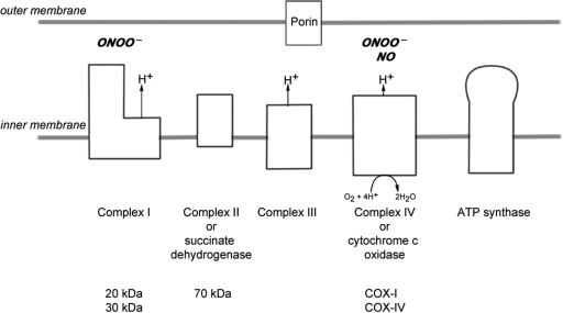
Multiple sclerosis is a chronic inflammatory disease, which leads to focal plaques of demyelination and tissue injury in the CNS. The structural and immunopathological patterns of demyelination suggest that different immune mechanisms may be involved in tissue damage. In a subtype of lesions, which are mainly found in patients with acute fulminant multiple sclerosis with Balo's type concentric sclerosis and in a subset of early relapsing remitting multiple sclerosis, the initial myelin changes closely resemble those seen in white matter stroke (WMS), suggesting a hypoxia-like tissue injury. Since mitochondrial injury may be involved in the pathogenesis of such lesions, we analysed a number of mitochondrial respiratory chain proteins in active lesions from acute multiple sclerosis and from WMS using immunohistochemistry. Functionally important defects of mitochondrial respiratory chain complex IV [cytochrome c oxidase (COX)] including its catalytic component (COX-I) are present in Pattern III but not in Pattern II multiple sclerosis lesions. The lack of immunohistochemically detected COX-I is apparent in oligodendrocytes, hypertrophied astrocytes and axons, but not in microglia. The profile of immunohistochemically detected mitochondrial respiratory chain complex subunits differs between multiple sclerosis and WMS. The findings suggest that hypoxia-like tissue injury in Pattern III multiple sclerosis lesions may be due to mitochondrial impairment.
Overview
The claims and statistics of this paper haven’t been checked yet.
Free: it reads the open-access text and checks the first 5 claims that cite a source we can check. Create a free account to check every claim.
Integrity signals
Registry
Cites
Statistics
Figures


Verification
Claims the paper makes
The claims and statistics of this paper haven’t been checked yet.
Free: it reads the open-access text and checks the first 5 claims that cite a source we can check. Create a free account to check every claim.
Claims other papers make about this one
Each is checked against this paper’s own text: does it say what the citing paper says it does?
The source backs part of the claim, not all of it — or backs it for a different population, condition or comparison.
In the citing paper
“Oxidative damage and mitochondrial dysfunction are key features of MS pathology [277,278,279,280].”
From Ageing, Cellular Senescence and Neurodegenerative Disease (2018) · cites this paper as [279]
Experimental — accuracy not yet measuredThe source backs part of the claim, not all of it — or backs it for a different population, condition or comparison.
In the citing paper
“Other features common to both cuprizone-induced and type 3 MS lesions are mitochondrial stress, selective loss of myelin-associated glycoprotein (MAG), lesions with indistinct borders that are not perivascular, pronounced glial activation, and lymphocyte infiltration [43, 46, 65, 69–71].”
From Oligodendrocyte death and myelin loss in the cuprizone model: an updated overview of the intrinsic and extrinsic causes of cuprizone demyelination (2022) · cites this paper as [69]
Experimental — accuracy not yet measured
Self-check
Percentages, sums and reported differences re-computed from the paper’s own numbers.
The self-check needs the paper’s full text.
Citations
Cites
Mitochondrial DNA mutations in human disease
Taylor, R. W., & Turnbull, D. M. (2005) · Nature Reviews Genetics · doi:10.1038/nrg1606 · Cited by 1,852 · cited 5×
“…c oxidase Abbreviations: CNPase=cyclic nucleotide phosphodiesterase; COX=cytochrome c oxidase; LFB=Luxol fast blue; MAG=myelin associated glycoprotein; MBP=myelin basic protein; mtDNA=mitochondrial DNA; NO=nitric oxide; NWM=normal white matter; PLP=proteolipid protein; WMS=white matter stroke”
BackgroundMethodInfluentialSelf-citationMultiple Sclerosis — The Plaque and Its Pathogenesis
Frohman, E. M., Racke, M. K., & Raine, C. S. (2006) · New England Journal of Medicine · doi:10.1056/nejmra052130 · Cited by 1,740 · cited 1×
“Multiple sclerosis is an inflammatory demyelinating disease of the central nervous system with axonal degeneration and astrogliosis (Frohman et al., 2006).”
BackgroundMitochondrial Respiratory-Chain Diseases
DiMauro, S., & Schon, E. A. (2003) · New England Journal of Medicine · doi:10.1056/nejmra022567 · Cited by 1,661 · cited 5×
“Keywords: multiple sclerosis; Pattern III lesion; mitochondria; cytochrome c oxidase Abbreviations: CNPase=cyclic nucleotide phosphodiesterase; COX=cytochrome c oxidase; LFB=Luxol fast blue; MAG=myelin associated glycoprotein; MBP=myelin basic protein; mtDNA=mitochondrial DNA; NO=nitric oxide;…”
BackgroundInfluentialvon Büdingen, H.-C., Palanichamy, A., Lehmann-Horn, K., Michel, B. A., & Zamvil, S. S. (2015) · European Neurology · doi:10.1159/000377675 · Cited by 1,463
Cleeter, M. W. J., Cooper, J. M., Darley-Usmar, V. M., Moncada, S., & Schapira, A. H. V. (1994) · FEBS Letters · doi:10.1016/0014-5793(94)00424-2 · Cited by 1,241 · cited 1×
“NO competitively inhibits the binding of oxygen to complex IV (Brown and Cooper, 1994; Cleeter et al., 1994).”
BackgroundHIF-1 Regulates Cytochrome Oxidase Subunits to Optimize Efficiency of Respiration in Hypoxic Cells
Fukuda, R., Zhang, H., Kim, J.-W., Shimoda, L., Dang, C. V., & Semenza, G. L. (2007) · Cell · doi:10.1016/j.cell.2007.01.047 · Cited by 1,225 · cited 5×
“Widespread defects of mitochondrial respiratory chain complexes inWMS lesions The transcription of mtDNA and nuclear DNA encoded subunits of the mitochondrial respiratory chain complexes are modulated by hypoxia (Vijayasarathy et al., 2003; Piruat and Lopez-Barneo, 2005; Fukuda et al., 2007).”
BackgroundInfluentialMicrodomains of Intracellular Ca2+: Molecular Determinants and Functional Consequences
Rizzuto, R., & Pozzan, T. (2006) · Physiological Reviews · doi:10.1152/physrev.00004.2005 · Cited by 1,206 · cited 1×
“Mitochondria also play an important role in apoptosis and calcium homeostasis (Rizzuto and Pozzan, 2006; Kalman et al., 2007).”
BackgroundRelapsing and remitting multiple sclerosis: Pathology of the newly forming lesion
Barnett, M. H., & Prineas, J. W. (2004) · Annals of Neurology · doi:10.1002/ana.20016 · Cited by 1,185 · cited 5×
“Mitochondrial defects in acute multiple sclerosis lesions Don Mahad,1 Iryna Ziabreva,1Hans Lassmann2 and DouglasTurnbull1 1The Mitochondrial Research Group, University of Newcastle uponTyne, UK and 2Centre for Brain Research, Medical University of Vienna, Austria Correspondence to: Prof. Dr Hans Lassmann, Center for B…”
BackgroundInfluentialBrown, G. C., & Cooper, C. E. (1994) · FEBS Letters · doi:10.1016/0014-5793(94)01290-3 · Cited by 1,055 · cited 2×
“…c oxidase Abbreviations: CNPase=cyclic nucleotide phosphodiesterase; COX=cytochrome c oxidase; LFB=Luxol fast blue; MAG=myelin associated glycoprotein; MBP=myelin basic protein; mtDNA=mitochondrial DNA; NO=nitric oxide; NWM=normal white matter; PLP=proteolipid protein; WMS=white matter stroke”
BackgroundSystemic infections and inflammation affect chronic neurodegeneration
Perry, V. H., Cunningham, C., & Holmes, C. (2007) · Nature Reviews Immunology · doi:10.1038/nri2015 · Cited by 1,027 · cited 1×
“These activated microglia, found in close proximity to apoptotic oligodendrocytes, may release a range of mediators (Barnett and Prineas, 2004; Perry et al., 2007).”
Background
Cited by
Items 301–400 of 424
Mather, M. L., Jeffries, M. A., & Wood, T. L. (2022) · Current Opinion in Pharmacology · doi:10.1016/j.coph.2022.102193 · Cited by 8
Burtscher, J., Motl, R. W., Berek, K., Ehrenreich, H., Kopp, M., & Hohenauer, E. (2025) · Redox Biology · doi:10.1016/j.redox.2025.103666 · Cited by 8
“Dysfunction of mitochondrial respiration (particularly due to complex IV deficits) may be distinctive for a specific pathological pattern described for MS, pattern III, which is characterized by moderate inflammation and the disappearance of myelin proteins in distal oligodendrocyte extensions [77].”
InfluentialMitochondrial DNA G13708A variation and multiple sclerosis: Is there an association?
Andalib, S., Talebi, M., Sakhinia, E., Farhoudi, M., Sadeghi‐Bazargani, H., Emamhadi, M., Masoodian, N., Balaghi-Inalou, M., Vafaee, M. S., & GJEDDE, A. H. (2017) · Revue Neurologique · doi:10.1016/j.neurol.2017.02.002 · Cited by 7
Demirdöğen, F., Akdağ, T., Gündüz, Z. B., & Odabaş, F. Ö. (2022) · Multiple Sclerosis and Related Disorders · doi:10.1016/j.msard.2022.103948 · Cited by 7
Opposite Roles of Co-enzyme Q10 and Formaldehyde in Neurodegenerative Diseases
Xu, J., Jin, X., Ye, Z., Wang, D., Zhao, H., & Tong, Z. (2022) · American Journal of Alzheimer s Disease & Other Dementias® · doi:10.1177/15333175221143274 · Cited by 7
Tagge, I., Anderson, V. C., SPRINGER, C. S. J., Sammi, M. K., BOURDETTE, D. N., Spain, R., & Rooney, W. D. (2021) · Journal of Neuroimaging · doi:10.1111/jon.12912 · Cited by 7
López-Muguruza, E., Peiró-Moreno, C., Pérez‐Cerdá, F., Matute, C., & Ruiz, A. (2025) · Frontiers in Neuroanatomy · doi:10.3389/fnana.2025.1557214 · Cited by 6
Orchestrating Stress Responses in Multiple Sclerosis: A Role for Astrocytic IFNγ Signaling
Habean, M. L., Kaiser, K. E., & Williams, J. L. (2024) · International Journal of Molecular Sciences · doi:10.3390/ijms25147524 · Cited by 6
“…an enhanced expression of mitochondrial protein and density in active and chronic active MS lesions [79] in addition to the defective expression of proteins associated with mitochondrial respiratory chain complexes, specifically cyclooxygenase 1, in astrocytes, axons, and oligodendrocytes [79,80].”
BackgroundClinical and Pathological Topics of Multiple Sclerosis
Lassmann, H. (2009) · Rinsho Shinkeigaku · doi:10.5692/clinicalneurol.49.715 · Cited by 7
Self-citationOverlaps between multiple system atrophy and multiple sclerosis: A novel perspective
Jellinger, K. A., & Wenning, G. K. (2016) · Movement Disorders · doi:10.1002/mds.26870 · Cited by 6
Pathology of Multiple Sclerosis
Dunham, J., & Mahajan, K. R. (2021) · Springer eBooks · doi:10.1007/978-3-030-61883-4_8 · Cited by 7
Nezamzadeh, F., Masroor, A., Esmailkhani, A., & Bialvaei, A. Z. (2025) · Neuroscience · doi:10.1016/j.neuroscience.2025.07.024 · Cited by 6
Oxidative Stress in Multiple Sclerosis Pathology and Therapeutic Potential of Nrf2 Activation
de Vries, H. E., Schreibelt, G., & van Horssen, J. (2010) · Humana Press eBooks · doi:10.1007/978-1-60327-514-9_5 · Cited by 5
Reactive Oxygen Species: Its Effects on various Diseases
Marwal, A., Verma, S. S., Trivedi, S., & Prajapat, R. (2013) · JOURNAL OF ADVANCES IN BIOTECHNOLOGY · doi:10.24297/jbt.v3i1.1684 · Cited by 7
Pelzers, E., Goethem, J. W. V., van den Hauwe, L., Degryse, H., Geens, K., & Parizel, P. M. (2008) · PubMed · Cited by 5
How does Nogo-A signalling influence mitochondrial function during multiple sclerosis pathogenesis?
Nheu, D., & Petratos, S. (2024) · Neuroscience & Biobehavioral Reviews · doi:10.1016/j.neubiorev.2024.105767 · Cited by 4
“In certain types of lesions exhibiting hypoxic characteristics, there is evidence of reduced levels in key-adenosine triphosphate (ATP) producing mitochondrial complexes, D. Nheu and S. Petratos cytochrome c oxidase subunit (COX) 1 and 4, in axons and oligodendrocytes (Mahad et al., 2008).”
Salgueiro, A. M., Ferreira‐Marques, M., Ribeiro, R. F. N., Lopes, S. M., Pereira, D., Costa, D. G., Santana, M. M., de Almeida, L. P., & Cavadas, C. (2026) · Translational Neurodegeneration · doi:10.1186/s40035-026-00557-1 · Cited by 4
“In addition to immune-mediated myelin damage, mitochondrial dysfunction and impaired energy metabolism have emerged as central contributors to axonal degeneration [109].”
Azzam, S., Broadwater, L., Li, S., Freeman, E. J., McDonough, J. A., & Gregory, R. B. (2013) · Proteome Science · doi:10.1186/1477-5956-11-19 · Cited by 4
Lewis, P. A., & Spillane, J. E. (2018) · Elsevier eBooks · doi:10.1016/b978-0-12-811069-0.00007-0 · Cited by 6
Metabolic support of axons by oligodendrocytes: Implications for multiple sclerosis
Campbell, G. R., & Mahad, D. (2013) · Multiple Sclerosis and Related Disorders · doi:10.1016/j.msard.2013.06.008 · Cited by 4
Self-citationShahzadeh Fazeli, S. A. A. H., Sanati, M. H., Nasrabadi, D., Pouya, A., Baharvand, H., & S.Gh., H. S. (2009) · DOAJ (DOAJ: Directory of Open Access Journals) · Cited by 4
Li, A., Liao, B., Wu, K., Fan, R., Zhu, B., Sun, X., & Liu, L. (2025) · Molecular Carcinogenesis · doi:10.1002/mc.70047 · Cited by 4
“Under aerobic conditions, the primary source of energy for normal cells was the transfer of metabolites through the respiratory complexes (CI ‐ CIV) on the inner mitochondrial membrane, ultimately synthesizing ATP via ATP synthase [8].”
[Pathogenic mechanisms of neuronal damage in multiple sclerosis].
Flores-Alvarado, L. J., Gabriel-Ortíz, G., Pacheco-Mois, F. P., & Bitzer-Quintero, K. (2015) · PubMed · Cited by 5
CNS neuroimmunology seen by a neuropathologist
Lassmann, H. (2014) · Revue Neurologique · doi:10.1016/j.neurol.2014.07.001 · Cited by 3
Self-citationCoyle, P. K. (2019) · Current clinical neurology · doi:10.1007/978-3-030-24436-1_3 · Cited by 3
Demirdöğen, F., Akdağ, T., Gündüz, Z. B., & Odabaş, F. Ö. (2022) · Multiple Sclerosis and Related Disorders · doi:10.1016/j.msard.2022.103999 · Cited by 3
Metabolomics Profile of the Secretome of Space-Flown Oligodendrocytes
Vergnes, L., Foucaud, B., Cepeda, C., & Monteros, A. E. D. L. (2023) · Cells · doi:10.3390/cells12182249 · Cited by 3
Bradl, M., & Lassmann, H. (2011) · Neuropathology · doi:10.1111/j.1440-1789.2011.01282.x · Cited by 4
Self-citationImmune Response in the Human Central Nervous System in Multiple Sclerosis and Stroke
Lassmann, H. (2015) · Wiley eBooks · doi:10.1002/9781118732748.ch1 · Cited by 4
Self-citationMelittin and hyaluronidase compound derived from bee venom for the treatment of multiple sclerosis
Khoei, N. S., Atashpaz, S., Ghabili, K., Khoei, N. S., & Omidi, Y. (2009) · Cited by 5
Dying oligodendrocytes persist without mitochondriaPreprint
Bame, X., Gilani, S. Z., Kamen, Y., & Hill, R. (2026) · bioRxiv (Cold Spring Harbor Laboratory) · doi:10.64898/2026.01.19.699752 · Cited by 3
Illés, Z., Lassmann, H., & Gallyas, F. (2013) · Springer eBooks · doi:10.1007/978-1-4614-7953-6_15 · Cited by 2
Self-citationMetabolic Reprogramming of Oligodendrocytes in Intrauterine Growth Restriction
Peters, H., Fung, C. M., Dettman, R. W., Dizon, M. L. V., & Chang, J. (2025) · Developmental Neuroscience · doi:10.1159/000548455 · Cited by 2
“Impaired OXPHOS, specifically ETC complex I and IV defects, has been reported in OPCs in MS lesions [57, 59].”
An inflammation-associated lncRNA induces neuronal damage via mitochondrial dysfunction
Olazagoitia‐Garmendia, A., Márquez, H. R., Trobisch, T., Moreno‐Castro, C., Etxebarria, A. R., Mentxaka, J., Tripathi, A. K., Yang, B., Ruíz, I. M., Anguíta, J., et al. (2025) · Molecular Therapy — Nucleic Acids · doi:10.1016/j.omtn.2025.102533 · Cited by 2
“39,41,46 Neurons are particularly affected by mitochondrial dysfunction, where in pathological conditions dysregulated mitochondrial dynamics increase negative signals for the cell.”
InfluentialNeurodegenerative Mechanisms of Multiple Sclerosis
Xu, H. (2014) · Cited by 2
Zainal, I. G., & Al-Gebori, A. M. (2012) · Engineering and Technology Journal · doi:10.30684/etj.30.14.9 · Cited by 2
The Use of Mesenchymal Stem Cells for Treating Neurodegenerative Diseases
Kemp, K. C., Redondo, J., Mallam, E., Scolding, N. J., & Wilkins, A. (2015) · Stem cells and cancer stem cells · doi:10.1007/978-94-017-7233-4_1 · Cited by 2
Anemia and dysregulation of iron metabolism in multiple sclerosis
Belova, A. N., Solovieva, V. S., & Boyko, A. N. (2018) · S S Korsakov Journal of Neurology and Psychiatry · doi:10.17116/jnevro201811808210 · Cited by 2
Impact of Curcumin on Aging: Its Manifestations and Limitations
Banji, D., & Banji, O. J. F. (2023) · Springer eBooks · doi:10.1007/978-981-99-7731-4_13 · Cited by 3
Coyle, P. K. (2011) · Humana Press eBooks · doi:10.1007/978-1-60327-860-7_3 · Cited by 2
Modulation of monocyte responses by progressive multiple sclerosis therapy.
Beyers, C. Z. (2020) · Open Access Victoria University of Wellington | Te Herenga Waka (Figshare) · doi:10.26686/wgtn.17148233.v1 · Cited by 2
Proteomics and Management of Multiple Sclerosis
Vo, A., Wong, K., Abram, A., Le, T. T. K., & Ho, L. (2024) · Springer eBooks · doi:10.1007/978-3-031-64021-6_11 · Cited by 1
Mitochondrial-Immune Dysfunction in MS: Therapeutic Potential of EV-Mediated Transfer
Navazi, P., Fereidouni, M., & Erfanian, N. (2026) · Cellular and Molecular Neurobiology · doi:10.1007/s10571-026-01716-8 · Cited by 1
“These alterations result in diminished ATP production, excessive reactive oxygen species (ROS) generation, and neuronal apoptosis, collectively driving irreversible axonal damage (Mahad et al. 2008 ; Sadeghian et al. 2016 ; Barcelos et al. 2019 ; Liu et al. 2025 ).”
Vasilev, G. V., Ivanova, S., & Milanov, I. G. (2026) · Neurology International · doi:10.3390/neurolint18050086 · Cited by 1
“Unlike acute peripheral inflammatory activity, these processes occur behind a relatively intact blood–brain barrier and are less visible to conventional inflammatory markers [51,53,57].”
Free Radicals in Neurodegenerative Diseases: Modulation by Palladium α-Lipoic Acid Complex
Chirakkal, V., Garnett, M., & Antonawich, F. (2011) · InTech eBooks · doi:10.5772/28706 · Cited by 1
Serum Bilirubin Level Changes in Multiple Sclerosis Patients
Barzegar, M., Shaygannejad, V., Mirmosayyeb, O., Valipour, S., Akbari, R., Vesal, S., Ashtar, F., Dehghani, L., Mohammadi, A., & Esmaeil, N. (2018) · Caspian Journal of Neurological Sciences · doi:10.29252/nirp.cjns.4.12.30 · Cited by 1
Harding's Syndrome – Clinical Case Report and Literature Review
Jesus, J., Soares, R. A., Matias, M. J., Geraldes, R., Brandão, E., da Silva, R., & Chibante, J. (2020) · Revista Sociedade Portuguesa de Oftalmologia · doi:10.48560/rspo.20526 · Cited by 1
Mitochondrial abnormalities in neurological disorders
Ferreira, A. F. F., Singulani, M. P., & de Britto, L. R. G. (2021) · Elsevier eBooks · doi:10.1016/b978-0-12-822416-8.00012-9 · Cited by 1
Genetics of Multiple Sclerosis
Mescheriakova, J. Y., Kreft, K. L., & Hintzen, R. Q. (2013) · Springer eBooks · doi:10.1007/978-1-4614-7953-6_10 · Cited by 1
Bradl, M., & Lassmann, H. (2012) · Oxford University Press eBooks · doi:10.1093/med/9780199794591.003.0061 · Cited by 1
Self-citationGoyal, A., Gopika, S., & Agrawal, N. (2022) · Protein and Peptide Letters · doi:10.2174/0929866529666220622124253 · Cited by 1
Neurotrophic Factors in Multiple Sclerosis: a Review of the Mechanism
Azizpour, S., & Moghadasi, V. (2025) · Neurology Letters · doi:10.61186/nl.4.1.7 · Cited by 1
Multiple Sklerose und andere autoimmune ZNS-Erkrankungen
Kroner, A., Kleinschnitz, C., Stadelmann, C., Raab, P., Stangel, M., Trebst, C., Voß, E. V., & Cordes, A.-L. (2011) · Springer eBooks · doi:10.1007/978-3-642-20477-7_1 · Cited by 4
Axonal Loss and Neurodegeneration in Multiple Sclerosis
Dutta, R., Chen, J. M., Ohno, N., Ontaneda, D., & Trapp, B. D. (2017) · Wiley eBooks · doi:10.1002/9781118661895.ch20 · Cited by 1
Teo, W. (2015) · Libraries and Cultural Resources (University of Calgary) · doi:10.11575/prism/26864 · Cited by 1
Fortune, A. J., King, N. E., Gélinas-Marion, A., Moloney, R. A., Cashion, J. M., Burdon, K. P., Taylor, B. V., Sutherland, B. A., Fletcher, J. L., Blackburn, N. B., et al. (2025) · medRxiv · doi:10.1101/2025.07.15.25331537 · Cited by 1
The role of hypoxia in neuroinflammatory disease
Desai, R. (2013) · UCL Discovery (University College London) · Cited by 1
Mechanisms and Patterns of Axonal Loss in Multiple Sclerosis
Mayo, Z., & Jacob, A. (2012) · InTech eBooks · doi:10.5772/35703
Axonal Protection with Sodium Channel Blocking Agents in Models of Multiple Sclerosis
Black, J. A., Smith, K. J., & Waxman, S. G. (2012) · Springer eBooks · doi:10.1007/978-1-4614-2218-1_8
Neue therapeutische Konzepte bei der Multiplen Sklerose: Ein Ausblick
Aktaş, O., & Hartung, H. (2010) · Nervenheilkunde · doi:10.1055/s-0038-1628764
Mohammed, A. M., Al-Youzbaki, A. P. D. W. B., & Meena, M. (2014) · Global Journal of Medical Research
Mitochondria and Multiple Sclerosis
Hirano, M. (2008) · Journal watch · doi:10.1056/jn200809300000003
Emerging Oral Medications for Multiple Sclerosis
Fredrikson, S., & Hillert, J. A. (2012) · doi:10.2217/9781780840482
REGULATION OF MITOCHONDRIAL GENE EXPRESSION IN MULTIPLE SCLEROSIS CORTEX
Pandit, A. (2012) · OhioLink ETD Center (Ohio Library and Information Network)
Douglas, J. N. (2016) · doi:10.21007/etd.cghs.2016.0402
“In MS patients this increased energy demand cannot be met because of the impaired mitochondrial energy production in the CNS.17,28,36 Impaired mitochondrial energy production leaves neurons in a depleted energy state, which has been shown to reduce the ability of Na+/K+ ATPase function.37 Depleted mitochondrial energy…”
BackgroundGiannetti, P. (2015) · Spiral (Imperial College London) · doi:10.25560/34680
Pathological Heterogeneity in Multiple Sclerosis
Tham, M. (2016) · University Library - University of Saskatchewan (University of Saskatchewan)
Templeton, N. (2017) · Open Access Victoria University of Wellington | Te Herenga Waka (Figshare) · doi:10.26686/wgtn.17060486.v1 · Cited by 1
Popova, E. V., Bryukhov, V. V., & Кротенкова, М. В. (2017) · INTERNATIONAL NEUROLOGICAL JOURNAL · doi:10.22141/2224-0713.7.93.2017.116553
Role of astroglial α7 nicotinic acetylcholine receptors in neuroinflammation and oxidative stress
Patel (2017) · Digital Repository Service (Northeastern University) · doi:10.17760/d20274193
Potential drug targets for oral medications
Luessi, F., & Zipp, F. (2012) · doi:10.2217/ebo.12.32
Sclérose en plaques : les nouvelles approches physiopathologiques
Zéphir, H. (2019) · Pratique Neurologique - FMC · doi:10.1016/j.praneu.2019.02.011
Studies of the Biology of Intrathecal Treatment in Progressive MS
Bergman, J. (2020) · DiVA at Umeå University (Umeå University)
Vorob'eva, A. A., Fominykh, V., Vtorova, N. V., Gulyaeva, N. V., Zigangirova, N. A., & Zakharova, M. (2012) · Medical academic journal · doi:10.17816/maj12426-28
Luessi, F. (2021) · Neuromethods · doi:10.1007/978-1-0716-1213-2_18
White Matter Damage in Multiple Sclerosis
Sánchez‐Gómez, M. V., Pérez‐Cerdá, F., & Matute, C. (2013) · Springer eBooks · doi:10.1007/978-1-4614-9123-1_17
Jamali, M., Taherianfard, M., Aminlari, M., & Jalaiee, J. (2021) · Physiology and Pharmacology · doi:10.52547/phypha.25.4.8
(2009) · Current Opinion in Neurology · doi:10.1097/wco.0b013e32832cf9cb
Charakterisierung von Peroxiredoxin 2 im zentralen Nervensystem bei Multipler Sklerose
Winkler, D. A. (2021) · eDiss (Georg-August-Universität Göttingen) · doi:10.53846/goediss-8882
Jia, X., Baranzini, S. E., & Hauser, S. L. (2020) · Elsevier eBooks · doi:10.1016/b978-0-12-813866-3.00018-7
Γνωσιακή έκπτωση και εγκεφαλική ατροφία στη σκλήρυνση κατά πλάκας
Ανδραβίζου, Α. (2020) · doi:10.12681/eadd/48167
Hametner, S., Silvaieh, S., Thurnher, M. M., Dal‐Bianco, A., Çetin, H., Ponleitner, M., Zebenholzer, K., Pemp, B., Trattnig, S., Rössler, K., et al. (2022) · Acta Neuropathologica Communications · doi:10.1186/s40478-022-01462-0
Self-citationNunes, B. G. (2021) · Digital Library of Theses and Dissertations (Universidade de São Paulo) · doi:10.11606/d.42.2021.tde-29042022-152032
Górka, M., Dębiec, M., Białoń, N., Bieczek, D., Suszyńśki, K., & Górka, D. (2023) · Postępy Biochemii · doi:10.18388/pb.2021_492
Modern ideas about concentric sclerosis Balo: a clinical case
Mnatsakanyan, G., Raevskaya, A. I., Андреевна, В. И., & Karpov, S. M. (2024) · Vrach · doi:10.29296/25877305-2024-03-08
Endocannabinoid modulation of astroglial cells in multiple sclerosis
García, Á. M. (2022) · theses.fr (ABES) · doi:10.70675/5208d72bzd194z4486z8bcazb4b776f1984d · Cited by 1
Jia, X., Oksenberg, J. R., Hauser, S. L., & Baranzini, S. E. (2024) · Elsevier eBooks · doi:10.1016/b978-0-443-19176-3.00021-2
A rare case of the late onset of Balo concentric sclerosis
Шидловский, В. А., Gorkova, E. A., Yakovlev, A. A., Lalayan, Т. V., & Смочилин, А. Г. (2024) · The Scientific Notes of the Pavlov University · doi:10.24884/1607-4181-2024-31-1-70-76
Disease Modifying Agents in the Treatment of Multiple Sclerosis
Rizvi, S. (2011) · Humana Press eBooks · doi:10.1007/978-1-60327-860-7_7
Πολλαπλή σκλήρυνση: μηχανισμοί και θεραπεία, νέα δεδομένα
Γερογιάννης, Σ. (2019) · University of Thessaly Institutional Repository (University of Thessaly) · doi:10.26253/heal.uth.7904
Kieran, N. W., Suresh, R., Dorion, M.-F., MacDonald, A., Blain, M., Wen, D., Fuh, S.-C., Ryan, F., Diaz, R. J., Stratton, J. A., et al. (2022) · Figshare · doi:10.6084/m9.figshare.17982578
Kieran, N. W., Suresh, R., Dorion, M.-F., MacDonald, A., Blain, M., Wen, D., Fuh, S.-C., Ryan, F., Diaz, R. J., Stratton, J. A., et al. (2022) · Figshare · doi:10.6084/m9.figshare.17982599
Kieran, N. W., Suresh, R., Dorion, M.-F., MacDonald, A., Blain, M., Wen, D., Fuh, S.-C., Ryan, F., Diaz, R. J., Stratton, J. A., et al. (2022) · Figshare · doi:10.6084/m9.figshare.17982563
Cortical hyperactivity beyond immune attack : pivotal role of TNF-alpha in early Multiple Sclerosis
Pramanik, G. K. (2017) · Gutenberg Open Science · doi:10.25358/openscience-835
Kieran, N. W., Suresh, R., Dorion, M.-F., MacDonald, A., Blain, M., Wen, D., Fuh, S.-C., Ryan, F., Diaz, R. J., Stratton, J. A., et al. (2022) · Figshare · doi:10.6084/m9.figshare.17982590
Kieran, N. W., Suresh, R., Dorion, M.-F., MacDonald, A., Blain, M., Wen, D., Fuh, S.-C., Ryan, F., Diaz, R. J., Stratton, J. A., et al. (2022) · Figshare · doi:10.6084/m9.figshare.17982584
Metadata
- Authors
- Mahad, D., Ziabreva, I., Lassmann, H., & Turnbull, D.
- Affiliations
- Medical University of Vienna (AT)
- Corresponding author
- Lassmann, Hans
- Venue
- Brain
- Publisher
- Oxford University Press (OUP)
- Published
- 2008-05-30
- Volume
- 131
- Issue
- 7
- Pages
- 1722-1735
- ISSN
- 0006-8950 (ISSN-L)
- 1460-2156
- PMC
- PMC2442422
- OpenAlex
- W2149715738
- Cited by
- 406
- References
- 50
- Citations by year
- 2026: 8 · 2025: 8 · 2024: 22 · 2023: 18 · 2022: 33 · 2021: 25 · 2020: 30 · 2019: 26 · 2018: 13 · 2017: 23 · 2016: 25 · 2015: 28 · 2014: 28 · 2013: 30 · 2012: 24
- FWCI
- 15.42
- Type
- journal article
- Language
- en
- Open access
- hybrid
- doi.org
- Licence
- cc-by-nc
- Locations
- Brain (publishedVersion, cc-by-nc)
- Europe PMC (PubMed Central) (publishedVersion, cc-by-nc)
- PubMed Central (publishedVersion, cc-by-nc)
- PubMed (publishedVersion)
- Edinburgh Research Explorer (University of Edinburgh) (submittedVersion, cc-by-nc)
- Newcastle University ePrints (Newcastle Univesity)
- Funders
- Wellcome Trust (078415)
- Austrian Science Fund (P 19854)
- Topics
- Health Sciences › Medicine › Clinical Neurology › Multiple Sclerosis Research Studies
- Life Sciences › Biochemistry, Genetics and Molecular Biology › Molecular Biology › Mitochondrial Function and Pathology
- Health Sciences › Medicine › Clinical Neurology › Parkinson's Disease Mechanisms and Treatments
- Keywords
- multiple sclerosis, mitochondrial dysfunction, cytochrome c oxidase, coxI, mitochondrial respiratory chain, Balo's concentric sclerosis, demyelination, oligodendrocytes, astrocytes, microglia, relapsing-remitting multiple sclerosis
- MeSH terms
- Acute Disease, Adult, Aged, Aged, 80 and over, Astrocytes, Axons, Diffuse Cerebral Sclerosis of Schilder, Electron Transport Complex IV, Electron Transport, Female, Humans, Male, Middle Aged, Mitochondria, Multiple Sclerosis, Oligodendroglia, Mitochondrial Encephalomyopathies, Mitochondrial Proteins, Mitochondrial Diseases
About these checks
Registry, retraction, Cites and statistics results are computed, not judged, with one exception: a printed reference that no index lists is read once by a model before it counts as a citation error. Claims are extracted by a model and kept only when their quote appears verbatim in the text; each cited source is then checked, with a model’s help, for whether it says what the claim relies on. Those verdicts can be wrong — read the claim’s quote and the cited source before relying on one.
Statistics and arithmetic are recomputed, not judged.
Confidence bands are shown only for fields calibrated on our annotated test set. None is calibrated yet, so no band is shown.
What counts as a claim ›How we measure confidence ›How we check this ›
Are you an author, or do you see a problem with this page? Request a review
Data sources
Bibliographic data: Semantic Scholar, OpenAlex, Crossref.
Contains information from the Semantic Scholar Academic Graph Datasets, made available under the ODC Attribution License (ODC-BY 1.0).
Semantic Scholar’s data is described in: Kinney, R., et al. (2023). The Semantic Scholar Open Data Platform. arXiv:2301.10140.
