

Visual dysfunction in Parkinson’s disease
Open accessThe DOI matches its Crossref record; 233 of the 233 references it cites were found in a registry or an index.
Abstract
Patients with Parkinson's disease have a number of specific visual disturbances. These include changes in colour vision and contrast sensitivity and difficulties with complex visual tasks such as mental rotation and emotion recognition. We review changes in visual function at each stage of visual processing from retinal deficits, including contrast sensitivity and colour vision deficits to higher cortical processing impairments such as object and motion processing and neglect. We consider changes in visual function in patients with common Parkinson's disease-associated genetic mutations including GBA and LRRK2 . We discuss the association between visual deficits and clinical features of Parkinson's disease such as rapid eye movement sleep behavioural disorder and the postural instability and gait disorder phenotype. We review the link between abnormal visual function and visual hallucinations, considering current models for mechanisms of visual hallucinations. Finally, we discuss the role of visuo-perceptual testing as a biomarker of disease and predictor of dementia in Parkinson's disease.
Overview
The claims and statistics of this paper haven’t been checked yet.
Free: it reads the open-access text and checks the first 5 claims that cite a source we can check. Create a free account to check every claim.
Integrity signals
Registry
Cites
Statistics
Figures
Verification
Claims the paper makes
The claims and statistics of this paper haven’t been checked yet.
Free: it reads the open-access text and checks the first 5 claims that cite a source we can check. Create a free account to check every claim.
Self-check
Percentages, sums and reported differences re-computed from the paper’s own numbers.
The self-check needs the paper’s full text.
Citations
Cites
The precuneus: a review of its functional anatomy and behavioural correlates
Cavanna, A. E., & Trimble, M. R. (2006) · Brain · doi:10.1093/brain/awl004 · Cited by 5,128 · cited 2×
“Finally, in a structure from motion task, where volunteers detect an object that is formed from the contrast of moving dots, patients with Parkinson’s disease show higher thresholds for correctly identifying objects (Uc et al., 2005).”
BackgroundApplication of fourier analysis to the visibility of gratings
Campbell, F. W., & Robson, J. G. (1968) · The Journal of Physiology · doi:10.1113/jphysiol.1968.sp008574 · Cited by 3,019 · cited 3×
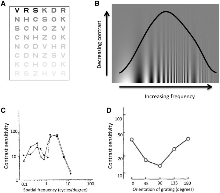
“This is more suggestive of a cortical rather than retinal or geniculate site of disruption as cortical neurons show stronger selectivity for different spatial frequencies than neurons in the retina or thalamus (Campbell and Robson, 1968; De Valois et al., 1982; Regan, 1982).”
BackgroundStages in the development of Parkinson’s disease-related pathology
Braak, H., Ghebremedhin, E., Rüb, U., Bratzke, H., & Del Tredici, K. (2004) · Cell and Tissue Research · doi:10.1007/s00441-004-0956-9 · Cited by 2,689 · cited 1×
“One theory of disease progression in Parkinson’s disease has suggested that the cerebral cortex is not damaged until the later stages of the disease (Braak et al., 2004).”
BackgroundThe Sydney multicenter study of Parkinson's disease: The inevitability of dementia at 20 years
Hely, M. A., Reid, W. G. J., Adena, M. A., Halliday, G. M., & Morris, J. G. L. (2008) · Movement Disorders · doi:10.1002/mds.21956 · Cited by 2,246 · cited 1×
“Visual hallucinations are also common in Parkinson’s disease, with a reported prevalence as high as 74% after 20 years of disease (Fenelon et al., 2000; Hely et al., 2008).”
BackgroundA direct demonstration of functional specialization in human visual cortex
Zeki, S., Watson, J., Lueck, C., Friston, K., Kennard, C., & Frackowiak, R. (1991) · The Journal of Neuroscience · doi:10.1523/jneurosci.11-03-00641.1991 · Cited by 1,716 · cited 2×
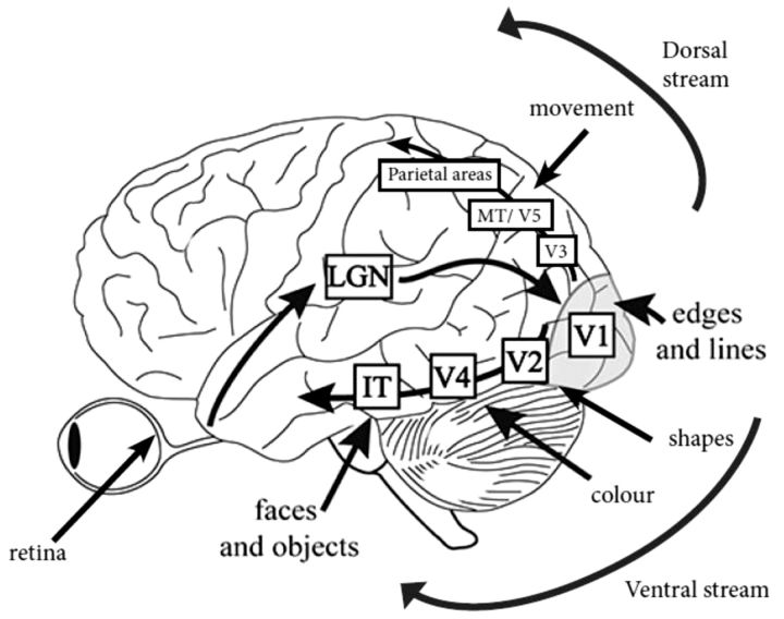
“IT = infero-temporal region; MT/V5 = motion processing region.”
BackgroundPharmacological Treatment of Parkinson Disease: A Review
Connolly, B. S., & Lang, A. E. (2014) · JAMA · doi:10.1001/jama.2014.3654 · Cited by 1,498 · cited 2×
“General advice is to consider visuo-perceptual ability and if there is any doubt, to refer the patient for a formal driving assessment via the national driving authority (Crizzle et al., 2012).”
BackgroundSpatial frequency selectivity of cells in macaque visual cortex
De Valois, R. L., Albrecht, D. G., & Thorell, L. G. (1982) · Vision Research · doi:10.1016/0042-6989(82)90113-4 · Cited by 1,489 · cited 2×
“This is more suggestive of a cortical rather than retinal or geniculate site of disruption as cortical neurons show stronger selectivity for different spatial frequencies than neurons in the retina or thalamus (Campbell and Robson, 1968; De Valois et al., 1982; Regan, 1982).”
Functional analysis of human MT and related visual cortical areas using magnetic resonance imaging
Tootell, R., Reppas, J., Kwong, K., Malach, R., Born, R., Brady, T., Rosen, B., & Belliveau, J. (1995) · The Journal of Neuroscience · doi:10.1523/jneurosci.15-04-03215.1995 · Cited by 1,415 · cited 2×
“IT = infero-temporal region; MT/V5 = motion processing region.”
BackgroundMazzulli, J. R., Xu, Y.-H., Sun, Y., Knight, A. L., McLean, P. J., Caldwell, G. A., Sidransky, E., Grabowski, G. A., & Krainc, D. (2011) · Cell · doi:10.1016/j.cell.2011.06.001 · Cited by 1,341 · cited 1×
“Both the GBA and SNCA genes are linked with the lysosomal pathway (Mazzulli et al., 2011), suggesting the visual deficits associated with these genes may share a pathophysiological mechanism, with lysosomal dysfunction associated with cortical visual dysfunction.”
BackgroundTHE DESIGN OF A NEW LETTER CHART FOR MEASURING CONTRAST SENSITIVITY
Pelli, D. G., Robson, J. G., & Wilkins, A. J. (1988) · Cited by 1,308
Research works printed but not linked: 3
- Ogden, 1990, Deficits on visuospatiaol tests involving forward planning in high-functioning Parkinsonians, Neuropsychiatry Neuropsychol Behav Neurol, 3, 125
- 10.1037/0033-295X.89.4.407 · doi:10.1037/0033-295x.89.4.407
- 10.1097/00006324-199404000-00002 · doi:10.1097/00006324-199404000-00002
Cited by
Items 101–200 of 479
Neural correlates of early cognitive dysfunction in Parkinson's disease
Weil, R. S., Winston, J. S., Leyland, L., Pappa, K., Mahmood, R., Morris, H. R., & Rees, G. E. (2019) · Annals of Clinical and Translational Neurology · doi:10.1002/acn3.767 · Cited by 27
Self-citationDan, R., Růžička, F., Bezdíček, O., Roth, J., Růžička, E., Vymazal, J., Goelman, G., & Jech, R. (2019) · European Neuropsychopharmacology · doi:10.1016/j.euroneuro.2019.09.003 · Cited by 32
Haarsma, J., Deveci, N., Corbin, N., Callaghan, M. F., & Kok, P. (2023) · The Journal of Neuroscience · doi:10.1523/jneurosci.0998-23.2023 · Cited by 26
“Elucidating the various ways perception can go awry is crucial as it sheds new light on false perceptual inferences as seen in psychiatric disorders like psychosis and neurologic disorders like Parkinson ’ s disease (Weil et al., 2016).”
BackgroundA conceptual model of the visual control of posture
Bronstein, A. M. (2019) · Progress in brain research · doi:10.1016/bs.pbr.2019.04.023 · Cited by 32
Haghshomar, M., Shobeiri, P., Seyedi, S. A., Abbasi-Feijani, F., Poopak, A., Sotoudeh, H., Kamali, A., & Aarabi, M. H. (2022) · The Cerebellum · doi:10.1007/s12311-021-01355-3 · Cited by 28
“Color perception is often involved in the blue-green axis [130].”
BackgroundOptical coherence tomography in neurodegenerative disorders
Cunha, L. P., Pires, L. A., Cruzeiro, M. M., Almeida, A. L. M., MARTINS, L. C., Martins, P. N., Shigaeff, N., & Vale, T. C. (2022) · Arquivos de Neuro-Psiquiatria · doi:10.1590/0004-282x-anp-2021-0134 · Cited by 32
Liu, S., Chen, S., Ren, J., Li, B., & Qin, B. (2017) · Neuropeptides · doi:10.1016/j.npep.2017.11.007 · Cited by 27
Hou, Y., Wei, Q., Ou, R., Zhang, L., Yuan, X., Gong, Q., & Shang, H. (2021) · BMC Neurology · doi:10.1186/s12883-021-02360-z · Cited by 28
Stalin, A., & Dalton, K. (2020) · Investigative Ophthalmology & Visual Science · doi:10.1167/iovs.61.6.21 · Cited by 30
“IOVS 2016;57(12):ARVO E-Abstract 5161).”
Yalçın, M., Malhan, D., Basti, A., Peralta, A. R., Ferreira, J. J., & Relógio, A. (2021) · Cancers · doi:10.3390/cancers13235978 · Cited by 24
“play a role in circadian changes, given the fundamental role of light as an entrainment stimulus [53].”
Undetected ophthalmological disorders in Parkinson’s disease
Borm, C. D. J. M., Werkmann, M., de Graaf, D., Visser, F., Hofer, A., Peball, M., Śmiłowska, K., Putz, D., Seppi, K., Poewe, W. H., et al. (2022) · Journal of Neurology · doi:10.1007/s00415-022-11014-0 · Cited by 28
Parkinson’s disease does not alter automatic visual-motor coupling in postural control
Cruz, C. F., Piemonte, M. E. P., Okai-Nóbrega, L. A., Okamoto, E., Fortaleza, A. C. D. S., Mancini, M., Horak, F. B., & Barela, J. Â. (2018) · Neuroscience Letters · doi:10.1016/j.neulet.2018.08.050 · Cited by 23
Differential Temporal Perception Abilities in Parkinson’s Disease Patients Based on Timing Magnitude
Bernardinis, M., Atashzar, S. F., Jog, M., & Patel, R. V. (2019) · Scientific Reports · doi:10.1038/s41598-019-55827-y · Cited by 26
Illuminating and Sniffing Out the Neuromodulatory Roles of Dopamine in the Retina and Olfactory Bulb
Korshunov, K. S., Blakemore, L. J., & Trombley, P. Q. (2020) · Frontiers in Cellular Neuroscience · doi:10.3389/fncel.2020.00275 · Cited by 31
Xu, N., Zhou, Y., Patel, A. C., Zhang, N., & Liu, Y. (2022) · Neuroscience · doi:10.1016/j.neuroscience.2022.11.022 · Cited by 25
Dry eye in Parkinson's disease: a narrative review
Ungureanu, L., Chaudhuri, К. R., Diaconu, Ș., & Falup‐Pecurariu, C. (2023) · Frontiers in Neurology · doi:10.3389/fneur.2023.1236366 · Cited by 23
“For example, dopamine depletion and alpha-synuclein aggregation in the cell layers of the intra-retinal region have been shown to lead to a dysfunction in visual processing with impairment in color discrimination, contrast sensitivity, visual acuity, object, and motion perception (2, 3); double vision has been associa…”
BackgroundDemmin, D. L., Netser, R., Roché, M. W., Thompson, J. L., & Silverstein, S. M. (2019) · Schizophrenia Research · doi:10.1016/j.schres.2019.07.024 · Cited by 23
Patterns of Sulcal depth and cortical thickness in Parkinson’s disease
Wang, E., Jia, Y., Ya, Y., Xu, J., Mao, C., Luo, W., Fan, G., & Jiang, Z. (2021) · Brain Imaging and Behavior · doi:10.1007/s11682-020-00428-x · Cited by 23
“Visual dysfunction is common in PD, including deficits in visual acuity, contrast sensitivity, color vision, visuospatial construction, motion perception, spatial neglect, and visual hallucinations (Weil et al. 2016).”
BackgroundCummings, L. (2020) · Cambridge University Press eBooks · doi:10.1017/9781108587921 · Cited by 24
Alterations in cortical volume and complexity in Parkinson's disease with depression
Yuan, J., Liu, Y., Liao, H., Tan, C., Cai, S., Shen, Q., Liu, Q., Wang, M., Tang, Y., Li, X., et al. (2024) · CNS Neuroscience & Therapeutics · doi:10.1111/cns.14582 · Cited by 24
“27 Therefore, we speculate that the extensive deposition of Lewy bodies, which damage cortical neurons, leads to structural and functional impairments in the temporal–occipital cortex, consequently affecting the visual processing of emotional information.”
BackgroundMisperceptions and Parkinson's disease
Friedman, J. H. (2017) · Journal of the Neurological Sciences · doi:10.1016/j.jns.2016.12.059 · Cited by 24
The Neuro-Ophthalmological Assessment in Parkinson’s Disease
Borm, C. D. J. M., Śmiłowska, K., de Vries, N. M., Bloem, B. R., & Theelen, T. (2019) · Journal of Parkinson s Disease · doi:10.3233/jpd-181523 · Cited by 29
“We will not address how specific visual disorders may assist in the differential diagnosis of parkinsonism; other recent papers have addressed this [1, 19, 20].”
Suicide in Parkinson’s Disease: A Systematic Review
Berardelli, I., Belvisi, D., Nardella, A., Falcone, G., Lamis, D. A., Fabbrini, G., Berardelli, A., Girardi, P., & Pompili, M. (2019) · CNS & Neurological Disorders - Drug Targets · doi:10.2174/1871527318666190703093345 · Cited by 23
Diplopia in Parkinson’s disease: visual illusion or oculomotor impairment?
Visser, F., Vlaar, A. M. M., Borm, C. D. J. M., Apostolov, V., Lee, Y. X., Notting, I. C., Weinstein, H. C., & Berendse, H. W. (2019) · Journal of Neurology · doi:10.1007/s00415-019-09430-w · Cited by 21
Stereopsis and Eye Movement Abnormalities in Parkinson’s Disease and Their Clinical Implications
Ba, F., Sang, T. T., He, W., Fatehi, J., Mostofi, E., & Zheng, B. (2022) · Frontiers in Aging Neuroscience · doi:10.3389/fnagi.2022.783773 · Cited by 20
Berg, S. Z., & Berg, J. (2023) · Frontiers in Immunology · doi:10.3389/fimmu.2023.1228530 · Cited by 23
Hunt, D., Stuart, S., Nell, J. M., Hausdorff, J. M., Galna, B., Rochester, L., & Alcock, L. (2018) · Behavioural Brain Research · doi:10.1016/j.bbr.2018.03.003 · Cited by 20
Cucca, A., Acosta, I., Berberian, M., Lemen, A. C., Rizzo, J., Ghilardi, M. F., Quartarone, A., Feigin, A., Rocco, A. D., & Biagioni, M. C. (2018) · Complementary Therapies in Medicine · doi:10.1016/j.ctim.2018.07.011 · Cited by 25
Overconfidence in visual perception in parkinson's disease
Halperin, O., Karni, R., Israeli‐Korn, S., Hassin‐Baer, S., & Zaidel, A. (2021) · European Journal of Neuroscience · doi:10.1111/ejn.15093 · Cited by 24
“, 1987), deterioration of spatiotemporal and color sensitivity (Montse et al., 2001; Weil et al., 2016).”
BackgroundAbnormal visual gain control and excitotoxicity in early-onset Parkinson’s disease Drosophila models
Himmelberg, M. M., West, R. J. H., Elliott, C. J. H., & Wade, A. R. (2017) · Journal of Neurophysiology · doi:10.1152/jn.00681.2017 · Cited by 20
“Human patients also show a reduction in retinal dopamine and report a range of low-level visual deficits, including poor contrast sensitivity and reduced light sensitivity (Archibald et al. Burn 2011; Beitz 2014; Chaudhuri and Scha- pira 2009; Weil et al. 2016).”
BackgroundLes symptômes non-moteurs de la maladie de Parkinson de la physiopathologie au diagnostic précoce
Aubignat, M., Tir, M., & Krystkowiak, P. C. (2020) · La Revue de Médecine Interne · doi:10.1016/j.revmed.2020.06.019 · Cited by 30
Oscillation-specific nodal alterations in early to middle stages Parkinson’s disease
Guan, X., Guo, T., Zeng, Q., Wang, J., Zhou, C., Liu, C., Wei, H., Zhang, Y., Xuan, M., Gu, Q., et al. (2019) · Translational Neurodegeneration · doi:10.1186/s40035-019-0177-5 · Cited by 21
Ulusoy, E. K., & Ulusoy, D. M. (2020) · International Journal of Neuroscience · doi:10.1080/00207454.2020.1761353 · Cited by 19
Stuart, S., Hickey, A., Galna, B., Lord, S., Rochester, L., & Godfrey, A. (2016) · Physiological Measurement · doi:10.1088/1361-6579/38/1/n16 · Cited by 19
“Visual and cognitive impairments are common in Parkinson’s disease (PD) compared to healthy older adults [3, 4], and are associated with reduced mobility and falls [7, 8].”
Cortical thickness as predictor of response to exercise in people with Parkinson's disease
Silva‐Batista, C., Ragothaman, A., Mancini, M., Carlson‐Kuhta, P., Harker, G. R., Jung, S. H., Nutt, J. G., Fair, D. A., Horak, F. B., & Miranda-Domínguez, Ó. (2020) · Human Brain Mapping · doi:10.1002/hbm.25211 · Cited by 23
“Visualmotor abnormalities are more common in freezers (Davidsdottir, Cronin-Golomb, & Lee, 2005; Weil et al., 2016) and correlated with the severity of gait impairment (Uc et al., 2005).”
BackgroundThe Cerebellum Is a Common Key for Visuospatial Execution and Attention in Parkinson’s Disease
Sako, W., Abe, T., Matsumoto, Y., Nakamura, K., Haji, S., Osaki, Y., Harada, M., & Izumi, Y. (2021) · Diagnostics · doi:10.3390/diagnostics11061042 · Cited by 18
“BA37, also involved in the attention-domain-related network, is part of the ventral stream of visual processing [56,57], which is key for the performance of visuospatial tasks.”
BackgroundAssociation between gene expression and functional‐metabolic architecture in Parkinson's disease
Zang, Z., Zhang, X., Song, T., Li, J., Nie, B., Mei, S., Hu, Z., Zhang, Y., & Lu, J. (2023) · Human Brain Mapping · doi:10.1002/hbm.26443 · Cited by 17
“The visual functional impairment was a major non-motor aspect of PD as a majority of PD patients reported visual symptoms (Weil et al., 2016).”
Lateralization of cochlear dysfunction as a specific biomarker of Parkinson’s disease
Sisto, R., Viziano, A., Stefani, A., Moleti, A., Cerroni, R., Liguori, C., Garasto, E., & Pierantozzi, M. (2020) · Brain Communications · doi:10.1093/braincomms/fcaa144 · Cited by 22
“However, in this framework, the possible involvement of sensory systems has been scarcely investigated, although earlier studies have reported the impairment of visual and auditory systems in patients with Parkinson’s disease (Pisani et al., 2015; Vitale et al., 2016; Weil et al., 2016).”
ResultRetinal Neurovascular Coupling: From Mechanisms to a Diagnostic Window into Brain Disorders
Shen, W. (2025) · Cells · doi:10.3390/cells14221798 · Cited by 23
“Such alterations may contribute to impair visual function, including deficits in motion perception and contrast detection, which are commonly reported in Parkinson’s disease patients [101].”
Hawkins, K. E., Chiarovano, E., Paul, S. S., Burgess, A. M., MacDougall, H. G., & Curthoys, I. S. (2021) · Journal of Vestibular Research · doi:10.3233/ves-201626 · Cited by 22
“Additionally, PwPD are known to have impaired contrast sensitivity [29], and the use of a LED lightbox as the background for the fixation target may help keep the gaze in the correct position for VC testing.”
Sensitivity and Specificity of the ECAS in Parkinson’s Disease and Progressive Supranuclear Palsy
Foley, J. A., Niven, E., Paget, A., Bhatia, K. P., Farmer, S. F., Jarman, P., Limousin, P. D., Warner, T. T., Morris, H. R., Bak, T. H., et al. (2018) · Parkinson s Disease · doi:10.1155/2018/2426012 · Cited by 21
Self-citationA role for DJ-1 against oxidative stress in the mammalian retina
Martín‐Nieto, J., Uribe, M. L., Esteve-Rudd, J., Herrero, M. T., & Campello, L. (2019) · Neuroscience Letters · doi:10.1016/j.neulet.2019.134361 · Cited by 17
“in retinal structure and function, in addition to disorders of cortical visual processing [9,10].”
BackgroundSanches, E. S., Boia, R., Leitão, R. A., Madeira, M. H., Ribeiro, C., Ambrósio, A. F., Fernandes, R., & Silva, A. P. (2023) · Antioxidants · doi:10.3390/antiox12040937 · Cited by 19
“Interestingly, in pathological conditions characterized by an impairment in the dopaminergic system, such as Parkinson’s disease, visual acuity and color vision deficits have also been reported in both animal models [45,46] and PD patients [47].”
BackgroundLan, Y., Liu, X., Yin, C., Lyu, J., xiaoxaio, M., Cui, Z., Li, X., & Lou, X. (2023) · La radiologia medica · doi:10.1007/s11547-023-01673-y · Cited by 16
Altered Outer Retinal Structure, Electrophysiology and Visual Perception in Parkinson’s Disease
Tran, K. K. N., Lee, P. Y., Finkelstein, D. I., McKendrick, A. M., Nguyen, B. N., Bui, B. V., & Nguyen, C. T. O. (2023) · Journal of Parkinson s Disease · doi:10.3233/jpd-230293 · Cited by 21
Influence of WWOX/MAF genes on cognitive performance in patients with Parkinson's disease
Yuan, Y.-T., Hong, W., Tan, C., & Yu, R. (2025) · Neurobiology of Disease · doi:10.1016/j.nbd.2025.106887 · Cited by 19
“It is well-documented that visual disturbances are common non-motor symptoms in PD(Nieto-Escamez et al., 2023; Weil et al., 2016).”
Treatment of Visual Disorders in Parkinson Disease
Savitt, J. M., & Mathews, M. K. (2018) · Current Treatment Options in Neurology · doi:10.1007/s11940-018-0519-0 · Cited by 18
Ocular fixations and presaccadic potentials to explain pareidolias in Parkinson’s disease
Revankar, G. S., Hattori, N., Kajiyama, Y., Nakano, T., Mihara, M., Mori, E., & Mochizuki, H. (2020) · Brain Communications · doi:10.1093/braincomms/fcaa073 · Cited by 16
“As Parkinson’s disease patients have been reported to have problems with eye fixation as well as with oculomotor or saccadic pursuit of eye movements (Ziemssen and Reichmann, 2007; Matsumoto et al., 2011; Weil et al., 2016), we speculated that changes in eye-tracking are crucial when participants perform the NPT.”
BackgroundMarano, M., Rosati, J. D., Magliozzi, A., Casamassa, A., Rappa, A., Sergi, G., Iannizzotto, M., Yekutieli, Z., Vescovi, A. L., & Lazzaro, V. D. (2023) · Neurobiology of Sleep and Circadian Rhythms · doi:10.1016/j.nbscr.2023.100094 · Cited by 17
Huizinga, F., Heutink, J., de Haan, G. A., van der Lijn, I., van der Feen, F. E., Vrijling, A. C. L., Melis-Dankers, B., de Vries, S. M., Tucha, O. M., & Koerts, J. (2020) · PLoS ONE · doi:10.1371/journal.pone.0232232 · Cited by 16
Hawkins, K. E., Rey-Martínez, J. A., Chiarovano, E., Paul, S. S., Valldeperes, A., MacDougall, H. G., & Curthoys, I. S. (2021) · Experimental Brain Research · doi:10.1007/s00221-021-06107-7 · Cited by 19
“A common neurological condition involving both oculomotor (Weil et al. 2016) and central vestibular pathway dysfunction (Seidel et al.”
BackgroundZhou, H., Shen, B., Huang, Z., Zhu, S., Yang, W., Xie, F., Luo, Y., Yuan, F., Zhu, Z., Deng, C., et al. (2023) · npj Parkinson s Disease · doi:10.1038/s41531-023-00611-z · Cited by 15
Takamatsu, Y., Fujita, M., Ho, G. J., Wada, R., Sugama, S., Takenouchi, T., Waragai, M., Masliah, E., & Hashimoto, M. (2018) · Parkinson s Disease · doi:10.1155/2018/5789424 · Cited by 15
Ghasemi, M., Foroutannia, A., & Babajani-Feremi, A. (2021) · Brain and Behavior · doi:10.1002/brb3.2101 · Cited by 15
“The links between the visual changes and other features of patients with PD suggested visual dysfunction as a marker of dementia in these patients (Weil et al., 2016).”
BackgroundAlfano, V., Federico, G., Mele, G., Garramone, F., Esposito, M., Aiello, M., Salvatore, M., & Cavaliere, C. (2022) · Diagnostics · doi:10.3390/diagnostics12040959 · Cited by 20
“However, most recent research links visual–spatial dysfunctions more clearly to dementia than depression and parietal dysfunctions, and much less with the fusiform gyrus [28,29].”
BackgroundToffoli, M., Chohan, H., Mullin, S., Jesuthasan, A., Yalkic, S., Koletsi, S., Menozzi, E., Rahall, S., Limbachiya, N., Loefflad, N., et al. (2023) · Neurobiology of Disease · doi:10.1016/j.nbd.2023.106343 · Cited by 14
Self-citationPenedo, T., Polastri, P. F., Rodrigues, S. T., Simieli, L., Baptista, A. M., Moretto, G. F., Imaizumi, L. F. I., Santinelli, F. B., & Barbieri, F. A. (2018) · Experimental Brain Research · doi:10.1007/s00221-018-5385-1 · Cited by 14
Visuoperceptual disturbances in Parkinson's disease
Kawabata, K., Ohdake, R., Watanabe, H., Bagarinao, E., Hara, K., Ogura, A., Masuda, M., Kato, T., Yokoi, T., Katsuno, M., et al. (2020) · Clinical Parkinsonism & Related Disorders · doi:10.1016/j.prdoa.2020.100036 · Cited by 15
“Earlier stages of visual deficits include visual acuity and contrast and color sensitivity, and these can be partly due to retinal dopaminergic deficiency [1].”
BackgroundInfluentialPrestimulus Low-Alpha Frontal Networks Are Associated with Pareidolias in Parkinson's Disease
Revankar, G. S., Kajiyama, Y., Hattori, N., Shimokawa, T., Nakano, T., Mihara, M., Mori, E., & Mochizuki, H. (2021) · Brain Connectivity · doi:10.1089/brain.2020.0992 · Cited by 16
“Visual mis-perceptual phenomena are strongly influenced by time of the day (‘‘sundowning’’), age-related vision problems, and mood that fluctuate during the day (Ffytche et al., 2017; Suzuki et al., 2011; Watanabe et al., 2018; Weil et al., 2016).”
Brown, G. L., Camacci, M. L., Kim, S. D., Grillo, S. L., Nguyen, J. V., Brown, D., Ullah, S. P., Lewis, M. M., Du, G., Kong, L., et al. (2021) · Journal of Parkinson s Disease · doi:10.3233/jpd-212676 · Cited by 14
“Although the exact cause is unknown, some of the visual deficits in PD are due, at least in part, to retinal dopamine deficiency [4].”
BackgroundLiebermann‐Jordanidis, H., Roheger, M., Boosfeld, L., Franklin, J. G., & Kalbe, E. (2022) · Journal of Parkinson s Disease · doi:10.3233/jpd-223238 · Cited by 16
The utility and application of electrophysiological methods in the study of visual hallucinations
Morgan, K. D., Elder, G. J., Ffytche, D. H., Collerton, D., & Taylor, J. (2018) · Clinical Neurophysiology · doi:10.1016/j.clinph.2018.08.019 · Cited by 18
“Similar abnormalities have been observed in PD, which in turn have been linked to dysfunction of the dopaminergic retinal system, decreases in surround inhibition in the eye, and have further been associated with the formation of VH (Diederich et al. 2005; Onofrj et al. 2006; Weil et al. 2016).”
BackgroundBoring, M. J., Jessen, Z. F., Wozny, T. A., Ward, M., Whiteman, A. C., Richardson, R. M., & Ghuman, A. S. (2019) · NeuroImage · doi:10.1016/j.neuroimage.2019.05.080 · Cited by 15
“Because previous studies have demonstrated that object recognition is intact in movement disorders (Weil et al., 2016), even in advanced Parkinson’s with visual hallucinations (Meppelink et al.”
ResultBernardin, F., Schwitzer, T., Angioï-Duprez, K., Giersch, A., Ligier, F., Bourion-Bédès, S., Jansen, C., Schwan, R., & Laprévote, V. (2021) · Psychiatry Research · doi:10.1016/j.psychres.2021.113780 · Cited by 14
Association of visual impairment with risk for future Parkinson's disease
Zhu, Z., Hu, W., Liao, H., Tan, Z., Chen, Y., Shi, D., Shang, X., Zhang, X., Huang, Y., Yu, H., et al. (2021) · EClinicalMedicine · doi:10.1016/j.eclinm.2021.101189 · Cited by 13
MRI Assessment of Intrinsic Neural Timescale and Gray Matter Volume in Parkinson's Disease
Wei, Y., Zhang, C., Peng, Y., Chen, C., Han, S., Wang, W., Zhang, Y., Lu, H., & Cheng, J. (2023) · Journal of Magnetic Resonance Imaging · doi:10.1002/jmri.28864 · Cited by 14
“13 Nonmotor symptoms, such as visual dysfunction (retinal de fi cits, facial emotion recognition de fi cits, and visual hallucinations) 14 and cognitive impairment, 15 also occur frequently in PD. Imaging biomarkers in PD are increasingly important for monitoring progression in clinical trials.”
BackgroundDiagnosis of Parkinson’s disease by eliciting trait-specific eye movements in multi-visual tasks
Jiang, M., Liu, Y., Cao, Y., Xia, S., Teng, F., Zhao, W., Lin, Y., & Liu, W. (2025) · Journal of Translational Medicine · doi:10.1186/s12967-024-06044-3 · Cited by 19
“In this work, we focus on the importance of eye movements in the diagnosis of PD.Previous research has indicated that abnormal eye movement is a significant clinical symptom in PD patients, and is a sensitive and specific marker for PD patients and high-risk populations [10–13].”
Efficient assessment of the time course of perceptual sensitivity change
Zhao, Y., Lesmes, L., & Lu, Z. (2018) · Vision Research · doi:10.1016/j.visres.2018.10.009 · Cited by 13
“, 2016), hearing loss (Moore, 1996), reading and language disorders (Demb, Boynton, & Heeger, 1998; Wright, Bowen, & Zecker, 2000), Parkinson’s disease (Archibald, Clarke, Mosimann, & Burn, 2009; Weil et al., 2016), schizophrenia (Butler et al.”
Spatial judgment in Parkinson’s disease: Contributions of attentional and executive dysfunction.
Salazar, R. D., Moon, K. L. M., Neargarder, S. A., & Cronin‐Golomb, A. (2019) · Behavioral Neuroscience · doi:10.1037/bne0000329 · Cited by 15
“…and visual perception (Amick, Cronin-Golomb, & Gilmore, 2003; Archibald, Clarke, Mosimann, & Burn, 2011; Bodis-Wollner, Glazman, & Yerram, 2013; Bodis-Wollner et al., 1987; Castelo-Branco et al., 2009; Diederich, Fénelon, Stebbins, & Goetz, 2009; Matsui et al., 2006; Pieri, Diederich, Raman, &…”
Flash Electroretinography Parameters and Parkinson’s Disease
Netser, R., Demmin, D. L., Dobkin, R. D., Goldstein, A., Roché, M. W., Zernik, A. N., & Silverstein, S. M. (2020) · Journal of Parkinson s Disease · doi:10.3233/jpd-191830 · Cited by 13
Visual search and target detection during simulated driving in Parkinson’s disease
Ranchet, M., Morgan, J. C., Akinwuntan, A., & Devos, H. (2019) · Accident Analysis & Prevention · doi:10.1016/j.aap.2019.105328 · Cited by 13
Effective connectivity in subcortical visual structures in de novo Patients with Parkinson’s Disease
Bellot, E., Kauffmann, L., Coizet, V., Meoni, S., Moro, E., & Michel Dojat, D. (2021) · NeuroImage Clinical · doi:10.1016/j.nicl.2021.102906 · Cited by 13
Radlicka-Borysewska, A., Jabłońska, J., Lenarczyk, M., Szumiec, Ł., Harda, Z., Bagińska, M., Barut, J., Pera, J., Kreiner, G., Wojcik, D. K., et al. (2024) · Frontiers in Neuroscience · doi:10.3389/fnins.2024.1375265 · Cited by 13
“Additionally, the effects of time were significant in several of the parameters (Supplementary Table S1) that describe hind paw print size by print area (males: days: F(3, 51) = 44.”
BackgroundInfluentialGao, Y., Liu, H., Zheng, Q. R., Li, Y., Cao, G., Yuan, J., Hu, S., & Li, Z. (2024) · Nano Today · doi:10.1016/j.nantod.2024.102392 · Cited by 14
Early detection of Parkinson's disease: Retinal functional impairments as potential biomarkers
Linan, V. S., Rioux, V., Peralta, M. R., Dupré, N., Hébert, M., & Lévesque, M. (2025) · Neurobiology of Disease · doi:10.1016/j.nbd.2025.106872 · Cited by 13
“These disturbances may originate from retinal dysfunction (Kaur et al., 2015; Weil et al., 2016) and make electroretinography (ERG) a promising tool for detecting early-stage PD.”
Lin, J., Finkelstein, D. I., Anderson, A. J., Lee, P. Y., Bui, B. V., Wijeratne, T., Alty, J., & Nguyen, C. T. O. (2025) · Journal of Neurology · doi:10.1007/s00415-025-13080-6 · Cited by 13
“Overall, similarities between the retina and brain in terms of embry-ology, structure, and PD-related pathology [14, 33, 34] make the retina a logical and promising avenue as a site to search for early PD biomarkers.”
Lee, B., Zhu, X., Woo, M. K., Adriany, G., Schillak, S., & Chen, W. (2017) · NMR in Biomedicine · doi:10.1002/nbm.3842 · Cited by 13
Wu, H., Zhou, C., Bai, X., Liu, X., Chen, J., Wen, J., Guo, T., Wu, J., Guan, X., Gao, T., et al. (2021) · Human Brain Mapping · doi:10.1002/hbm.25768 · Cited by 9
“well as higher-order visual deficits (Weil et al., 2016).”
BackgroundThe Functional Vision Protection Effect of Danshensu via Dopamine D1 Receptors: In Vivo Study
Chen, Y., Huang, Y.-P., Wu, P., Chiang, W., Wang, P.-H., & Chen, B.-Y. (2021) · Nutrients · doi:10.3390/nu13030978 · Cited by 12
Petzold, A. (2021) · Journal of Neuro-Ophthalmology · doi:10.1097/wno.0000000000001173 · Cited by 12
“There is more to vision in PD than OCT alone (62) and cognitive impairment is relevant (63).”
BackgroundVision, attention, and driving
Anderson, D. E., Ghate, D. A., & Rizzo, M. (2021) · Handbook of clinical neurology · doi:10.1016/b978-0-12-821377-3.00017-9 · Cited by 16
Eye movements in Parkinson's disease during visual search
Beylergil, S. B., Kilbane, C., Shaikh, A. G., & Ghasia, F. F. (2022) · Journal of the Neurological Sciences · doi:10.1016/j.jns.2022.120299 · Cited by 13
Evidence From Parkinson's Disease That the Superior Colliculus Couples Action and Perception
Pretegiani, E., Vanegas‐Arroyave, N., FitzGibbon, E. J., Hallett, M., & Optican, L. M. (2019) · Movement Disorders · doi:10.1002/mds.27861 · Cited by 14
“Several visual deficits have been described in PD, but their neural substrates are not always clear.(21,22) Increased basal ganglia inhibition on the SC, a crucial structure for both voluntary and reflexive saccades, might cause eye movement abnormalities.”
BackgroundSystematic Evaluation of Levodopa Effect on Visual Improvement in Amblyopia: A Meta-analysis
Su-ping, W., Li, Q.-X., & Li, S. (2019) · Clinical Neuropharmacology · doi:10.1097/wnf.0000000000000372 · Cited by 11
“The patients with PD may also develop several visual impairments during the disease.(23) Earlier studies reported that the contrast sensitivity was increased significantly in PD patients after levodopa treatment.”
BackgroundChoi, J.-H., Kim, J., Yang, H. K., Lee, H., Shin, C. M., Jeong, S. J., Kim, W., Han, J. W., Yoon, I., Song, Y. S., et al. (2020) · Journal of Korean Medical Science · doi:10.3346/jkms.2020.35.e230 · Cited by 12
Diplopia in Parkinson's disease: Indication of a cortical phenotype with cognitive dysfunction?
Naumann, W., Gogarten, J. H., Schönfeld, S., Klostermann, F., Marzinzik, F., & Schindlbeck, K. A. (2021) · Acta Neurologica Scandinavica · doi:10.1111/ane.13479 · Cited by 12
Dopamine and eye movement control in Parkinson’s disease: deficits in corollary discharge signals?
Railo, H., Olkoniemi, H., Eeronheimo, E., Pääkkönen, O., Joutsa, J., & Kaasinen, V. (2018) · PeerJ · doi:10.7717/peerj.6038 · Cited by 17
“Advanced PD patients have many visual symptoms (Weil et al., 2016) and impaired internal monitoring of movements could lead to deficits in experienced visual stability in later stages of PD.”
BackgroundIs contrast sensitivity a physiological marker in attention-deficit hyperactivity disorder?
Dönmez, Y. E., Özcan, Ö., Çankaya, C., Berker, M., ULUCAN, P. B., Güntürkün, P. N., & Ceylan, O. M. (2020) · Medical Hypotheses · doi:10.1016/j.mehy.2020.110326 · Cited by 14
Wen, J., Guo, T., Wu, J., Bai, X., Zhou, C., Wu, H., Liu, X., Chen, J., Cao, Z., Gu, L., et al. (2022) · Journal of Parkinson s Disease · doi:10.3233/jpd-223372 · Cited by 12
Dynamic functional connectivity assesses the progression of Parkinson’s disease
Li, Z., Chen, W., Zeng, X., Ni, J., Guo, Y., Zhang, H., Yang, L., Ma, Y., & Meng, F. (2023) · The Innovation Medicine · doi:10.59717/j.xinn-med.2023.100027 · Cited by 11
Exploring Bottom-Up Visual Processing and Visual Hallucinations in Parkinson's Disease With Dementia
Murphy, N., Killen, A., Gupta, R. K., Graziadio, S., Rochester, L., Firbank, M., Baker, M. R., Allan, C. L., Collerton, D., Taylor, J., et al. (2021) · Frontiers in Neurology · doi:10.3389/fneur.2020.579113 · Cited by 12
Optical coherence tomography in patients with Parkinson’s disease
Elkattan, M., Esmat, S. M., Esmail, E. H., Deraz, H. A., & Ismail, R. S. (2022) · The Egyptian Journal of Neurology Psychiatry and Neurosurgery · doi:10.1186/s41983-021-00421-1 · Cited by 10
Wei, X., Shen, Q., Litvan, I., Huang, M., Lee, R. R., & Harrington, D. L. (2022) · Frontiers in Aging Neuroscience · doi:10.3389/fnagi.2022.853029 · Cited by 12
“First, we analyzed VN internetwork connections with all other networks (aDMN, pDMN, LFPN, RFPN, SN), as the VN has been poorly studied in PD despite disturbances in visual cognition (Weil et al., 2016), which correlate with occipital thinning (Garcia-Diaz et al.”
BackgroundMethodInfluentialRamírez‐Sanz, J. M., Garrido‐Labrador, J. L., Olivares‐Gil, A., García‐Bustillo, Á., González, Á. A., Díez-Pastor, J.-F., Jahouh, M., González‐Santos, J., Gonzalez-Bernal, J. J., Allende‐Río, M., et al. (2023) · Healthcare · doi:10.3390/healthcare11040507 · Cited by 13
“In consequence, even non-demented patients with mild to moderate PD may encounter difficulties with planning, organizational skills, and concentration while performing daily activities and walking [10].”
BackgroundExpression of α‐Synuclein in the mouse retina is confined to inhibitory presynaptic elements
Yang, Q., Lin, X., Xiao, J., Zhong, W., Wang, F., Tan, H., Rao, B., Qu, J., & Zhang, J. (2023) · The Journal of Comparative Neurology · doi:10.1002/cne.25481 · Cited by 10
“Functionally, visual dysfunctions such as loss of contrast sensitivity, impaired visual acuity, complex visual hallucina- tions, altered electroretinogram (ERG), and disturbed color vision have been reported in patients (Weil et al., 2016, Lee et al., 2014, Price et al., 1992, Archibald et al., 2011, Langheinrich et a…”
BackgroundInfluentialLight Therapy Promoting Dopamine Release by Stimulating Retina in Parkinson Disease—Reply
Videnović, A., Klerman, E. B., & Zee, P. C. (2017) · JAMA Neurology · doi:10.1001/jamaneurol.2017.1909 · Cited by 10
Double vision in Parkinson’s Disease: a systematic review
Śmiłowska, K., Wowra, B., & Sławek, J. W. (2020) · Neurologia i Neurochirurgia Polska · doi:10.5603/pjnns.a2020.0092 · Cited by 10
“[2017] [11] PD with diplopia n = 37 70.”
BackgroundInfluentialMorgia, C. L., Carelli, V., & Sadun, A. A. (2021) · Handbook of clinical neurology · doi:10.1016/b978-0-12-819975-6.00020-0 · Cited by 11
Engel, D., Student, J., Schwenk, J. C. B., Morris, A. P., Waldthaler, J., Timmermann, L., & Bremmer, F. (2021) · Journal of Neurophysiology · doi:10.1152/jn.00183.2021 · Cited by 8
Self-Reported Visual Complaints in People with Parkinson’s Disease: A Systematic Review
van der Lijn, I., de Haan, G. A., Huizinga, F., van der Feen, F. E., Rutgers, A. W. F., Stellingwerf, C., van Laar, T., & Heutink, J. (2022) · Journal of Parkinson s Disease · doi:10.3233/jpd-202324 · Cited by 13
Metadata
- Authors
- Weil, R. S., Schrag, A. E., Warren, J. D., Crutch, S. J., Lees, A. J., & Morris, H. R.
- Affiliations
- Royal Free London NHS Foundation Trust (GB)
- National Hospital for Neurology and Neurosurgery (GB)
- UCL Queen Square Institute of Neurology (GB)
- Corresponding author
- Weil, Rimona S.
- ORCID
- Venue
- Brain
- Publisher
- Oxford University Press (OUP)
- Published
- 2016-07-13
- Volume
- 139
- Issue
- 11
- Pages
- 2827-2843
- ISSN
- 0006-8950 (ISSN-L)
- 1460-2156
- PMC
- PMC5091042
- OpenAlex
- W2474900144
- Cited by
- 481
- References
- 232
- Citations by year
- 2026: 41 · 2025: 55 · 2024: 54 · 2023: 48 · 2022: 52 · 2021: 55 · 2020: 66 · 2019: 37 · 2018: 34 · 2017: 28 · 2016: 2
- FWCI
- 4.67
- Type
- journal article
- Language
- en
- Open access
- green
- www.ncbi.nlm.nih.gov
- Licence
- cc-by
- Locations
- Brain (publishedVersion)
- PubMed Central (publishedVersion, cc-by)
- PubMed (publishedVersion)
- UCL Discovery (University College London) (submittedVersion)
- HighWire Press Open Archive (submittedVersion)
- Funders
- National Institute for Health and Care Research
- Parkinson's UK (K-1501, J‐0804)
- Academy of Medical Sciences (AMS-SGCL13-Weil)
- Engineering and Physical Sciences Research Council (EP/M006093/1)
- Economic and Social Research Council (ES/L001810/1)
- Topics
- Health Sciences › Medicine › Clinical Neurology › Parkinson's Disease Mechanisms and Treatments
- Life Sciences › Neuroscience › Cognitive Neuroscience › Visual perception and processing mechanisms
- Life Sciences › Neuroscience › Cognitive Neuroscience › Hallucinations in medical conditions
- Keywords
- Parkinson's disease, visual dysfunction, contrast sensitivity, mental rotation, emotion recognition, visual neglect, motion processing, object processing, GBA mutations, LRRK2 mutations, REM sleep behavior disorder, postural instability, gait disturbance, visual hallucinations, biomarkers, dementia prediction
- MeSH terms
- Leucine-Rich Repeat Serine-Threonine Protein Kinase-2, beta-Glucosidase, Humans, Parkinson Disease, Vision Disorders, Visual Pathways, Visual Perception
About these checks
Registry, retraction, Cites and statistics results are computed, not judged, with one exception: a printed reference that no index lists is read once by a model before it counts as a citation error. Claims are extracted by a model and kept only when their quote appears verbatim in the text; each cited source is then checked, with a model’s help, for whether it says what the claim relies on. Those verdicts can be wrong — read the claim’s quote and the cited source before relying on one.
Statistics and arithmetic are recomputed, not judged.
Confidence bands are shown only for fields calibrated on our annotated test set. None is calibrated yet, so no band is shown.
What counts as a claim ›How we measure confidence ›How we check this ›
Are you an author, or do you see a problem with this page? Request a review
Data sources
Bibliographic data: Semantic Scholar, OpenAlex, Crossref.
Contains information from the Semantic Scholar Academic Graph Datasets, made available under the ODC Attribution License (ODC-BY 1.0).
Semantic Scholar’s data is described in: Kinney, R., et al. (2023). The Semantic Scholar Open Data Platform. arXiv:2301.10140.
