

Visual dysfunction in Parkinson’s disease
Open accessThe DOI matches its Crossref record; 233 of the 233 references it cites were found in a registry or an index.
Abstract
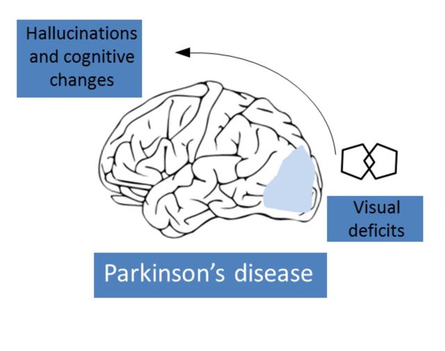
Patients with Parkinson's disease have a number of specific visual disturbances. These include changes in colour vision and contrast sensitivity and difficulties with complex visual tasks such as mental rotation and emotion recognition. We review changes in visual function at each stage of visual processing from retinal deficits, including contrast sensitivity and colour vision deficits to higher cortical processing impairments such as object and motion processing and neglect. We consider changes in visual function in patients with common Parkinson's disease-associated genetic mutations including GBA and LRRK2 . We discuss the association between visual deficits and clinical features of Parkinson's disease such as rapid eye movement sleep behavioural disorder and the postural instability and gait disorder phenotype. We review the link between abnormal visual function and visual hallucinations, considering current models for mechanisms of visual hallucinations. Finally, we discuss the role of visuo-perceptual testing as a biomarker of disease and predictor of dementia in Parkinson's disease.
Overview
The claims and statistics of this paper haven’t been checked yet.
Free: it reads the open-access text and checks the first 5 claims that cite a source we can check. Create a free account to check every claim.
Integrity signals
Registry
Cites
Statistics
Figures
Verification
Claims the paper makes
The claims and statistics of this paper haven’t been checked yet.
Free: it reads the open-access text and checks the first 5 claims that cite a source we can check. Create a free account to check every claim.
Self-check
Percentages, sums and reported differences re-computed from the paper’s own numbers.
The self-check needs the paper’s full text.
Citations
Cites
The precuneus: a review of its functional anatomy and behavioural correlates
Cavanna, A. E., & Trimble, M. R. (2006) · Brain · doi:10.1093/brain/awl004 · Cited by 5,128 · cited 2×
“Finally, in a structure from motion task, where volunteers detect an object that is formed from the contrast of moving dots, patients with Parkinson’s disease show higher thresholds for correctly identifying objects (Uc et al., 2005).”
BackgroundApplication of fourier analysis to the visibility of gratings
Campbell, F. W., & Robson, J. G. (1968) · The Journal of Physiology · doi:10.1113/jphysiol.1968.sp008574 · Cited by 3,019 · cited 3×
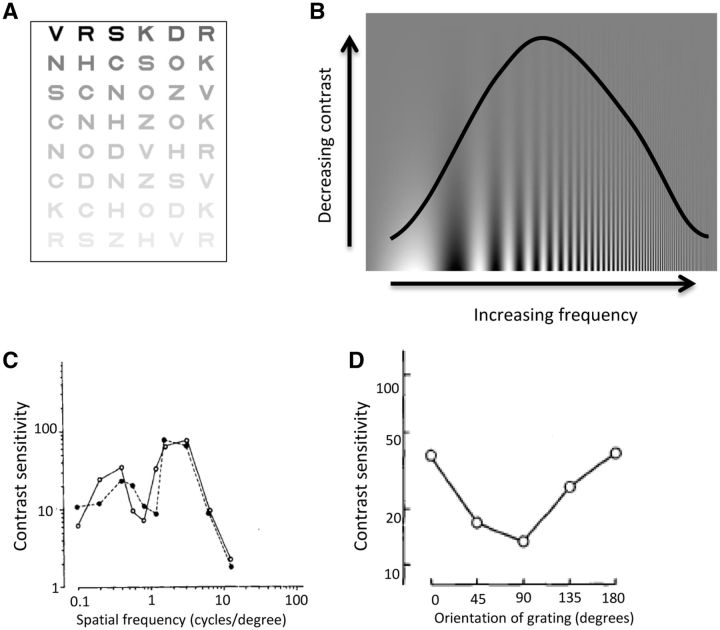
“This is more suggestive of a cortical rather than retinal or geniculate site of disruption as cortical neurons show stronger selectivity for different spatial frequencies than neurons in the retina or thalamus (Campbell and Robson, 1968; De Valois et al., 1982; Regan, 1982).”
BackgroundStages in the development of Parkinson’s disease-related pathology
Braak, H., Ghebremedhin, E., Rüb, U., Bratzke, H., & Del Tredici, K. (2004) · Cell and Tissue Research · doi:10.1007/s00441-004-0956-9 · Cited by 2,689 · cited 1×
“One theory of disease progression in Parkinson’s disease has suggested that the cerebral cortex is not damaged until the later stages of the disease (Braak et al., 2004).”
BackgroundThe Sydney multicenter study of Parkinson's disease: The inevitability of dementia at 20 years
Hely, M. A., Reid, W. G. J., Adena, M. A., Halliday, G. M., & Morris, J. G. L. (2008) · Movement Disorders · doi:10.1002/mds.21956 · Cited by 2,246 · cited 1×
“Visual hallucinations are also common in Parkinson’s disease, with a reported prevalence as high as 74% after 20 years of disease (Fenelon et al., 2000; Hely et al., 2008).”
BackgroundA direct demonstration of functional specialization in human visual cortex
Zeki, S., Watson, J., Lueck, C., Friston, K., Kennard, C., & Frackowiak, R. (1991) · The Journal of Neuroscience · doi:10.1523/jneurosci.11-03-00641.1991 · Cited by 1,716 · cited 2×
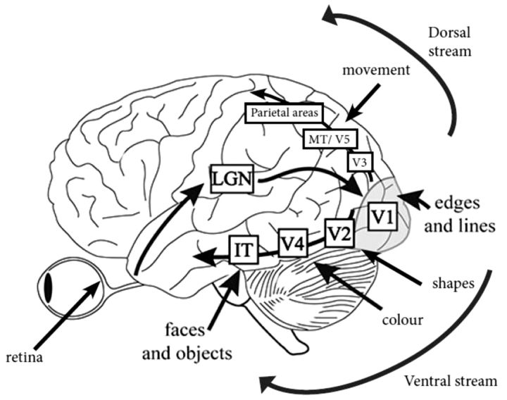
“IT = infero-temporal region; MT/V5 = motion processing region.”
BackgroundPharmacological Treatment of Parkinson Disease: A Review
Connolly, B. S., & Lang, A. E. (2014) · JAMA · doi:10.1001/jama.2014.3654 · Cited by 1,498 · cited 2×
“General advice is to consider visuo-perceptual ability and if there is any doubt, to refer the patient for a formal driving assessment via the national driving authority (Crizzle et al., 2012).”
BackgroundSpatial frequency selectivity of cells in macaque visual cortex
De Valois, R. L., Albrecht, D. G., & Thorell, L. G. (1982) · Vision Research · doi:10.1016/0042-6989(82)90113-4 · Cited by 1,489 · cited 2×
“This is more suggestive of a cortical rather than retinal or geniculate site of disruption as cortical neurons show stronger selectivity for different spatial frequencies than neurons in the retina or thalamus (Campbell and Robson, 1968; De Valois et al., 1982; Regan, 1982).”
Functional analysis of human MT and related visual cortical areas using magnetic resonance imaging
Tootell, R., Reppas, J., Kwong, K., Malach, R., Born, R., Brady, T., Rosen, B., & Belliveau, J. (1995) · The Journal of Neuroscience · doi:10.1523/jneurosci.15-04-03215.1995 · Cited by 1,415 · cited 2×
“IT = infero-temporal region; MT/V5 = motion processing region.”
BackgroundMazzulli, J. R., Xu, Y.-H., Sun, Y., Knight, A. L., McLean, P. J., Caldwell, G. A., Sidransky, E., Grabowski, G. A., & Krainc, D. (2011) · Cell · doi:10.1016/j.cell.2011.06.001 · Cited by 1,341 · cited 1×
“Both the GBA and SNCA genes are linked with the lysosomal pathway (Mazzulli et al., 2011), suggesting the visual deficits associated with these genes may share a pathophysiological mechanism, with lysosomal dysfunction associated with cortical visual dysfunction.”
BackgroundTHE DESIGN OF A NEW LETTER CHART FOR MEASURING CONTRAST SENSITIVITY
Pelli, D. G., Robson, J. G., & Wilkins, A. J. (1988) · Cited by 1,308
Research works printed but not linked: 3
- Ogden, 1990, Deficits on visuospatiaol tests involving forward planning in high-functioning Parkinsonians, Neuropsychiatry Neuropsychol Behav Neurol, 3, 125
- 10.1037/0033-295X.89.4.407 · doi:10.1037/0033-295x.89.4.407
- 10.1097/00006324-199404000-00002 · doi:10.1097/00006324-199404000-00002
Cited by
Items 201–300 of 479
Xu, T., Liu, X., Lin, X., Xiao, J., Zhang, D., Ye, F., Lü, F., Qu, J., Zhang, J., & Chen, J. (2024) · American Journal Of Pathology · doi:10.1016/j.ajpath.2024.01.017 · Cited by 10
Wong, P., Chen-Wei, H. U. N. G., Yang, Y., Yeh, N.-C., Cheng, S.-J., Liao, Y.-Y., & Wang, R. (2024) · European Journal of Physical and Rehabilitation Medicine · doi:10.23736/s1973-9087.24.08261-3 · Cited by 10
Beard, K., Gauff, A. K., Pennington, A. M., Marion, D. W., Smith, J. M., & Sloley, S. S. (2024) · Journal of Neurotrauma · doi:10.1089/neu.2024.0136 · Cited by 11
Convergence insufficiency and Parkinson's disease progression
Herrero‐Gracia, A., Hernández‐Andrés, R., Cobija, M. J. L., Ciuffreda, K. J., & Díez-Ajenjo, M. A. (2025) · Parkinsonism & Related Disorders · doi:10.1016/j.parkreldis.2025.107341 · Cited by 12
Retinal Changes in Parkinson's Disease
Mailankody, P., Battu, R., Lenka, A., Shereef, P. M. M., Thennarasu, K., Yadav, R., & Pal, P. K. (2022) · Neurology India · doi:10.4103/0028-3886.349658 · Cited by 9
Huang, Y.-C., Hong, C., Chi, W., Yen, C.-F., Liao, H. F., Liou, T., & Chan, L. (2024) · Archives of Gerontology and Geriatrics · doi:10.1016/j.archger.2024.105366 · Cited by 10
Mannone, M., Fazio, P., Kurths, J., Ribino, P., & Marwan, N. (2024) · The European Physical Journal Special Topics · doi:10.1140/epjs/s11734-024-01345-6 · Cited by 11
Eye as an object of investigation of cognitive impairment in Parkinson's disease
Litvinenko, I., Дынин, П. С., Trufanov, A. G., Gimadutdinov, R. F., & Maltsev, D. S. (2018) · S S Korsakov Journal of Neurology and Psychiatry · doi:10.17116/jnevro2018118062105 · Cited by 9
Yıldız, D., Pekel, N. B., Yener, N. P., Seferoğlu, M., Güneş, A., & Sığırlı, D. (2019) · Annals of Indian Academy of Neurology · doi:10.4103/aian.aian_424_17 · Cited by 14
Dik, D., Halliday, G. M., Sytnyk, V., & Shepherd, C. E. (2025) · Brain · doi:10.1093/brain/awaf305 · Cited by 11
Verghese, S., Berkowitz, S. T., Shah, V. M., Shah, P. K., Priya, S., Saravanan, V., Narendran, V., & Selvan, V. (2022) · Indian Journal of Ophthalmology · doi:10.4103/ijo.ijo_1409_21 · Cited by 8
“[19] Deficits in visual acuity, color, and contrast sensitivity are at least partly due to retinal dopamine deficiency in patients with PD as they have reduced dopamine innervations around the fovea.[20] In a study by Kaur et al.”
BackgroundNagai, K., Kaneko, Y., Suzuki, M., Teramoto, H., Morita, A., Kamei, S., Watanabe, Y., Okada, M., & Uchiyama, M. (2019) · Neuroscience Research · doi:10.1016/j.neures.2019.11.003 · Cited by 8
Impaired sensory evidence accumulation and network function in Lewy body dementia
O’Callaghan, C., Firbank, M., Tomassini, A., Schumacher, J., O'Brien, J. T., & Taylor, J. (2021) · Brain Communications · doi:10.1093/braincomms/fcab089 · Cited by 10
“Visual dysfunction is common, ranging from deficits in early processes such as contrast and colour discrimination, to impairments in higher-order object perception.(18,19) Likewise, the co-ordination of brain networks subserving externally versus internally driven orienting processes is impaired.”
BackgroundRetinal Diseases and Parkinson Disease: A Population-Based Study
Chen, P.-C., Chung, C., Cheng, Y.-Y., Chen, W.-T., Hong, C., Chan, L., & Chien, L. (2021) · Frontiers in Neuroscience · doi:10.3389/fnins.2021.679092 · Cited by 8
“To minimize the potential of identifying retinal diseases secondary to glaucoma or optic nerve disease, a prevalent disease in the elderly population, we analyzed the risk of glaucoma and optic nerve diseases in parallel for the purpose of comparison (Weinreb et al., 2014).”
BackgroundChiew, W. A., Mathew, D., Kumar, C. M., Seet, E. C. P., Imani, F., & Khademi, S.-H. (2023) · Anesthesiology and Pain Medicine · doi:10.5812/aapm-136093 · Cited by 9
Non-Motor Symptoms in Parkinson's Disease: The Other Side of the Disease
El‐Mansoury, B., Maloui, A. B., Draoui, A., Hamdan, Y. A., Oukhrib, M., Smimih, K., Elmourid, A., Khiat, A. E., Boulbaroud, S., Meftah, I., et al. (2023) · Advances in medical diagnosis, treatment, and care (AMDTC) book series · doi:10.4018/978-1-6684-5156-4.ch003 · Cited by 10
Visual Hallucinations and Illusions
Liu, G. T., Volpe, N. J., & Galetta, S. L. (2018) · Elsevier eBooks · doi:10.1016/b978-0-323-34044-1.00012-2 · Cited by 8
Leodori, G., Santilli, M., Modugno, N., D’Avino, M., Bartolo, M. I. D., Fabbrini, A., Rocchi, L., Conte, A., Fabbrini, G., & Belvisi, D. (2023) · Brain Sciences · doi:10.3390/brainsci13091243 · Cited by 7
“However, patients were tested with eyes closed, and since an abnormal visual control of balance is thought to contribute to postural instability in PD, performing the tests with the eyes closed may have limited the BSS sensitivity to probe postural control in this population [32].”
BackgroundImpaired pre-synaptic plasticity and visual responses in auxilin-knockout mice
Cheng, X., Tang, Y. J., Vidyadhara, D. J., Li, B., Zimmerman, M., Pak, A., Nareddula, S., Edens, P. A., Chandra, S. S., & Chubykin, A. A. (2023) · iScience · doi:10.1016/j.isci.2023.107842 · Cited by 8
Pan, Y., Qu, H., Zhao, Y., Qin, Z., Liu, J., & Wang, W. (2025) · Brain Research Bulletin · doi:10.1016/j.brainresbull.2025.111460 · Cited by 7
“Studies have suggested that VIS dysfunction in patients with PD may lead to specific visual disturbances (Hou et al., 2021; Weil et al., 2016), affecting a range of motor and non-motor behaviors, from visual-spatial functions to sensorimotor functions (Cucca et al., 2021; Hou et al., 2021),…”
Olmos, J., Antoine Manzanera, B., & Martínez, F. (2023) · Pattern Recognition Letters · doi:10.1016/j.patrec.2023.09.012 · Cited by 8
He, G., Huang, X., Sun, H.-H., Xing, Y., Gu, S., Ren, J., Liu, W., & Lu, M. (2024) · CNS Neuroscience & Therapeutics · doi:10.1111/cns.14867 · Cited by 7
“45,46 50 Such alterations could impair the visual processing of emotional cues and the management (SWS).”
BackgroundDynamics of retinal changes in early-stage Parkinson’s disease
Murueta‐Goyena, A., Teijeira‐Portas, S., Martín, E. B., Vázquez-Picón, R., Bajo, B. R., Portillo, J. B., Molina, J. S., Dias, P., Croitoru, I. M., Rodríguez-Agirretxe, I., et al. (2025) · Acta Neuropathologica Communications · doi:10.1186/s40478-025-01936-x · Cited by 7
Use of a Modified STROOP Test to Assess Color Discrimination Deficit in Parkinson's Disease
Langston, R. G., & Virmani, T. (2018) · Frontiers in Neurology · doi:10.3389/fneur.2018.00765 · Cited by 7
“Although several underlying mechanisms have been proposed, the connection between iPD pathogenesis and decline in visual capabilities remains incompletely understood (7).”
BackgroundAnoop, U. R., & Verma, K. (2021) · BMJ Innovations · doi:10.1136/bmjinnov-2019-000393 · Cited by 6
Validation of a non-motor fluctuations questionnaire in Parkinson's disease
Faggianelli, F., Loundou, A., Baumstarck, K., Nathalie, S., Auquier, P., Eusébio, A., Defebvre, L., Brefel‐Courbon, C., Houéto, J., Maltête, D., et al. (2021) · Revue Neurologique · doi:10.1016/j.neurol.2021.06.013 · Cited by 8
Student, J., Engel, D., Timmermann, L., Bremmer, F., & Waldthaler, J. (2022) · Frontiers in Human Neuroscience · doi:10.3389/fnhum.2022.762380 · Cited by 7
van der Lijn, I., de Haan, G. A., van der Feen, F. E., Huizinga, F., Fuermaier, A. B. M., van Laar, T., & Heutink, J. (2022) · PLoS ONE · doi:10.1371/journal.pone.0272559 · Cited by 7
Das, J., Morris, R., Barry, G., Vitório, R., Oman, P., McDonald, C., Walker, R. W., & Stuart, S. (2022) · PLoS ONE · doi:10.1371/journal.pone.0275738 · Cited by 10
“NMS are difficult to treat and include autonomic dysfunction, pain, cognitive dysfunction and sensory deficits including visual dysfunction [4].”
BackgroundUse of artificial intelligence algorithms to predict systemic diseases from retinal images
Khan, R., Surya, J., Roy, M., Priya, M. N. S., Mohan, S., Raman, S., Raman, A., Vyas, A., & Raman, R. (2023) · Wiley Interdisciplinary Reviews Data Mining and Knowledge Discovery · doi:10.1002/widm.1506 · Cited by 9
Tamilselvam, Y. K., Jog, M., & Patel, R. V. (2023) · IEEE Transactions on Neural Systems and Rehabilitation Engineering · doi:10.1109/tnsre.2023.3299884 · Cited by 9
“visual [7], haptic [8] and auditory perception [9]) in PD patients.”
Contrast Acuity With Different Colors in Parkinson's Disease
Gupta, H., Zhang, N., Driver‐Dunckley, E. D., Mehta, S. H., Beach, T. G., & Adler, C. H. (2019) · Movement Disorders Clinical Practice · doi:10.1002/mdc3.12821 · Cited by 9
“perception, and visuospatial construction have been described in PD.(1) Normal visual function requires several components, such”
BackgroundInfluentialWilson, D., Hallett, M., & Anderson, T. (2021) · Movement Disorders Clinical Practice · doi:10.1002/mdc3.13317 · Cited by 7
Levodopa therapy affects brain functional network dynamics in Parkinson's disease
Liu, X., Zhang, Y., Weng, Y., Zhong, M., Wang, L., Gao, Z., Hu, H., Zhang, Y., Huang, B., & Huang, R. (2024) · Progress in Neuro-Psychopharmacology and Biological Psychiatry · doi:10.1016/j.pnpbp.2024.111169 · Cited by 6
Grass, L., Grimaldi, S., & Damier, P. (2025) · Revue Neurologique · doi:10.1016/j.neurol.2025.06.012 · Cited by 9
Camacho-Ordóñez, A., Cervantes‐Arriaga, A., Rodríguez‐Violante, M., Hernandez-Medrano, A. J., Somilleda-Ventura, S. A., Pérez-Cano, H. J., Nava-Castañeda, Á., & Guerrero-Berger, Ó. (2023) · European Journal of Ophthalmology · doi:10.1177/11206721231173725 · Cited by 9
“10,11 The retina is considered an extension of the central nervous system and contains dopaminergic amacrine cells in the inner nuclear retinal layer.”
BackgroundShen, Q., Liao, H., Cai, S., Liu, Q., Wang, M., Song, C., Zhou, F., Liu, Y., Yuan, J., Tang, Y., et al. (2023) · Frontiers in Aging Neuroscience · doi:10.3389/fnagi.2023.1241516 · Cited by 6
“These alterations have been considered to be linked to impaired visual processing in these regions and visual-cognitive deficits in diagnosed PD (Weil et al., 2016).”
BackgroundEEG-Based Methods for Diagnosing Color Vision Deficiency: A Comprehensive Review
AlEssa, G. N., & Alzahrani, S. I. (2024) · Applied Sciences · doi:10.3390/app14177579 · Cited by 6
Better understanding fall risk: AI-based computer vision for contextual gait assessment
Moore, J., McMeekin, P. J., Stuart, S., Morris, R., Celik, Y., Walker, R. W., Hetherington, V., & Godfrey, A. (2024) · Maturitas · doi:10.1016/j.maturitas.2024.108116 · Cited by 7
Visual hallucinations in Parkinson's disease are associated with deficits in social perception
Albert, L., Vehar, N., Potheegadoo, J., Bernasconi, F., & Blanke, O. (2025) · Journal of Parkinson s Disease · doi:10.1177/1877718x251336196 · Cited by 6
“The involvement of these brain areas and networks in NEH and VH in PD also seems of particular relevance, as recent brain imaging work revealed involvement of premotor and prefrontal regions in PD patients with minor hallucinations that generally precede VH. 96 Overall, we showed that the overestimation bias is an imp…”
InfluentialFlickering Stimuli Do Not Reliably Induce Visual Hallucinations in Parkinson’s Disease
Zarkali, A., Lees, A. J., & Weil, R. S. (2019) · Journal of Parkinson s Disease · doi:10.3233/jpd-191635 · Cited by 10
“No participants experienced typical formed hallucinations of people or animals that characterise PD-associated hallucinations [1, 20].”
BackgroundSelf-citationAlcock, L., Galna, B., Hausdorff, J. M., Lord, S., & Rochester, L. (2020) · Neuroscience · doi:10.1016/j.neuroscience.2020.03.032 · Cited by 6
Emotional valence may influence memory performance for visual artworks in Parkinson’s disease
Ciccarelli, N., Tella, S. D., Monaco, M. R. L., Carfì, A., Serafini, E., Donne, V. D., & Silveri, M. C. (2019) · Neurological Sciences · doi:10.1007/s10072-019-03939-8 · Cited by 6
Disorders of music processing in dementia
Benhamou, E., & Warren, J. D. (2020) · Elsevier eBooks · doi:10.1016/b978-0-12-817422-7.00004-3 · Cited by 7
Self-citationStability of Action and Kinesthetic Perception in Parkinson’s Disease
Ricotta, J. M., & Latash, M. L. (2021) · Journal of Human Kinetics · doi:10.2478/hukin-2021-0006 · Cited by 8
Bonnet, C. T., Delval, A., Singh, T., & Defebvre, L. (2021) · European Journal of Neuroscience · doi:10.1111/ejn.15351 · Cited by 6
“PD patients also have visual impairments (e.g., altered contrast sensitivity and double vision; Ekker et al., 2017; Weil et al., 2016) and cognitive impairments (Yang et al.”
BackgroundVisual Dysfunction and Parkinson's Disease
Weil, R. S. (2020) · Movement Disorders · doi:10.1002/mds.28212 · Cited by 7
“with loss of visual acuity and color vision as well as higher order visual deficits.(1) A related observation is”
BackgroundSelf-citationStampacchia, S., Bernasconi, F., Van De Ville, D., Amico, E., Pagonabarraga, J., Kulisevsky, J., & Blanke, O. (2025) · doi:10.1101/2025.04.17.649310 · Cited by 5
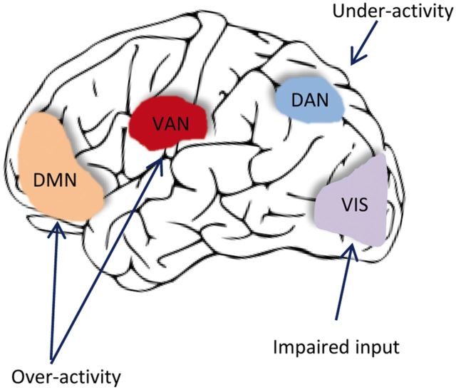
“…in PD arise from decreased activation of the dorsal attentional network (DAN) when processing ambiguous visual stimuli arising from altered bottom-up processing in early visual areas 27 , leading to over-reliance on top-down signals from default mode (DMN) and/or salience (SAL) networks.”
Lu, W., Song, T., Zang, Z., Li, J., Zhang, Y., & Lu, J. (2024) · NeuroImage · doi:10.1016/j.neuroimage.2024.120943 · Cited by 5
Churchill, L., Ignatavicius, A., Konuri, A., Anderson, J., Taylor, N. L., Lewis, S. J. G., & Matar, E. (2025) · Movement Disorders · doi:10.1002/mds.70025 · Cited by 9
“52 Dysfunction within the visual network, primarily composed of regions within the occipital lobe, has been well-documented in both PD and DLB, 53,54 where it is associated with cognitive decline and dementia development.”
Fraza, C., Bučková, B. R., Johansson, M. E., Helmich, R. C. G., Marquand, A. F., & Beckmann, C. F. (2025) · Scientific Reports · doi:10.1038/s41598-025-20484-x · Cited by 7
Biassoni, E., Kreshpa, W., Massa, F., D’Amico, F., Bauckneht, M., Arnaldi, D., Pardini, M., Orso, B., Girtler, N. G., Brugnolo, A., et al. (2023) · Parkinsonism & Related Disorders · doi:10.1016/j.parkreldis.2023.105371 · Cited by 6
Rong, D., Chuan-Peng, H., Yang, J., Guo, Z., Liu, W., & Yu, M. (2024) · Brain Research Bulletin · doi:10.1016/j.brainresbull.2024.110933 · Cited by 6
“Several signs of a dysfunctional visual pathway emerge in the retina (Ahn et al., 2018) during the early stages of PD (Weil et al., 2016) and progress along with the disease to higher visual brain areas (Bellot et al., 2022; Marinus et al., 2018).”
BackgroundXiao, K., Li, J., Zhou, L., Liu, X., Xiao, Z., He, R., Chu, H., Tang, Y., Liu, P., & Lu, X. (2024) · Neuroscience · doi:10.1016/j.neuroscience.2024.11.073 · Cited by 7
The Many Faces of Blurry Vision in Parkinson’s Disease: An Illustrative Case Series
Borm, C. D. J. M., Bloem, B. R., Hoyng, C. B., de Vries, N. M., & Theelen, T. (2022) · Case Reports in Neurology · doi:10.1159/000523987 · Cited by 5
Functional topologies of spatial cognition predict cognitive and motor progression in Parkinson’s
Harrington, D. L., Shen, Q., Wei, X., Litvan, I., Huang, M., & Lee, R. R. (2022) · Frontiers in Aging Neuroscience · doi:10.3389/fnagi.2022.987225 · Cited by 6
“Since the frontal cortex, posterior cortex, and the basal ganglia govern different facets of spatial cognition (Helmich et al., 2007; Weil et al., 2016; Garcia-Diaz et al., 2018), PCA was conducted separately for angle-modulated connectivity features of seeds within each of these areas to characterize their strength a…”
BackgroundMethodPatil, S. A., Grossman, S. N., Kenney, R., Balcer, L. J., & Galetta, S. L. (2022) · Neurology · doi:10.1212/wnl.0000000000201490 · Cited by 8
“OCT and OCT Angiography in Neurodegenerative Disease A growing literature is investigating roles for OCT and OCT angiography measurements as potential markers for AD and PD progression.(7) Measures investigated include RNFL and GCIPL thickness as well as foveal and parafoveal macular thickness, with comparisons with c…”
BackgroundKim, S., Choi, J.-H., Woo, K. A., Joo, J. Y., Jeon, B., & Lee, J. (2023) · Journal of Neural Transmission · doi:10.1007/s00702-023-02724-4 · Cited by 6
“…between abnormalities in color vision and cognitive dysfunction, reporting that performance of the Farns-worth–Munsell 100 hue test, a measure to evaluate color discrimination abilities, correlated with measures of visuospatial and executive function (Weil et al. 2016; Bertrand et al. 2012).”
Gonçalves, A., Mendes, A., Damásio, J., Vila‐Chã, N., Boleixa, D., Leal, B., & Cavaco, S. (2024) · Journal of Parkinson s Disease · doi:10.3233/jpd-230292 · Cited by 7
A deep learning model of dorsal and ventral visual streams for DVSD
Zareh, M., Toulabinejad, E., Manshaei, M. H., & Zahabi, S. J. (2024) · Scientific Reports · doi:10.1038/s41598-024-78304-7 · Cited by 9
PANDA: Parkinson's Assistance and Notification Driving Aid
Wen, T., Zhang, X., Wan, Z., Zhao, J., Zhu, Y., Su, N., Peng, X., Huang, J., Sun, W. G., Tian, F., et al. (2025) · CHI Conference on Human Factors in Computing Systems · doi:10.1145/3706598.3713920 · Cited by 5
“These systems rely on diverse data sources, including vehicle status parameters such as steering wheel input, pedal activity, speed, and lane-keeping data, accessed via the CAN bus [20, 41, 45].”
Clinical Significance and Pathogenesis of Visual Impairment in Parkinson’s Disease
Alenicova, O. A. (2017) · Journal of Neurology & Stroke · doi:10.15406/jnsk.2017.07.00266 · Cited by 5
“Studies based on these tests have revealed color discrimination disorders, mostly in the blue-green spectrum, in moderate and severe stages of PD, also a relationship has been found between the CV disturbances and deteriorating visuospatial orientation and cognitive functions [13].”
BackgroundNew insight into Parkinson’s disease‐related impairment of the automatic control of upright stance
Bonnet, C. T., Delval, A., Singh, T., Kechabia, Y., & Defebvre, L. (2020) · European Journal of Neuroscience · doi:10.1111/ejn.14870 · Cited by 5
“The most important difference between white fixation and structured fixation is that PD patients were potentially distracted by peripheral objects, which interfered with the main (fixation) task (Weil et al., 2016).”
BackgroundOhdake, R., Kawabata, K., Watanabe, H., Hara, K., Ogura, A., Kato, T., Masuda, M., Katsuno, M., & Sobue, G. (2020) · Journal of Neural Transmission · doi:10.1007/s00702-020-02248-1 · Cited by 6
Increasing Contrast Improves Object Perception in Parkinson's Disease with Visual Hallucinations
Díaz‐Santos, M., Monge, Z. A., Salazar, R. D., Gilmore, G. C., Neargarder, S. A., & Cronin‐Golomb, A. (2020) · Movement Disorders Clinical Practice · doi:10.1002/mdc3.13104 · Cited by 5
Veréb, D., Kovács, M. A., Antal, S. I., Kocsis, K., Szabó, N., Kincses, B., Bozsik, B., Faragó, P., Tóth, E. M., Király, A., et al. (2022) · Frontiers in Neurology · doi:10.3389/fneur.2022.927481 · Cited by 5
“The trajectory of some of these changes not only corresponds to increasing motor deficits, in line with dopaminergic neuron loss (2), but also non-axial symptoms (5) that highlights their heterogeneous nature.”
BackgroundLai, H., Zhang, B.-R., Shen, T., Pu, J., Jiang, Y., Yue, Y.-M., He, T., Qu, B., Zhao, S., & Yan, Y. (2022) · Neural Regeneration Research · doi:10.4103/1673-5374.355764 · Cited by 7
“Furthermore, higher visual processing regions, including the occipital and parietal regions, are affected in PD (Weil et al., 2016).”
Chen, S., Zhong, H., & Mei, G. (2022) · PsyCh Journal · doi:10.1002/pchj.525 · Cited by 5
Thomas, G. E. C., Zeidman, P., Sultana, T., Zarkali, A., Razi, A., & Weil, R. S. (2022) · bioRxiv (Cold Spring Harbor Laboratory) · doi:10.1101/2022.03.25.485621 · Cited by 5
Self-citationInvestigation of symptom-specific functional connectivity patterns in Parkinson’s disease
Kıçık, A., Bayram, A., Erdoğdu, E., Kurt, E., Arslan, D. B., Cengiz, S. D., Bılgıç, B., HANAĞASI, H. A., Öztürk-Işık, E., Gurvit, H. I., et al. (2025) · Neurological Sciences · doi:10.1007/s10072-025-08287-4 · Cited by 4
Hatsuta, H., Mizutani, M., Yagita, K., Sano, T., & Takao, M. (2025) · Parkinsonism & Related Disorders · doi:10.1016/j.parkreldis.2025.107935 · Cited by 4
Visuospatial impairment in Parkinson's Disease: why we should keep an eye on it
Oliveira, H. S. D., Camargos, S., Cardoso, F., & Caramelli, P. (2025) · Parkinsonism & Related Disorders · doi:10.1016/j.parkreldis.2025.108064 · Cited by 7
Dan, T., Ding, J., & Guorong Wu, A. (2025) · Nature Communications · doi:10.1038/s41467-025-64471-2 · Cited by 5
“The resulting “ visual belt ” mirrors the ‘ β / γ ’ -band hypersynchrony and tremor-related cerebello-thalamo-visual loop repeatedly reported in PPMI cohorts 41 .”
Ophey, A., Farrher, E., Pagel, N., Seger, A., Doppler, C. E. J., Shah, N. J., Kalbe, E., Fink, G. R., & Sommerauer, M. (2023) · European Journal of Neurology · doi:10.1111/ene.15818 · Cited by 4
“Hence, recognizing visuo- cognitive dysfunction operationalized by early metabolic alterations may serve as a potential prognostic marker of PD- D [9].”
BackgroundInfluentialSheldrake, E., McGrath, C., Rudman, D. L., & Holmes, J. (2021) · Physical & Occupational Therapy In Geriatrics · doi:10.1080/02703181.2021.1989545 · Cited by 4
Maio, L. G. D., Cennamo, G., Montorio, D., Michele, G. D., Palmieri, G. R., Baratto, L., Perillo, S., Giglio, A., Filla, A., Morra, V. B., et al. (2024) · Acta Neurologica Scandinavica · doi:10.1155/2024/8555083 · Cited by 5
“Furthermore, several retinal visual disturbances are complained of by PD subjects, such as reduced visual acuity, visual fi eld defects, color vision impairment, and reduced contrast sensitivity [3, 4].”
Ocular disorders in Parkinson’s disease: A review
Bansal, Y., & Grover, R. (2024) · Journal of Clinical Ophthalmology and Research · doi:10.4103/jcor.jcor_111_23 · Cited by 4
Jiang, M., Liu, Y., Cao, Y., Liu, Y., Wang, J., Li, P., Xia, S., Li, P., & Liu, W. (2024) · Neuroscience Letters · doi:10.1016/j.neulet.2024.137956 · Cited by 6
Revankar, G. S., Ozono, T., Suzuki, M., Kanemoto, H., Furuya, K., Shigenobu, K., Yoshiyama, K., Yamamoto, Y., Ogasawara, I., Yoshida, N., et al. (2024) · Heliyon · doi:10.1016/j.heliyon.2024.e40254 · Cited by 4
Brown, T., Kanel, P., Griggs, A., Carli, G., Vangel, R., Albin, R. L., & Bohnen, N. I. (2024) · Parkinsonism & Related Disorders · doi:10.1016/j.parkreldis.2024.107229 · Cited by 4
Cortical myelin content mediates differences in affective temperaments
Qiu, Y., She, S., Zhang, S., Wu, F., Liang, Q., Peng, Y., Yuan, H., Ning, Y., Wu, H., & Huang, R. (2021) · Journal of Affective Disorders · doi:10.1016/j.jad.2021.01.038 · Cited by 4
Genoves, G. G., Cruz, C. F., Doná, F., Andrade, T., Ferraz, H., & Barela, J. (2021) · Neurophysiologie clinique · doi:10.1016/j.neucli.2021.03.005 · Cited by 4
Retrograde Procedural Memory in Parkinson’s Disease: A Cross-Sectional, Case-Control Study
Pauly, L., Pauly, C., Hansen, M., Schröder, V. E., Rauschenberger, A., Leist, A. K., & Krüger, R. (2022) · Journal of Parkinson s Disease · doi:10.3233/jpd-213081 · Cited by 10
“In comparison with the control group, the patient group presented impaired visuo-constructive functions, in line with previous findings on Parkinson’s disease [39].”
ResultFernández-Baizán, C., García, M. P. F., Diaz-Caceres, E., Menéndez‐González, M., Arias, J. L., & Méndez, M. (2022) · Topics in Geriatric Rehabilitation · doi:10.1097/tgr.0000000000000376 · Cited by 6
Intrasession Repeatability of OCT Angiography Parameters in Neurodegenerative Disease
Akrobetu, D., Robbins, C. B., Ma, J. P., Soundararajan, S., Quist, M. S., Stinnett, S. S., Moore, K. P. L., Johnson, K. G., Liu, A., Grewal, D. S., et al. (2023) · Ophthalmology Science · doi:10.1016/j.xops.2023.100275 · Cited by 4
Susceptibility to geometrical visual illusions in Parkinson’s disorder
Wincza, R., Hartley, C., Readman, M. R., Linkenauger, S. A., & Crawford, T. J. (2024) · Frontiers in Psychology · doi:10.3389/fpsyg.2023.1289160 · Cited by 5
Tran, K. K. N., Wong, V. H. Y., Vessey, K. A., Finkelstein, D. I., Bui, B. V., & Nguyen, C. T. O. (2024) · Biomedicines · doi:10.3390/biomedicines12010130 · Cited by 5
“Parkinson’s disease is well known for its motor manifestations [16,17]; however, patients can manifest a multitude of non-motor symptoms [18–21], ranging from olfactory desensitisation [22] to autonomic dysfunction [23] to changes in vision [24] by way of PD’s complex pathogenesis profile.”
BackgroundUrsachen visueller Halluzinationen bei der Parkinson-Krankheit
Diederich, N. J. (2021) · Der Nervenarzt · doi:10.1007/s00115-021-01165-2 · Cited by 4
Impact of Visual Impairment on Vision-Related Quality of Life in Parkinson’s Disease
Pengo, M., Murueta‐Goyena, A., Teijeira‐Portas, S., Acera, M., Pino, R. D., Sáez-Atxukarro, O., Díez‐Cirarda, M., Tijero, B., Fernández‐Valle, T., Gómez‐Esteban, J. C., et al. (2022) · Journal of Parkinson s Disease · doi:10.3233/jpd-213143 · Cited by 4
Effects of Levodopa on Impairments to High-Level Vision in Parkinson's Disease
Anderson, S., & Stegemöller, E. L. (2020) · Frontiers in Neurology · doi:10.3389/fneur.2020.00708 · Cited by 7
“Deficits in low level visual processing are evident in persons with PD, including impairments in visual acuity (2, 3), contrast sensitivity (4), and line discrimination (4–6).”
BackgroundNon-motor symptoms of Parkinson's disease
El‐Mansoury, B., Khiat, A. E., Skaou, A., Hiba, O. E., Jayakumar, A. R., Hamdan, Y. A., Bitar, A., & Ferssiwi, A. (2024) · Elsevier eBooks · doi:10.1016/b978-0-443-15702-8.00017-8 · Cited by 4
Exploring ocular disorders in Parkinson's disease: A comprehensive review and future perspectives
Thacker, M., Wong, K. Y., Zhou, L., Liu, J., & Wong, M. (2024) · Experimental Eye Research · doi:10.1016/j.exer.2024.110225 · Cited by 3
Nikparast, F., Sharak, N. A., Ganji, Z., Shoeibi, A., Shoeibi, N., & Zare, H. (2025) · Photodiagnosis and Photodynamic Therapy · doi:10.1016/j.pdpdt.2025.105310 · Cited by 3
Albert, L., Bernasconi, F., Potheegadoo, J., & Blanke, O. (2024) · Parkinsonism & Related Disorders · doi:10.1016/j.parkreldis.2024.107195 · Cited by 4
Alterations in center-surround contrast suppression in patients with major depressive disorder
Nickel, K., Heinrich, S. P., Beringer, M., Endres, D., Runge, K., Küchlin, S., Maier, S. J., Bach, M., Domschke, K., van Elst, L. T., et al. (2024) · Scientific Reports · doi:10.1038/s41598-024-78584-z · Cited by 6
Das, J., Barry, G., Walker, R. W., Vitório, R., Çelik, Y., McDonald, C., Storey, B., Oman, P., Morris, R., & Stuart, S. (2024) · PLOS Digital Health · doi:10.1371/journal.pdig.0000696 · Cited by 4
Ma, Y., Wang, L., Li, T., Zhang, J., Funahashi, S., Wu, J., Wang, X., Zhang, K., Liu, T., & Yan, T. (2025) · Brain Structure and Function · doi:10.1007/s00429-025-02909-5 · Cited by 3
“…application relies on a clearer understanding of the pathological mechanism of PD. Neuroimaging evidence suggests that PD patients experience functional alterations in both the primary sensory regions and higher-order cognitive networks (Li et al. 2023a; Melzer et al. 2011; Weil et al. 2016).”
Schweyer, K., Mantel, T., Knier, B., Aly, L., Kirschke, J. S., Meindl, T., & Haslinger, B. A. (2025) · Brain and Behavior · doi:10.1002/brb3.70509 · Cited by 3
“In consideration of the fact that different domains of visual processing capacities seem to be affected, it has been suggested that both retinal and higher order neuronal dysfunction is involved (Weil et al. 2016).”
Data-driven characterization of distinct cognitive subtypes in Parkinson’s disease dementia
Labrador‐Espinosa, M. Á., Silva‐Rodríguez, J., Mir, P., & Grothe, M. J. (2025) · npj Parkinson s Disease · doi:10.1038/s41531-025-00970-9 · Cited by 4
“This fi nding is in line with previous research showing that PD patients carrying GBA variants exhibit a distinct cognitive pro fi le characterized by relatively greater impairments in visuospatial abilities 41 – 43 .”
Visual Hallucinations in Serotonergic Psychedelics and Lewy Body Diseases
Heller, N. H., Barrett, F. S., Buchborn, T., Collerton, D., Dupuis, D., Halberstadt, A. L., Jardri, R., Noorani, T. N., Preller, K. H., Taylor, J., et al. (2025) · Schizophrenia Bulletin · doi:10.1093/schbul/sbaf068 · Cited by 3
“65 This could confound comparisons of simple hallucinations in LBDs and SPs.”
Metadata
- Authors
- Weil, R. S., Schrag, A. E., Warren, J. D., Crutch, S. J., Lees, A. J., & Morris, H. R.
- Affiliations
- Royal Free London NHS Foundation Trust (GB)
- National Hospital for Neurology and Neurosurgery (GB)
- UCL Queen Square Institute of Neurology (GB)
- Corresponding author
- Weil, Rimona S.
- ORCID
- Venue
- Brain
- Publisher
- Oxford University Press (OUP)
- Published
- 2016-07-13
- Volume
- 139
- Issue
- 11
- Pages
- 2827-2843
- ISSN
- 0006-8950 (ISSN-L)
- 1460-2156
- PMC
- PMC5091042
- OpenAlex
- W2474900144
- Cited by
- 481
- References
- 232
- Citations by year
- 2026: 41 · 2025: 55 · 2024: 54 · 2023: 48 · 2022: 52 · 2021: 55 · 2020: 66 · 2019: 37 · 2018: 34 · 2017: 28 · 2016: 2
- FWCI
- 4.67
- Type
- journal article
- Language
- en
- Open access
- green
- www.ncbi.nlm.nih.gov
- Licence
- cc-by
- Locations
- Brain (publishedVersion)
- PubMed Central (publishedVersion, cc-by)
- PubMed (publishedVersion)
- UCL Discovery (University College London) (submittedVersion)
- HighWire Press Open Archive (submittedVersion)
- Funders
- National Institute for Health and Care Research
- Parkinson's UK (K-1501, J‐0804)
- Academy of Medical Sciences (AMS-SGCL13-Weil)
- Engineering and Physical Sciences Research Council (EP/M006093/1)
- Economic and Social Research Council (ES/L001810/1)
- Topics
- Health Sciences › Medicine › Clinical Neurology › Parkinson's Disease Mechanisms and Treatments
- Life Sciences › Neuroscience › Cognitive Neuroscience › Visual perception and processing mechanisms
- Life Sciences › Neuroscience › Cognitive Neuroscience › Hallucinations in medical conditions
- Keywords
- Parkinson's disease, visual dysfunction, contrast sensitivity, mental rotation, emotion recognition, visual neglect, motion processing, object processing, GBA mutations, LRRK2 mutations, REM sleep behavior disorder, postural instability, gait disturbance, visual hallucinations, biomarkers, dementia prediction
- MeSH terms
- Leucine-Rich Repeat Serine-Threonine Protein Kinase-2, beta-Glucosidase, Humans, Parkinson Disease, Vision Disorders, Visual Pathways, Visual Perception
About these checks
Registry, retraction, Cites and statistics results are computed, not judged, with one exception: a printed reference that no index lists is read once by a model before it counts as a citation error. Claims are extracted by a model and kept only when their quote appears verbatim in the text; each cited source is then checked, with a model’s help, for whether it says what the claim relies on. Those verdicts can be wrong — read the claim’s quote and the cited source before relying on one.
Statistics and arithmetic are recomputed, not judged.
Confidence bands are shown only for fields calibrated on our annotated test set. None is calibrated yet, so no band is shown.
What counts as a claim ›How we measure confidence ›How we check this ›
Are you an author, or do you see a problem with this page? Request a review
Data sources
Bibliographic data: Semantic Scholar, OpenAlex, Crossref.
Contains information from the Semantic Scholar Academic Graph Datasets, made available under the ODC Attribution License (ODC-BY 1.0).
Semantic Scholar’s data is described in: Kinney, R., et al. (2023). The Semantic Scholar Open Data Platform. arXiv:2301.10140.
