

Visual dysfunction in Parkinson’s disease
Open accessThe DOI matches its Crossref record; 233 of the 233 references it cites were found in a registry or an index.
Abstract
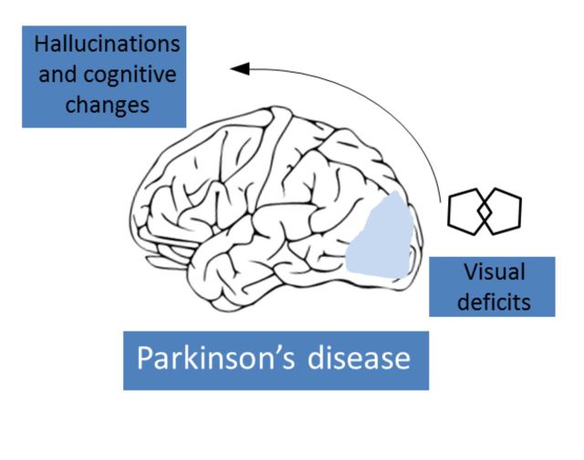
Patients with Parkinson's disease have a number of specific visual disturbances. These include changes in colour vision and contrast sensitivity and difficulties with complex visual tasks such as mental rotation and emotion recognition. We review changes in visual function at each stage of visual processing from retinal deficits, including contrast sensitivity and colour vision deficits to higher cortical processing impairments such as object and motion processing and neglect. We consider changes in visual function in patients with common Parkinson's disease-associated genetic mutations including GBA and LRRK2 . We discuss the association between visual deficits and clinical features of Parkinson's disease such as rapid eye movement sleep behavioural disorder and the postural instability and gait disorder phenotype. We review the link between abnormal visual function and visual hallucinations, considering current models for mechanisms of visual hallucinations. Finally, we discuss the role of visuo-perceptual testing as a biomarker of disease and predictor of dementia in Parkinson's disease.
Overview
The claims and statistics of this paper haven’t been checked yet.
Free: it reads the open-access text and checks the first 5 claims that cite a source we can check. Create a free account to check every claim.
Integrity signals
Registry
Cites
Statistics
Figures
Verification
Claims the paper makes
The claims and statistics of this paper haven’t been checked yet.
Free: it reads the open-access text and checks the first 5 claims that cite a source we can check. Create a free account to check every claim.
Self-check
Percentages, sums and reported differences re-computed from the paper’s own numbers.
The self-check needs the paper’s full text.
Citations
Cites
The precuneus: a review of its functional anatomy and behavioural correlates
Cavanna, A. E., & Trimble, M. R. (2006) · Brain · doi:10.1093/brain/awl004 · Cited by 5,128 · cited 2×
“Finally, in a structure from motion task, where volunteers detect an object that is formed from the contrast of moving dots, patients with Parkinson’s disease show higher thresholds for correctly identifying objects (Uc et al., 2005).”
BackgroundApplication of fourier analysis to the visibility of gratings
Campbell, F. W., & Robson, J. G. (1968) · The Journal of Physiology · doi:10.1113/jphysiol.1968.sp008574 · Cited by 3,019 · cited 3×
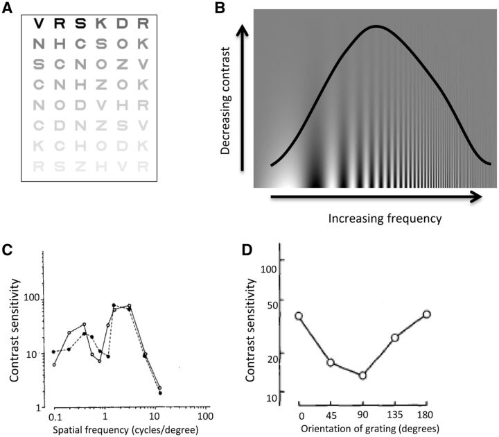
“This is more suggestive of a cortical rather than retinal or geniculate site of disruption as cortical neurons show stronger selectivity for different spatial frequencies than neurons in the retina or thalamus (Campbell and Robson, 1968; De Valois et al., 1982; Regan, 1982).”
BackgroundStages in the development of Parkinson’s disease-related pathology
Braak, H., Ghebremedhin, E., Rüb, U., Bratzke, H., & Del Tredici, K. (2004) · Cell and Tissue Research · doi:10.1007/s00441-004-0956-9 · Cited by 2,689 · cited 1×
“One theory of disease progression in Parkinson’s disease has suggested that the cerebral cortex is not damaged until the later stages of the disease (Braak et al., 2004).”
BackgroundThe Sydney multicenter study of Parkinson's disease: The inevitability of dementia at 20 years
Hely, M. A., Reid, W. G. J., Adena, M. A., Halliday, G. M., & Morris, J. G. L. (2008) · Movement Disorders · doi:10.1002/mds.21956 · Cited by 2,246 · cited 1×
“Visual hallucinations are also common in Parkinson’s disease, with a reported prevalence as high as 74% after 20 years of disease (Fenelon et al., 2000; Hely et al., 2008).”
BackgroundA direct demonstration of functional specialization in human visual cortex
Zeki, S., Watson, J., Lueck, C., Friston, K., Kennard, C., & Frackowiak, R. (1991) · The Journal of Neuroscience · doi:10.1523/jneurosci.11-03-00641.1991 · Cited by 1,716 · cited 2×
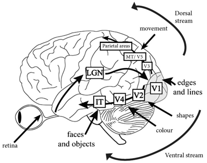
“IT = infero-temporal region; MT/V5 = motion processing region.”
BackgroundPharmacological Treatment of Parkinson Disease: A Review
Connolly, B. S., & Lang, A. E. (2014) · JAMA · doi:10.1001/jama.2014.3654 · Cited by 1,498 · cited 2×
“General advice is to consider visuo-perceptual ability and if there is any doubt, to refer the patient for a formal driving assessment via the national driving authority (Crizzle et al., 2012).”
BackgroundSpatial frequency selectivity of cells in macaque visual cortex
De Valois, R. L., Albrecht, D. G., & Thorell, L. G. (1982) · Vision Research · doi:10.1016/0042-6989(82)90113-4 · Cited by 1,489 · cited 2×
“This is more suggestive of a cortical rather than retinal or geniculate site of disruption as cortical neurons show stronger selectivity for different spatial frequencies than neurons in the retina or thalamus (Campbell and Robson, 1968; De Valois et al., 1982; Regan, 1982).”
Functional analysis of human MT and related visual cortical areas using magnetic resonance imaging
Tootell, R., Reppas, J., Kwong, K., Malach, R., Born, R., Brady, T., Rosen, B., & Belliveau, J. (1995) · The Journal of Neuroscience · doi:10.1523/jneurosci.15-04-03215.1995 · Cited by 1,415 · cited 2×
“IT = infero-temporal region; MT/V5 = motion processing region.”
BackgroundMazzulli, J. R., Xu, Y.-H., Sun, Y., Knight, A. L., McLean, P. J., Caldwell, G. A., Sidransky, E., Grabowski, G. A., & Krainc, D. (2011) · Cell · doi:10.1016/j.cell.2011.06.001 · Cited by 1,341 · cited 1×
“Both the GBA and SNCA genes are linked with the lysosomal pathway (Mazzulli et al., 2011), suggesting the visual deficits associated with these genes may share a pathophysiological mechanism, with lysosomal dysfunction associated with cortical visual dysfunction.”
BackgroundTHE DESIGN OF A NEW LETTER CHART FOR MEASURING CONTRAST SENSITIVITY
Pelli, D. G., Robson, J. G., & Wilkins, A. J. (1988) · Cited by 1,308
Research works printed but not linked: 3
- Ogden, 1990, Deficits on visuospatiaol tests involving forward planning in high-functioning Parkinsonians, Neuropsychiatry Neuropsychol Behav Neurol, 3, 125
- 10.1037/0033-295X.89.4.407 · doi:10.1037/0033-295x.89.4.407
- 10.1097/00006324-199404000-00002 · doi:10.1097/00006324-199404000-00002
Cited by
Items 301–400 of 479
Functional correlates of cognitive slowing in Parkinson's disease
Grolez, G., Viard, R., Lopes, R., Kuchcinski, G., Defebvre, L., Devos, D., Dujardin, K., & Moreau, C. (2020) · Parkinsonism & Related Disorders · doi:10.1016/j.parkreldis.2020.05.006 · Cited by 3
Cummings, L. (2020) · Cambridge University Press eBooks · doi:10.1017/9781108587921.001 · Cited by 4
The Association of Visually Guided Saccades and DAT-SPECT Findings in Parkinson’s Disease
Kobayashi, M., & Kuwabara, S. (2021) · European Neurology · doi:10.1159/000514246 · Cited by 3
Cummings, L. (2020) · Cambridge University Press eBooks · doi:10.1017/9781108587921.002 · Cited by 3
Oculomotor and Visual-Vestibular Disturbances in Parkinson’s Disease
Bronstein, A. M., Anderson, T., Kaski, D. N., MacAskill, M. R., & Shaikh, A. G. (2022) · Cambridge University Press eBooks · doi:10.1017/9781009039291.012 · Cited by 3
Piette, P., Fraudet, B., Vinatier, C., Leblong, É., & Galien, P. (2022) · International Journal of Physiotherapy and Research · doi:10.16965/ijpr.2022.121 · Cited by 2
“In addition, there are strong visual impairments in people with Parkinson’s [63].”
BackgroundOphthalmologic problems correlates with cognitive impairment in patients with Parkinson's disease
Zhang, C., Wu, Q., Hou, Y., Wang, Q., Zhang, G., Zhao, W., Wang, X., Wang, H., & Li, W. (2022) · Frontiers in Neuroscience · doi:10.3389/fnins.2022.928980 · Cited by 3
“The role of visual dysfunction as a biomarker of disease and a predictor of cognitive impairment in PD has been reported in previous studies (Weil et al., 2016).”
BackgroundResultInfluentialAn Oculomotor Digital Parkinson Biomarker from a Deep Riemannian Representation
Olmos, J., Antoine Manzanera, B., & Martínez, F. (2022) · Lecture notes in computer science · doi:10.1007/978-3-031-09037-0_55 · Cited by 4
Cold flow model testing of the HYTORT process retort design
Rex, R. C., Janka, J. C., & Knowlton, T. M. (1984) · OSTI OAI (U.S. Department of Energy Office of Scientific and Technical Information) · Cited by 3
Schizophrenia and visual perception disturbances
Torales, J. C., Meyer, E. D. D., & Fariña, E. D. (2020) · Revista Virtual de la Sociedad Paraguaya de Medicina Interna · doi:10.18004/rvspmi/2312-3893/2020.07.02.96 · Cited by 4
van der Lijn, I., de Haan, G. A., van der Feen, F. E., Huizinga, F., Stellingwerf, C., van Laar, T., & Heutink, J. (2023) · PLoS ONE · doi:10.1371/journal.pone.0283122 · Cited by 3
“Visual problems are increasingly recognized as part of the symptomatology of Parkinson’s disease (PD) [1].”
BackgroundMRI findings of visual system alterations in Parkinson’s disease
Arrigo, A., Calamuneri, A., Mormina, E., Aragona, P., Gaeta, M., & Quartarone, A. (2017) · Brain · doi:10.1093/brain/awx244 · Cited by 3
Sensorimotor hallucinations in Parkinson’s diseasePreprint
Bernasconi, F., Blondiaux, E., Potheegadoo, J., Stripeikyte, G., Pagonabarraga, J., Bejr‐Kasem, H., Bassolino, M., Akselrod, M., Martínez‐Horta, S., Sampedro, F., et al. (2020) · bioRxiv (Cold Spring Harbor Laboratory) · doi:10.1101/2020.05.11.054619 · Cited by 6
Gomes, M. T., Fernandes, H. M., & Cabral, J. (2020) · bioRxiv (Cold Spring Harbor Laboratory) · doi:10.1101/2020.11.04.368274 · Cited by 3
“Besides, people with PD and exhibiting ophthalmologic problems also present alterations in the VN [73].”
BackgroundRetrograde Procedural Memory in Parkinson's Disease
Pauly, L., Pauly, C., Hansen, M., Schröder, V. E., Rauschenberger, A., Leist, A. K., & Krüger, R. (2022) · Open Repository and Bibliography (University of Luxembourg) · Cited by 3
Color Vision Deficits and Binocular Vision Dysfunction in Parkinson’s Disease
Mleczek, J., Forjindam, A., Shaikh, A., & Ghasia, F. F. (2026) · Brain Sciences · doi:10.3390/brainsci16020213 · Cited by 2
“Parvocellular dysfunction is suggested to result from retinal structural changes and reduced retinal dopamine [4].”
Detailed Outer Retinal Assessment in Parkinson's Disease Using Directional OCT
Lin, J., Lee, P. Y., Tran, K. K. N., Finkelstein, D. I., Bui, B. V., & Nguyen, C. T. O. (2026) · Investigative Ophthalmology & Visual Science · doi:10.1167/iovs.67.3.59 · Cited by 2
“Moreover, increasing evidence of PD-related pathologies in the retina adds to the impetus to investigate OCT as a scalable biomarker for PD. 8,9 14–21 Discrepancy between studies may have arisen from differences in OCT scanning protocols, and in particular the likelihood that the Henle fiber layer…”
Visual Dysfunction and Occupational Performance in Persons With Parkinson’s Disease
Tester, N. J., Liu, C.-J., Shin, Y. C., & Shukla, A. W. (2023) · American Journal of Occupational Therapy · doi:10.5014/ajot.2023.050235 · Cited by 5
“Visual dysfunction in PwP ranges from dry eyes, blurriness, and double vision to deficits in contrast sensitivity, visual perception, smooth pursuits, and abnormal saccades (Armstrong, 2017; Rodnitzky, 2013; Weil et al., 2016).”
BackgroundCucca, A., Manara, C. V., Catalan, M., Liccari, M., Antonutti, L., Lombardo, T. M. I., Cenacchi, V., Rangan, S., Mingolo, S., Crisafulli, C., et al. (2023) · Frontiers in Neuroscience · doi:10.3389/fnins.2023.1256224 · Cited by 3
“Patients with PD may exhibit impairments on various visuospatial functions, including dynamic shape perception, orientation judgment, stereopsis, and motion perception (Koerts et al., 2010; Weil et al., 2016; Montagnese et al., 2022).”
BackgroundUematsu, T., Tsuboi, T., Hiraga, K., Tamakoshi, D., Fukushima, T., Sato, M., Nishida, K., Yokota, H., & Katsuno, M. (2024) · Parkinsonism & Related Disorders · doi:10.1016/j.parkreldis.2024.107116 · Cited by 2
A self-supervised deep Riemannian representation to classify parkinsonian fixational patterns
Sandoval, E., Olmos, J., & Martínez, F. (2024) · Artificial Intelligence in Medicine · doi:10.1016/j.artmed.2024.102987 · Cited by 2
Zhu, Y., Qi, X., Li, Y., Ding, G., Qian, Y., Lou, Y., Kong, X., Zhang, Y., Li, J., Li, X., et al. (2024) · Photodiagnosis and Photodynamic Therapy · doi:10.1016/j.pdpdt.2024.104400 · Cited by 4
Co-occurrence of Parkinson’s disease and Retinitis Pigmentosa: A genetic and in silico analysis
Dwivedi, A., Kumar, A., Faruq, M., Singh, V. K., Dwivedi, N., Singh, K., Hussain, I., Parida, S., Jha, G. K., Kumar, N., et al. (2024) · Neuroscience · doi:10.1016/j.neuroscience.2024.12.019 · Cited by 2
Relationship of foot pain with the increased risk of falls in patients with Parkinson’s disease
Jiménez‐Cebrián, A. M., Ruiz‐Sánchez, F. J., Losa‐Iglesias, M. E., Vallejo, R., López‐López, D., Montiel‐Luque, A., de Labra, C., García, M. S., & Navarro‐Flores, E. (2024) · Journal of Foot and Ankle Research · doi:10.1002/jfa2.70023 · Cited by 2
“In relation to this problem, Weil et al. [41] conducted a literature review on the visual disturbances present in Parkinson’s disease.”
Tamilselvam, Y. K., Ganguly, J., Jog, M., & Patel, R. V. (2024) · IEEE Transactions on Cognitive and Developmental Systems · doi:10.1109/tcds.2024.3520976 · Cited by 5
“2), including proprioception [97], vision [98], auditory [99], and tactile [25].”
Impact of Spatial Representation on the Quality of Prosthodontic Procedures
Wakam, R., Riom, S., Leroy, M., & Dehurtevent, M. (2024) · European Journal Of Dental Education · doi:10.1111/eje.13046 · Cited by 2
Scattering Improves Temporal Resolution of Vision: A Pilot Study on Brain Activity
Ávila, F. J. (2024) · Photonics · doi:10.3390/photonics12010023 · Cited by 2
“Similarly, reduced TCS functions are observed in Parkinson’s [10] and Alzheimer’s disease [11] patients who present with both retinal and cortical pathways affected.”
Lee, J., Kang, S. H., & Koh, S. (2025) · PLoS ONE · doi:10.1371/journal.pone.0317935 · Cited by 2
Yu, Y., Wang, J., Li, D., Lu, Y., Lu, L., & Qu, M. (2025) · Applied Neuropsychology Adult · doi:10.1080/23279095.2025.2478204 · Cited by 4
Malvasi, M., Pascale, E., Locuratolo, N., Fattapposta, F., Pauletti, C., Finoia, M. G., Conti, M. E., & Pacella, E. (2025) · European Journal of Ophthalmology · doi:10.1177/11206721251335060 · Cited by 3
“…to the brain as a result of α -synuclein aggregation, leading to dopamine depletion in the deep layers of the retina as well as in the brain in PD. 16,17 This is thought to occur through a mechanism of central nervous system (CNS)-activated retrograde degeneration towards the axon and associated…”
Gathering autistic adults’ visual experiences to inform adaptations: a qualitative interview study
Rattray, F., Ruane, M., Saliko, N., Absoud, M., & Happe, F. G. E. (2025) · Disability & Society · doi:10.1080/09687599.2025.2498415 · Cited by 4
“We did not ask participants to report on any co-occurring health or neurodevelopmental conditions; visual processing differences are widely reported in conditions such as Parkinson’s Disease and ADHD (Bellato et al. 2023; Weil et al. 2016).”
Exploration of prognostic predictors in Vikruteha vignaneeyam chapter of Ashtanga Sangraha
Mamidi, P., & Gupta, K. (2023) · Hospice and Palliative Medicine International Journal · doi:10.15406/hpmij.2023.06.00219 · Cited by 1
Østergaard, F. G., Skoven, C. S., Wade, A. R., Siebner, H. R., Laursen, B., Christensen, K. V., & Dyrby, T. B. (2021) · eNeuro · doi:10.1523/eneuro.0516-20.2021 · Cited by 2
“Most MRI-based studies of PD patients have focused on changes in brain structure (Niethammer et al., 2012; Weil et al., 2016).”
BackgroundKinugawa, K., Mano, T., Takatani, T., Kido, A., & Sugie, K. (2022) · European Neurology · doi:10.1159/000524365 · Cited by 2
David, F. J., Rivera, Y. M., Entezar, T., Arora, R., Drane, Q. H., Munoz, M. J., Rosenow, J. M., Sani, S., Pal, G. D., Verhagen-Metman, L., et al. (2022) · Frontiers in Neurology · doi:10.3389/fneur.2022.980935 · Cited by 2
“In persons with Parkinson’s disease (PD), motor deficits are known to be accompanied by visual deficits, including peripheral-visual deficits (8, 9), and somatosensory deficits (10, 11).”
BackgroundNg, K. S., Nordin, M. H., Tamilarsan, S. S., Hitam, W. H. W., & Sanihah, A. H. (2023) · Prague Medical Report · doi:10.14712/23362936.2023.32 · Cited by 2
Exploring Bottom-Up Visual Processing and Visual Hallucinations in Parkinson’s Disease with DementiaPreprint
Murphy, N., Killen, A., Graziadio, S., Rochester, L., Firbank, M., Baker, M. R., Allan, C. L., Collerton, D., Taylor, J., & Urwyler, P. (2020) · medRxiv · doi:10.1101/2020.06.28.20142042 · Cited by 2
de Andrade, A. L. C., Silva, R. P. F., Soares, P. R. L., Santos, T. P., Padilha, R. M. O., Bastos, P. E. D. S., & Cadena, P. G. (2023) · Research Square · doi:10.21203/rs.3.rs-2440652/v1 · Cited by 2
Resting State Network alterations during Deep Brain Stimulation in Parkinson’s DiseasePreprint
Kandemir, A. L., Schnitzler, A., & Florin, E. (2023) · medRxiv · doi:10.1101/2023.11.08.23298114 · Cited by 2
Mapping smartphone-based eye-tracking behavior across Japanese individuals on the pareidolia testPreprint
Revankar, G. S., Furuya, K., Mori, E., Suzuki, M., Nishio, Y., Ogasawara, I., Yamamoto, Y., Aradhya, A. M. S., Salian, A. C., Kajarekar, V. V., et al. (2024) · medRxiv · doi:10.1101/2024.06.20.24308648 · Cited by 2
Underlying control of gait and mobility
Alcock, L., Sigurdsson, H. P., Ray, N. J., Yarnall, A. J., & Rochester, L. (2025) · Elsevier eBooks · doi:10.1016/b978-0-443-21484-4.00001-6 · Cited by 1
Xu, T., Deng, Z., Yu, Y., Duan, W., Ma, Z., Liu, H., Li, L., Zhang, M., Zhou, S., Yang, P., et al. (2025) · Frontiers in Aging Neuroscience · doi:10.3389/fnagi.2025.1564754 · Cited by 1
“Structural changes in these regions may be associated with the occurrence of hallucinations in PD patients, a common non-motor symptom of the disease (Weil et al., 2016).”
Bozkurt, E., Muhafız, E., Erdoğan, C. E., Nizamoğulları, Ş., Toprak, G., & Meydan, B. (2025) · BMC Ophthalmology · doi:10.1186/s12886-025-04219-0 · Cited by 2
“…in PD include a marked reduction in contrast sensitivity, color vision abnormalities— particularly involving the blue cone pathway—deficits in visuospatial and visuoperceptual abilities, and oculo-motor disturbances such as impaired saccades, smooth pursuit, fixation, and dyskinesias [6–11].”
Integrative Review of Visuospatial Dysfunction in Parkinson’s Disease
Medeiros, K., & Leite, M. A. A. (2025) · Annals of Movement Disorders · doi:10.4103/aomd.aomd_22_25 · Cited by 1
Kechabia, Y., Defebvre, L., Delval, A., & Bonnet, C. T. (2025) · Scientific Reports · doi:10.1038/s41598-025-12878-8 · Cited by 1
“Parkinson’s disease (PD) is a common neurodegenerative disorder 1 that induces postural 2 visual 3,4 and attentional impairments 5 .”
Umar, S. M., Usman, Y., Atuadu, V., Shehu, K., Oyem, J. C., & Ibrahim, B. M. (2025) · The Journal of Nutritional Biochemistry · doi:10.1016/j.jnutbio.2025.110145 · Cited by 1
Balßuweit, K., Bublak, P., Finke, K., Ruiz‐Rizzo, A. L., Wagner, F., Klingner, C. M., & Brodoehl, S. (2025) · Frontiers in Neuroscience · doi:10.3389/fnins.2025.1689302 · Cited by 2
Contrast Sensitivity Function in Parkinson’s Disease: Effects of Illumination and Disease Stage
Herrero‐Gracia, A., Hernández‐Andrés, R., Muedra, C. P., Ciuffreda, K. J., & Díez-Ajenjo, M. A. (2026) · Ophthalmic and Physiological Optics · doi:10.1007/s44402-026-00091-7 · Cited by 3
“Notably, contrast sensitivity is often signi fi cantly affected [7 – 11].”
Raut, R. A., Polus, B., Ngo, Q. C., Pah, N. D., Clay‐Williams, R., & Kumar, D. K. (2026) · MethodsX · doi:10.1016/j.mex.2026.103935 · Cited by 1
“Including survey questions on color contrast preferences and visibility would have enabled developers to optimize interface color schemes for improved readability and accessibility [15].”
iPad Eye Tracking Reproduces Clinical Grade Oculomotor Differences in Parkinson’s Disease
Koerner, J., Zou, E., Karl, J. A., Poon, C., Malkani, R. G., Metman, L. V., Sodini, C. G., Sze, V., Heldt, T., & David, F. J. (2026) · npj Digital Medicine · doi:10.1038/s41746-026-02753-9 · Cited by 1
Hafsi, S., Marrakchi‐Kacem, L., Djaziri-Larbi, S., Mhamdi, F., Mahé, G., Loménie, N., Abida, Y., Atrous, A., Kacem, I., Gargouri, A., et al. (2026) · ACM Symposium on Eye Tracking Research and Applications (ETRA) · doi:10.1145/3797246.3803044 · Cited by 1
“These include abnormalities in saccades, smooth pursuit, and blinking, such as increased reaction times, hy-pometria, square saccades [F et al. 2005], reduced pursuit gain [Weil et al. 2016], and altered pupillary responses [Biousse et al. 2004].”
Munro, J., Lavigne, A.-A., Fecteau, S., Paolo, T. D., Hebert, M., & Parent, M. (2026) · npj Parkinson s Disease · doi:10.1038/s41531-026-01391-y · Cited by 1
Stampacchia, S., Bernasconi, F., Ville, D. V. D., Amico, E., Kohoutová, L., Taylor, J., Thomas, A. J., Pagonabarraga, J., Kulisevsky, J., & Blanke, O. (2026) · Nature Mental Health · doi:10.1038/s44220-026-00657-x · Cited by 8
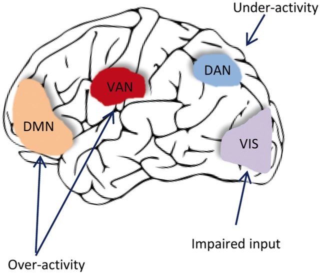
“This hypothesis posits that hallucinations are associated with overactivation of the DMN and SAL networks, whose interaction may lead to failures in stimulus-driven control and a bias towards internally generated perceptions 27 .”
Yap, T. P. (2026) · Journal of Clinical Medicine · doi:10.3390/jcm15124639 · Cited by 1
“Fourthly, this can impact the patient’s activities of daily living especially if the tasks require fine visual motor control [1,19,25].”
From Sgt. Pepper to Dreamachines: My Scientific Odyssey With William S. Burroughs
Lees, A. J. (2017) · JAMA · doi:10.1001/jama.2017.17914 · Cited by 1
Self-citationAdvice for those trying to find a cure for the shaking palsy
Lees, A. J. (2017) · The Lancet Neurology · doi:10.1016/s1474-4422(17)30430-1 · Cited by 1
Self-citationTransient Daily Episodes of Vision Loss Due to Parkinson’s Disease
Pelak, V. S., & Berman, B. D. (2018) · Neuro-Ophthalmology · doi:10.1080/01658107.2018.1494202 · Cited by 1
“Alterations in afferent visual pathway function, from the retina to the cerebral cortex, include diminished contrast sensitivity, colour vision impairment, and visuospatial and visuoperceptual impairment.(2) Increasing attention is being paid to non-motor symptoms (NMS) in PD, because they contribute to significant di…”
BackgroundEvaluation of the Visual Pathway in Parkinson’s Disease Patients
Nassar, M. H., Ragab, O. A. A., Al-Malt, A. M., Nawar, A. E., Rashed, K. H., & Tageldin, E. A. (2020) · Journal of Neurology and Experimental Neuroscience · doi:10.17756/jnen.2020-070 · Cited by 2
“Most PD disease patients had at least one visual symptom including; reading difficulties, sometimes with diplopia, disturbances of complex visual functions such as facial recognition, visuospatial orientation and visual hallucinations [3].”
Ocular manifestations of Parkinson disease
Al-Namaeh, M. (2020) · Medical Hypothesis Discovery & Innovation in Optometry · doi:10.51329/mehdioptometry101 · Cited by 2
Ophthalmological findings in movement disorders
Mehta, S., Takkar, A., Ray, S., & Lal, V. (2021) · Annals of Movement Disorders · doi:10.4103/aomd.aomd_35_20 · Cited by 1
Masitha, S. (2022) · Cerdika Jurnal Ilmiah Indonesia · doi:10.36418/cerdika.v2i8.423 · Cited by 1
“Akan tetapi dapat ditemukan perbedaan bahwa pada penelitian sebelumnya responden memiliki gangguan parkinson disease yang mungkin sangat bergantung pada penglihatan dengan defisit visual (Weil et al., 2016).”
Parkinson's disease: neurological manifestations and possibilities for neurosurgery
Malfera, F. B. C., Brandão, T. M., Cunha, A. T. D. A., de Carvalho, V. M., Hajj, G. A. V. E., Fernandes, A. C. S., Braga, J. P. N., Rodríguez, I. S. T., Figueiredo, L. G., Vilanova, R. R., et al. (2023) · Brazilian Journal of Implantology and Health Sciences · doi:10.36557/2674-8169.2023v5n5p6410-6422 · Cited by 1
New insights in non-motor symptoms in Parkinson’s disease
Falup‐Pecurariu, C., Fanciulli, A., Borgohain, R., Chaudhuri, К. R., & Metta, V. (2024) · Frontiers research topics · doi:10.3389/978-2-8325-4722-9 · Cited by 1
Kechabia, Y., Defebvre, L., Delval, A., & Bonnet, C. T. (2024) · European Journal of Neuroscience · doi:10.1111/ejn.16646 · Cited by 1
“…sway more than age-matched controls in all tasks (general standard deviation [SD] of both free-viewing and search tasks) and still in the search task (Figure 1A) because of the patients' various impairments (Figure 1A) (Ekker et al. 2017; Magrinelli et al. 2016; Rowe et al. 2002; Weil et al. 2016).”
Fahrkompetenz und Morbus Parkinson
Andelfinger, V., Weisbrod, M., Fritz, M., & Aschenbrenner, S. (2021) · Nervenheilkunde · doi:10.1055/a-1298-1151 · Cited by 1
Cummings, L. (2020) · Cambridge University Press eBooks · doi:10.1017/9781108587921.004 · Cited by 1
Visual disturbances in Parkinson’s disease
Nikitina, A. Y., Mel’nikova, N. V., Moshetova, L. K., & Levin, O. S. (2022) · S S Korsakov Journal of Neurology and Psychiatry · doi:10.17116/jnevro20221221125 · Cited by 2
Xu, N., Zhou, Y., Patel, A. C., zhang, N., & Liu, Y. (2022) · SSRN Electronic Journal · doi:10.2139/ssrn.4165341 · Cited by 1
Ahmadimehr, S. (2022) · Research Square · doi:10.21203/rs.3.rs-2318756/v1 · Cited by 1
Radlicka-Borysewska, A., Jabłońska, J., Lenarczyk, M., Szumiec, Ł., Harda, Z., Bagińska, M., Pera, J., Kreiner, G., Wojcik, D. K., & Parkitna, J. M. R. (2023) · bioRxiv (Cold Spring Harbor Laboratory) · doi:10.1101/2023.01.23.525182 · Cited by 1
Vignando, M., Ffytche, D., Mazibuko, N., Palma, G., Bhat, A., Montagnese, M., Dave, S., Tai, Y. F., Batzu, L., Leta, V., et al. (2026) · Nature Mental Health · doi:10.1038/s44220-026-00669-7
“We hypothesize reduced top-down and enhanced bottom-up connectivity at task in PD-VH, reflecting impaired prediction updating and reliance on compromised sensory information, as visual input is known to be defective in PD-VH 35 .”
Reply: MRI findings of visual system alterations in Parkinson’s disease
Weil, R. S., McColgan, P., Schrag, A. E., Warren, J. D., Crutch, S. J., Lees, A. J., & Morris, H. R. (2017) · Brain · doi:10.1093/brain/awx245
Self-citationdos Santos, D. D. C., Barbosa, E. R., & Cury, R. G. (2018) · JAMA Neurology · doi:10.1001/jamaneurol.2018.1066
Deterioro de la memoria visuoespacial en estadios 2 y 3 de la enfermedad de Parkinson
Sandoval, M. S., Clause, N., Cancino, M., Mora, P., Mundaca, P., Pantoja, C., Rojas, R., & Valdés, V. (2019) · Revista de Logopedia Foniatría y Audiología · doi:10.1016/j.rlfa.2018.09.003
da Silva, V. M. (2019) · Digital Library of Theses and Dissertations (Universidade de São Paulo) · doi:10.11606/d.100.2019.tde-22062019-221711
Tereshchenko, L. V., & Латанов, А. В. (2019) · Neuroscience and Behavioral Physiology · doi:10.1007/s11055-019-00847-4
Disturbances of visual perception in non-demented patients with Parkinson’s disease
Timofejavaitė, R., Vilda, V., LIVEIKIENE, A., & Lokominienė, R. K. (2019) · Neurologijos seminarai · doi:10.29014/ns.2019.21
Dumpala, S. (2020) · Queensland University of Technology · doi:10.5204/thesis.eprints.203959
Hardwick, M., Heath, D., Saha, R. A., & Cooper, S. A. (2020) · Neuro-Ophthalmology · doi:10.1080/01658107.2020.1835994 · Cited by 1
Stereopsis Deficits in Parkinson’s Disease and Their Clinical ImplicationsPreprint
Ba, F., Sang, T. T., Fatehi, J., He, W., Mostofi, E., & Zheng, B. (2020) · Research Square · doi:10.21203/rs.3.rs-106032/v1
The alphasynucleinopathies: Parkinson’s disease, Lewy body dementia, and multisystem atrophy
Hildebrandt, H. (2020) · Elsevier eBooks · doi:10.1016/b978-0-12-821072-7.00003-5
Modeling Visual Hallucination: A Generative Adversarial Network FrameworkPreprint
Zareh, M., Manshaei, M. H., Zahabi, S. J., & Krunz, M. (2021) · arXiv (Cornell University) · doi:10.48550/arxiv.2102.08209
“Visual hallucinations in Parkinson’s disease are caused by overactivity of the Default Mode Network (DMN) and Ventral Attention Network (VAN) and under-activity of the Dorsal Attention Network (DAN) 7 .”
InfluentialProgressive Supranuclear Palsy
Cummings, L. (2020) · Cambridge University Press eBooks · doi:10.1017/9781108587921.007
Cummings, L. (2020) · Cambridge University Press eBooks · doi:10.1017/9781108587921.005 · Cited by 1
Engel, D., Student, J., Schwenk, J. C. B., Morris, A. P., Waldthaler, J., Timmermann, L., & Bremmer, F. (2021) · bioRxiv (Cold Spring Harbor Laboratory) · doi:10.1101/2021.07.05.451110
Lipoic acid inhibits nerve cell apoptosis in rats with Parkinson's disease via Erk signaling pathway
Li, X., Li, W. S., Li, W., & Zhang, J. (2021) · Tropical Journal of Pharmaceutical Research · doi:10.4314/tjpr.v20i10.4
“Existing studies [5-7] have demonstrated that the destruction of dopaminergic neurons in the central nervous system is the main pathological mechanism of Parkinson's disease, which is closely associated with muscle stiffness, tremor and autonomic dysfunction in patients.”
BackgroundExplotaciones históricas de calizas de la Comunidad de Madrid:Colmenar de la Oreja
Riart, O. P., & Martínez, L. F. M. (1999) · Cited by 1
Zimmermann, N., Pontes, M. C., Fontana, R. D. S., Meira, I. D., Fonseca, R. P., & Delaere, F. J. (2022) · Applied Neuropsychology Adult · doi:10.1080/23279095.2022.2031200 · Cited by 1
“…for the evaluation of epilepsy (Corne et al., 2012), left temporal glioma (Verlut et al., 2015), stroke (Nys et al., 2005), Parkinson ’ s disease (Weil et al., 2016), schizophrenia (Girard et al., 2010), semantic dementia (Desgranges et al., 2007; Merck et al., 2013), and anterograde amnesia…”
Cummings, L. (2020) · Cambridge University Press eBooks · doi:10.1017/9781108587921.006
Cummings, L. (2020) · Cambridge University Press eBooks · doi:10.1017/9781108587921.012
Cummings, L. (2020) · Cambridge University Press eBooks · doi:10.1017/9781108587921.008
Cummings, L. (2020) · Cambridge University Press eBooks · doi:10.1017/9781108587921.011
Cummings, L. (2020) · Cambridge University Press eBooks · doi:10.1017/9781108587921.003
Cummings, L. (2020) · Cambridge University Press eBooks · doi:10.1017/9781108587921.015
(2020) · Cambridge University Press eBooks · doi:10.1017/9781108587921.013
Metadata
- Authors
- Weil, R. S., Schrag, A. E., Warren, J. D., Crutch, S. J., Lees, A. J., & Morris, H. R.
- Affiliations
- Royal Free London NHS Foundation Trust (GB)
- National Hospital for Neurology and Neurosurgery (GB)
- UCL Queen Square Institute of Neurology (GB)
- Corresponding author
- Weil, Rimona S.
- ORCID
- Venue
- Brain
- Publisher
- Oxford University Press (OUP)
- Published
- 2016-07-13
- Volume
- 139
- Issue
- 11
- Pages
- 2827-2843
- ISSN
- 0006-8950 (ISSN-L)
- 1460-2156
- PMC
- PMC5091042
- OpenAlex
- W2474900144
- Cited by
- 481
- References
- 232
- Citations by year
- 2026: 41 · 2025: 55 · 2024: 54 · 2023: 48 · 2022: 52 · 2021: 55 · 2020: 66 · 2019: 37 · 2018: 34 · 2017: 28 · 2016: 2
- FWCI
- 4.67
- Type
- journal article
- Language
- en
- Open access
- green
- www.ncbi.nlm.nih.gov
- Licence
- cc-by
- Locations
- Brain (publishedVersion)
- PubMed Central (publishedVersion, cc-by)
- PubMed (publishedVersion)
- UCL Discovery (University College London) (submittedVersion)
- HighWire Press Open Archive (submittedVersion)
- Funders
- National Institute for Health and Care Research
- Parkinson's UK (K-1501, J‐0804)
- Academy of Medical Sciences (AMS-SGCL13-Weil)
- Engineering and Physical Sciences Research Council (EP/M006093/1)
- Economic and Social Research Council (ES/L001810/1)
- Topics
- Health Sciences › Medicine › Clinical Neurology › Parkinson's Disease Mechanisms and Treatments
- Life Sciences › Neuroscience › Cognitive Neuroscience › Visual perception and processing mechanisms
- Life Sciences › Neuroscience › Cognitive Neuroscience › Hallucinations in medical conditions
- Keywords
- Parkinson's disease, visual dysfunction, contrast sensitivity, mental rotation, emotion recognition, visual neglect, motion processing, object processing, GBA mutations, LRRK2 mutations, REM sleep behavior disorder, postural instability, gait disturbance, visual hallucinations, biomarkers, dementia prediction
- MeSH terms
- Leucine-Rich Repeat Serine-Threonine Protein Kinase-2, beta-Glucosidase, Humans, Parkinson Disease, Vision Disorders, Visual Pathways, Visual Perception
About these checks
Registry, retraction, Cites and statistics results are computed, not judged, with one exception: a printed reference that no index lists is read once by a model before it counts as a citation error. Claims are extracted by a model and kept only when their quote appears verbatim in the text; each cited source is then checked, with a model’s help, for whether it says what the claim relies on. Those verdicts can be wrong — read the claim’s quote and the cited source before relying on one.
Statistics and arithmetic are recomputed, not judged.
Confidence bands are shown only for fields calibrated on our annotated test set. None is calibrated yet, so no band is shown.
What counts as a claim ›How we measure confidence ›How we check this ›
Are you an author, or do you see a problem with this page? Request a review
Data sources
Bibliographic data: Semantic Scholar, OpenAlex, Crossref.
Contains information from the Semantic Scholar Academic Graph Datasets, made available under the ODC Attribution License (ODC-BY 1.0).
Semantic Scholar’s data is described in: Kinney, R., et al. (2023). The Semantic Scholar Open Data Platform. arXiv:2301.10140.
