

Visual dysfunction in Parkinson’s disease
Open accessThe DOI matches its Crossref record; 233 of the 233 references it cites were found in a registry or an index.
Abstract
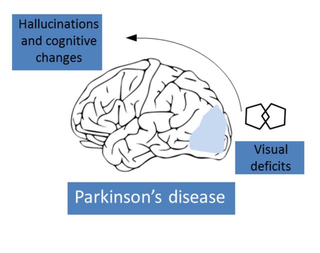
Patients with Parkinson's disease have a number of specific visual disturbances. These include changes in colour vision and contrast sensitivity and difficulties with complex visual tasks such as mental rotation and emotion recognition. We review changes in visual function at each stage of visual processing from retinal deficits, including contrast sensitivity and colour vision deficits to higher cortical processing impairments such as object and motion processing and neglect. We consider changes in visual function in patients with common Parkinson's disease-associated genetic mutations including GBA and LRRK2 . We discuss the association between visual deficits and clinical features of Parkinson's disease such as rapid eye movement sleep behavioural disorder and the postural instability and gait disorder phenotype. We review the link between abnormal visual function and visual hallucinations, considering current models for mechanisms of visual hallucinations. Finally, we discuss the role of visuo-perceptual testing as a biomarker of disease and predictor of dementia in Parkinson's disease.
Overview
The claims and statistics of this paper haven’t been checked yet.
Free: it reads the open-access text and checks the first 5 claims that cite a source we can check. Create a free account to check every claim.
Integrity signals
Registry
Cites
Statistics
Figures
Verification
Claims the paper makes
The claims and statistics of this paper haven’t been checked yet.
Free: it reads the open-access text and checks the first 5 claims that cite a source we can check. Create a free account to check every claim.
Self-check
Percentages, sums and reported differences re-computed from the paper’s own numbers.
The self-check needs the paper’s full text.
Citations
Cites
The precuneus: a review of its functional anatomy and behavioural correlates
Cavanna, A. E., & Trimble, M. R. (2006) · Brain · doi:10.1093/brain/awl004 · Cited by 5,128 · cited 2×
“Finally, in a structure from motion task, where volunteers detect an object that is formed from the contrast of moving dots, patients with Parkinson’s disease show higher thresholds for correctly identifying objects (Uc et al., 2005).”
BackgroundApplication of fourier analysis to the visibility of gratings
Campbell, F. W., & Robson, J. G. (1968) · The Journal of Physiology · doi:10.1113/jphysiol.1968.sp008574 · Cited by 3,019 · cited 3×
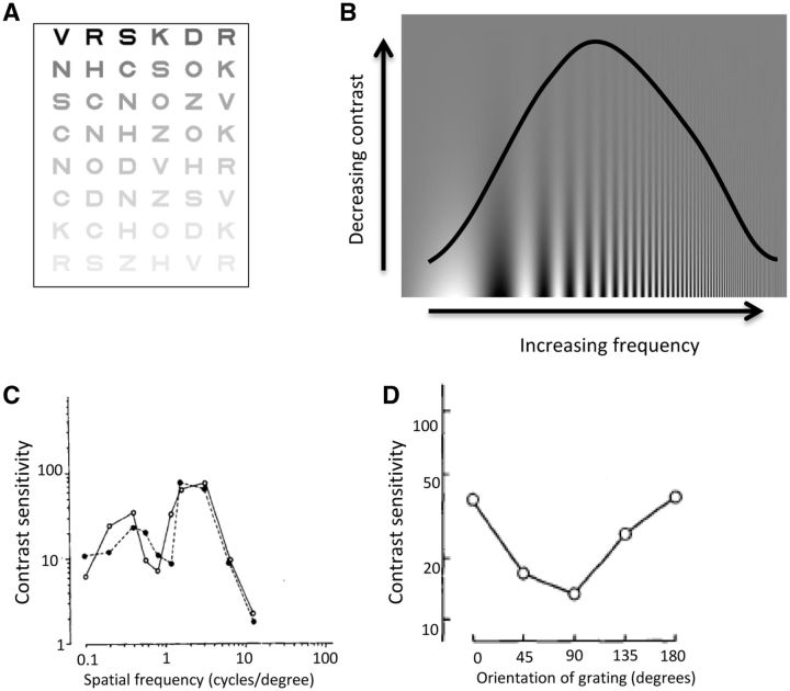
“This is more suggestive of a cortical rather than retinal or geniculate site of disruption as cortical neurons show stronger selectivity for different spatial frequencies than neurons in the retina or thalamus (Campbell and Robson, 1968; De Valois et al., 1982; Regan, 1982).”
BackgroundStages in the development of Parkinson’s disease-related pathology
Braak, H., Ghebremedhin, E., Rüb, U., Bratzke, H., & Del Tredici, K. (2004) · Cell and Tissue Research · doi:10.1007/s00441-004-0956-9 · Cited by 2,689 · cited 1×
“One theory of disease progression in Parkinson’s disease has suggested that the cerebral cortex is not damaged until the later stages of the disease (Braak et al., 2004).”
BackgroundThe Sydney multicenter study of Parkinson's disease: The inevitability of dementia at 20 years
Hely, M. A., Reid, W. G. J., Adena, M. A., Halliday, G. M., & Morris, J. G. L. (2008) · Movement Disorders · doi:10.1002/mds.21956 · Cited by 2,246 · cited 1×
“Visual hallucinations are also common in Parkinson’s disease, with a reported prevalence as high as 74% after 20 years of disease (Fenelon et al., 2000; Hely et al., 2008).”
BackgroundA direct demonstration of functional specialization in human visual cortex
Zeki, S., Watson, J., Lueck, C., Friston, K., Kennard, C., & Frackowiak, R. (1991) · The Journal of Neuroscience · doi:10.1523/jneurosci.11-03-00641.1991 · Cited by 1,716 · cited 2×
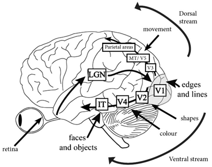
“IT = infero-temporal region; MT/V5 = motion processing region.”
BackgroundPharmacological Treatment of Parkinson Disease: A Review
Connolly, B. S., & Lang, A. E. (2014) · JAMA · doi:10.1001/jama.2014.3654 · Cited by 1,498 · cited 2×
“General advice is to consider visuo-perceptual ability and if there is any doubt, to refer the patient for a formal driving assessment via the national driving authority (Crizzle et al., 2012).”
BackgroundSpatial frequency selectivity of cells in macaque visual cortex
De Valois, R. L., Albrecht, D. G., & Thorell, L. G. (1982) · Vision Research · doi:10.1016/0042-6989(82)90113-4 · Cited by 1,489 · cited 2×
“This is more suggestive of a cortical rather than retinal or geniculate site of disruption as cortical neurons show stronger selectivity for different spatial frequencies than neurons in the retina or thalamus (Campbell and Robson, 1968; De Valois et al., 1982; Regan, 1982).”
Functional analysis of human MT and related visual cortical areas using magnetic resonance imaging
Tootell, R., Reppas, J., Kwong, K., Malach, R., Born, R., Brady, T., Rosen, B., & Belliveau, J. (1995) · The Journal of Neuroscience · doi:10.1523/jneurosci.15-04-03215.1995 · Cited by 1,415 · cited 2×
“IT = infero-temporal region; MT/V5 = motion processing region.”
BackgroundMazzulli, J. R., Xu, Y.-H., Sun, Y., Knight, A. L., McLean, P. J., Caldwell, G. A., Sidransky, E., Grabowski, G. A., & Krainc, D. (2011) · Cell · doi:10.1016/j.cell.2011.06.001 · Cited by 1,341 · cited 1×
“Both the GBA and SNCA genes are linked with the lysosomal pathway (Mazzulli et al., 2011), suggesting the visual deficits associated with these genes may share a pathophysiological mechanism, with lysosomal dysfunction associated with cortical visual dysfunction.”
BackgroundTHE DESIGN OF A NEW LETTER CHART FOR MEASURING CONTRAST SENSITIVITY
Pelli, D. G., Robson, J. G., & Wilkins, A. J. (1988) · Cited by 1,308
Research works printed but not linked: 3
- Ogden, 1990, Deficits on visuospatiaol tests involving forward planning in high-functioning Parkinsonians, Neuropsychiatry Neuropsychol Behav Neurol, 3, 125
- 10.1037/0033-295X.89.4.407 · doi:10.1037/0033-295x.89.4.407
- 10.1097/00006324-199404000-00002 · doi:10.1097/00006324-199404000-00002
Cited by
Items 401–479 of 479
Cummings, L. (2020) · Cambridge University Press eBooks · doi:10.1017/9781108587921.010
Cummings, L. (2020) · Cambridge University Press eBooks · doi:10.1017/9781108587921.014
Hou, Y., Wei, Q., Ou, R., Zhang, L., Yuan, X., Gong, Q., & Shang, H. (2020) · Parkinsonism & Related Disorders · doi:10.1016/j.parkreldis.2020.06.066
Cummings, L. (2020) · Cambridge University Press eBooks · doi:10.1017/9781108587921.009
Vieillissement cérébral pathologique
Gil, R., & Wager, M. (2021) · Elsevier eBooks · doi:10.1016/b978-2-294-76689-3.00027-9
Suciu, V.-I., Suciu, C.-I., Nicoară, S., & Perju‐Dumbravă, L. (2022) · Romanian Journal of Ophthalmology · doi:10.22336/rjo.2022.28
“It has also been suggested that the visual impairment in PD is linked to the general disability and gait disturbances of these patients [15].”
BackgroundAbnormal intrinsic neural timescale and reduced grey matter volume in Parkinson’s diseasePreprint
Wei, Y., Zhang, C., Peng, Y., Chen, C., Han, S., Wang, W., Zhang, Y., Lu, H., & Cheng, J. (2022) · bioRxiv (Cold Spring Harbor Laboratory) · doi:10.1101/2022.09.17.508074
Neural substrates in Parkinson’s Disease psychosis: A systematic reviewPreprint
Pisani, S., Gunasekera, B., Lu, Y., Vignando, M., Ffytche, D. H., Aarsland, D., Chaudhuri, К. R., Ballard, C. G., Lee, J., Kim, Y. K., et al. (2022) · medRxiv · doi:10.1101/2022.12.23.22283889
Cortical thickness and Subcortical volume changes differ between Parkinson disease subtypesPreprint
Huang, M., Yu, H., Guo, K., Yin, J., Lei, P., & Gao, B. (2023) · Research Square · doi:10.21203/rs.3.rs-2528215/v1
Habilidades visoperceptuales en el Parkinson idiopático y la demencia con cuerpos de Lewy
Abril, A., Flórez, M. F., Roa, E., Moncayo, J. F. O., & Pacific, E. J. O. (2023) · Ciencia y Tecnología para la Salud Visual y Ocular · doi:10.19052/sv.vol20.iss1.6
A Patient's Guide to Visual Perception Changes That Occur With Parkinson Disease
Walker, S., Colletta, K., Devos, H., Gaynes, B. I., Ellison, R. L., Alsakaji, R., Watters, K., Todfield, A., Chawla, J. P. S., & Kletzel, S. (2023) · Archives of Physical Medicine and Rehabilitation · doi:10.1016/j.apmr.2022.09.023
Gjika, F., Scopes, G., Çibuku, O., Rraklli, E., Stiljan, D., & Feraizi, E. (2023) · Acta Scientific Medical Sciences · doi:10.31080/asms.2023.07.1586
Olmos, J., Valenzuela, B., & Martínez, F. (2023) · Health and Technology · doi:10.1007/s12553-023-00782-y
“The most common symptoms include slowness of movements (Bradykinesia), rigidity, and tremor [3].”
BackgroundVisual Impairments in Parkinson’s Disease
Nikitina, A. Y., Melnikova, N. V., Moshetova, L. K., & Levin, O. S. (2023) · Neuroscience and Behavioral Physiology · doi:10.1007/s11055-023-01487-5
“Errors in drawing two intersecting pentagons at the onset of PD are an important predictor of the development of dementia [44].”
BackgroundDahmani, L., Bai, Y., Zhang, W., Ren, J., Li, S., Hu, Q., Fu, X., Ma, J., Wei, W., Wang, M., et al. (2024) · bioRxiv (Cold Spring Harbor Laboratory) · doi:10.1101/2024.01.31.578238
Deep Learning Approaches in the Early Diagnosis of Parkinson's Disease
El‐Mansoury, B., Hamdan, Y. A., Smimih, K., Khiat, A. E., Draoui, A., Hammani, M., Jayakumar, A. R., & Hiba, O. E. (2024) · Advances in medical diagnosis, treatment, and care (AMDTC) book series · doi:10.4018/979-8-3693-1281-0.ch007
Synsvansker ved Parkinsons sykdom
Dalbro, S. E. J., & Kerty, E. (2024) · Tidsskrift for Den norske legeforening · doi:10.4045/tidsskr.23.0716
Ödül Değerinde Kimyasal Dopamin ve İşlevleri
Pençe, M. E. (2024) · Akademisyen Kitabevi eBooks · doi:10.37609/akya.3130
Limb Speed Perception Accuracy in Neurodegenerative Disease
Brandmeir, C. L. (2024) · The Research Repository @ WVU (West Virginia University) · doi:10.33915/etd.12362 · Cited by 1
Analyse von Tränenflüssigkeit als Biomarkerquelle beim idiopathischen Parkinsonsyndrom
Paul, H. L. J. (2024) · eDiss (Georg-August-Universität Göttingen) · doi:10.53846/goediss-10491
Çekiç, S., Çınar, B. P., Demirel, E. A., Akdemir, M. O., Çelebi, U., Açıkgöz, M., & Atasoy, H. T. (2024) · Nöro Psikiyatri Arşivi · doi:10.29399/npa.28670 · Cited by 1
Disrupted functional brain network associated with presence of hallucinations in Parkinson’s disease
Montagnese, M., Mehta, M. A., Ffytche, D. H., Firbank, M., Lawson, R. A., Taylor, J., Bullmore, E. T., & Morgan, S. E. (2025) · Brain Communications · doi:10.1093/braincomms/fcaf185
“…more comprehensive measures across sensory modalities 54 ; (ii) visual testing was not conducted to assess potential low-level visual deficits, 55 and hallucination states could not be directly measured during scanning, limiting the ability to examine connections with active hallucinatory…”
Subthalamic Nucleus Deep Brain Stimulation Modulates Strabismus in Parkinson’s DiseasePreprint
Gupta, P., Beylergil, S. B., Kilbane, C., McIntyre, C. C., Noecker, A. M., Shaikh, A. G., & Ghasia, F. F. (2024) · medRxiv · doi:10.1101/2024.11.07.24316853
Neuro-Ophthalmology in Movement Disorders
Crotty, G. F., & Chwalisz, B. K. (2025) · Cambridge University Press eBooks · doi:10.1017/9781108989855.012
Hrizhymalska, K. Y., Komarovska, I. V., & Kukuruza, T. Y. (2024) · Archive of Ophthalmology and Maxillofacial Surgery of Ukraine · doi:10.22141/aomfs.1.2.2024.17
Effect of color visual stimulation by colored glass lenses on postural control
Zhang, W., Takada, H., Hettmannsperger, M., Négyesi, J., Wang, Z., & Nagatomi, R. (2025) · Scientific Reports · doi:10.1038/s41598-025-88102-4
“Interestingly in clinical studies, color perception impairment has been found in untreated patients with Parkinson’s disease (PD) in the early stages of the disease 31 .”
Aprosopsia: a novel metamorphopsia variant occurring in Parkinson’s disease psychosis
Reddy, A. B. S., Pisani, S., Kuruppu, S., Bhattacharyya, S., Ffytche, D. H., & Velayudhan, L. (2025) · Journal of Neurology · doi:10.1007/s00415-025-13015-1
“The symptoms are associated with a range of cognitive and visual processing deficits, including facial identity and emotional recognition deficits [2].”
Yang, J., & Wei, W. (2025) · Chinese Science Bulletin (Chinese Version) · doi:10.1360/tb-2024-1113
OCT in Parkinson’s Disease and Related Disorders
Zhou, J., Jin, K., Guan, X., Ye, J., & Grzybowski, A. E. (2025) · Springer eBooks · doi:10.1007/978-3-031-72156-4_12
Behavioural, biochemical and functional phenotyping of chronic exposure to chlordecone in micePreprint
Vulin, J., Chauveau, F., Dure, C., Humo, M., Valdebenito, M., Aufauvre, C., Vidal, L., Morignat, E., Verchère, J., Decesar, A., et al. (2025) · bioRxiv (Cold Spring Harbor Laboratory) · doi:10.1101/2025.06.16.659846
Guo, Z., Li, J., Xu, J., Chen, Y., Rong, D., Xing, Y., Ren, J., Yu, M., Liu, W., & Zheng, J. (2025) · SSRN Electronic Journal · doi:10.2139/ssrn.5347322
Zemp, D. D., Dinacci, D., & Galati, S. (2025) · Parkinson s Disease · doi:10.1155/padi/9272217
“…such as shorter duration of negative afterimages, disruption of processing at the level of the retina, oculomotor changes, reduced depth perception, inability to inhibit attention toward peripheral objects not relevant for the task, and more difculties for detecting the direction of motion [30].”
Dujardin, K., Péron, J., & Devignes, Q. (2025) · Elsevier eBooks · doi:10.1016/b978-2-294-78712-6.00008-7
Topical β-blockers for treatment of glaucoma and the risk of Parkinson’s disease
Meshi, A., Giladi, N., Zahavi, A., Gabbay, I. E., Gal‐Or, O., Dotan, A., Gazit, S., Patalon, T., Kozlov, Y., Geffen, N., et al. (2025) · British Journal of Ophthalmology · doi:10.1136/bjo-2024-326689
“4 No clear association has been found between PD and glaucoma in population-based studies, 5–7 although there are reports of a small increase in the risk of developing PD in older people with glaucoma.”
Impact of levodopa and deep brain stimulation on visual impairments in Parkinson’s disease
Aslam, S., Olson, M. C., Kovacs, M., Ansari, P., Karukote, A., Chan, J., Barbu, S., Felsen, S., Shill, H. A., & Adler, C. H. (2025) · Clinical Parkinsonism & Related Disorders · doi:10.1016/j.prdoa.2025.100390
“Among the nonmotor symptoms not discussed as frequently are visual changes affecting visuospatial processing, reading speed, and contrast acuity, all of which can impact quality of life [1].”
Validity of digital measurement of visual acuity and contrast sensitivity in Parkinson’s disease
Das, J., Barry, G., Vitório, R. R., Walker, R. W., Çelik, Y., McDonald, C., Morris, R., & Stuart, S. (2025) · BMC Digital Health · doi:10.1186/s44247-025-00195-z
“Despite increasing research on visual changes in PD, studies examining the correlation of visual function with PD features such as motor severity remain scarce [45, 46].”
Behavioural, biochemical and functional phenotyping of subchronic exposure to chlordecone in mice
Vulin, J., Chauveau, F., Duret, C., Humo, M., Valdebenito, M., Aufauvre, C., Vidal, L., Morignat, E., Verchère, J., Decesar, A., et al. (2025) · NeuroToxicology · doi:10.1016/j.neuro.2025.103334
Hallucinations at the interface of philosophy and the empirical sciences
Dolcini, N. (2025) · Frontiers in Psychology · doi:10.3389/fpsyg.2025.1699432
“…shows that impairments in lower-level visual processing and attention are more pronounced in Parkinson’s patients who experience visual hallucinations (Weil et al., 2016), and that hearing impairment or lesions in auditory pathways are associated with psychosis onset (Thewissen et al., 2005).”
Portaro, G., Giacomelli, M., Grisanti, S., Taruffi, L., Rauso, G. D., Fioravanti, V., Salomone, G., Argenziano, G., Sabadini, R., Toschi, G., et al. (2025) · Parkinsonism & Related Disorders · doi:10.1016/j.parkreldis.2025.108134
Phuenpathom, W., Sringean, J., Phumphid, S., Rattanapitak, N., Laithaisong, A., Srisang, T., Sanyawut, K., Anan, C., & Bhidayasiri, R. (2025) · Journal of Neural Transmission · doi:10.1007/s00702-025-03077-w
Liu, P., Sun, J., Hu, L., Chen, X., Han, J., Yu, L., Fan, S., Ye, R., Wang, K., Ji, G., et al. (2025) · iNew Medicine · doi:10.1002/inm3.70010
Boertien, J. M., van der Zee, S., Chrysou, A., Gerritsen, M. J. J., Jansonius, N. M., Spikman, J. M., & van Laar, T. (2020) · Figshare · doi:10.6084/m9.figshare.12479066
Yanbing, H., Wei, Q., Ru⁃wei, O., Zhang, L., Yuan, X., Gong, Q., & Shang, H. (2021) · Figshare · doi:10.6084/m9.figshare.16442066
Yanbing, H., Wei, Q., Ou, R., Zhang, L., Yuan, X., Gong, Q., & Shang, H. (2021) · Figshare · doi:10.6084/m9.figshare.16442063
Liley, D. T. J. (2026) · Journal of Neural Engineering · doi:10.1088/1741-2552/ae33f7
“…result as it suggests that perceptual sensitivity to the auditory stimuli, and possibly other types of sensory stimuli, may be enhanced in PD, contrary to the perceptual impairments that are typically reported (Bodis-Wollner 2003, Cecchini et al 2015, Weil et al 2016, Nieto-Escamez et al 2023).”
Compensatory hallucinogenesis across three neuropsychiatric disorders: a Bayesian account
Vin, R., Galbraith, J., Seabury, R. D., Yi, H. Y., Hernández-Busot, G., Oland, L., Epié, B., Trainer, A. E., Fredericks, C. A., & Powers, A. R. (2026) · Brain Communications · doi:10.1093/braincomms/fcag001 · Cited by 1
“…121,122 display subclinical impairments in visuoperception, visuospatial construction and colour vision starting 10–16 years prior to conversion, 88,100,123-127 and these predict progression to DLB over PD. 88,128 Greater impairment in visuospatial tasks that utilize the dorsal visual stream…”
InfluentialVisual misperceptions in favour of living beings in Parkinson's disease: A psychophysical study
de Langavant, L. C., Mouton, A., de Gardelle, V., Kouider, S., Bachoud‐Lévi, A., Fénelon, G., & Rémy, P. (2026) · Revue Neurologique · doi:10.1016/j.neurol.2025.12.005
Gao, Z., Lv, S., Ran, X., Du, C., Xia, M., Zhao, M., Xie, Y., Tian, P., Ren, W., Zhao, Z., et al. (2026) · Journal of NeuroEngineering and Rehabilitation · doi:10.1186/s12984-026-01882-2 · Cited by 2
“The degeneration of the dopaminergic system and dopamine deficiency in PD patients adversely affect neural pathways involved in visual processing, resulting in a reduced level of microstate B—associated with the visual network—in PD patients [38].”
Yang, F. X., Manohar, R. R., Luddy, A. C., Hung, A. Y., Wills, A.-M. A., Stephen, C. D., Schmahmann, J. D., & Gupta, A. S. (2026) · medRxiv · doi:10.64898/2026.02.03.26345521
Gupta, P., Kilbane, C., Shaikh, A. G., & Ghasia, F. F. (2026) · Contemporary clinical neuroscience · doi:10.1007/978-3-032-07539-0_26
Color Vision Deficits and Eye Movement Abnormalities in Parkinson’s Disease
Mleczek, J., Forjindam, A., Shaikh, A. G., & Ghasia, F. F. (2026) · Contemporary clinical neuroscience · doi:10.1007/978-3-032-07539-0_27
The influence of visual input and attention on gait initiation in people with Parkinson disease
Duppen, C., Surkin, J., Mahendar, S., Saunders, J., Cole, J. M., Browner, N., & Lewek, M. D. (2026) · PLoS ONE · doi:10.1371/journal.pone.0342661
“…stimulating) and insufficient (i.e., occluded) visual input during continuous walking can lead to shorter step lengths, slower gait speed, and episodes of freezing of gait (FOG) [13–17], reflecting a continued reliance on visual input despite impairments in vision and visual processing [18,19].”
Shoeibi, A., Shariati-Moghaddam, R., Ahmad, M. A., Ostadimoghaddam, H., Hashemi, H., Derakhshan, A., Hemmatian, Z., Yekta, A., & Khabazkhoob, M. (2026) · BMC Neurology · doi:10.1186/s12883-026-04822-8
“Furthermore, in patients with more advanced stages of PD, subtle alterations in the crystalline lens, such as reduced flexibility, may increase the likelihood of hyperopia, aligning with the borderline trend observed in this study [26].”
Damasse, J.-B. (2018) · theses.fr (ABES) · doi:10.70675/193e9600zb9f9z4bcazb4c1z4369b97dce0c
Self images: an empirical enquiry into Rembrandt’s self-portraits
Bettelheim, E. C., Liu, J., Dazzan, P., & Turkheimer, F. (2026) · Frontiers in Psychiatry · doi:10.3389/fpsyt.2026.1631373
Visual Diagnostics for Neuropsychologists
Schaefer, L. A., Pomeranz, H. D., & Chamoff, C. K. (2026) · Oxford University Press eBooks · doi:10.1093/9780197619537.003.0011
Corneal edema: an underrecognized safety risk of amantadine
Croteau, D. J., Tobenkin, A., Chan, V., Branagan, N., Goldrich, J., Freilich, E., & Brinker, A. D. (2026) · Journal of Neurology · doi:10.1007/s00415-026-13797-y
“Visual dysfunction associated with Parkinson’s disease includes impaired contrast sensitivity, reduced visual acuity, reduced color discrimination, dysfunction with depth perception, object perception, spatial neglect, and visual hallucinations, among other symptoms and signs [23, 24].”
Abdik, E., & Çakır, T. (2026) · npj Parkinson s Disease · doi:10.1038/s41531-026-01337-4
Savaş, M., & Beğen, S. K. (2026) · Journal of Speech Language and Hearing Research · doi:10.1044/2026_jslhr-25-00282
Zhang, Q., Qian, X., Peng, T., Hou, X., Ma, Z.-Y., Huang, J., Hu, L., & Luo, W. (2026) · Brain and Behavior · doi:10.1002/brb3.71489
“Visual impairment represents a prevalent nonmotor manifestation of PD, negatively impacting essential daily activities and contributing to reduced self-efficacy and overall quality of life (Weil et al. 2016 ; Hamedani et al. 2020 ).”
Cognitive effects of STN-DBS on mental rotation performance in Parkinson’s disease
Schoenfeld, M. J., Gulberti, A., David, N., Hamel, W., Pötter‐Nerger, M., Engel, A. K., & Moll, C. K. E. (2026) · Scientific Reports · doi:10.1038/s41598-026-52880-2
Effects of levodopa on the motor reserve network change with progression of Parkinson's disease
Rong, D., Huang, P., Yan, L., Yang, J., Liu, W., Shen, Y., Zhang, W., Yu, M., & Liu, W. (2026) · Progress in Neuro-Psychopharmacology and Biological Psychiatry · doi:10.1016/j.pnpbp.2026.111755
Effects of light therapy on sleep and anxiety in Parkinson's disease: A pilot trial
Shen, Y., Li, H., Dai, J., Zhou, Q.-J., Tao, M., Xie, W.-Y., Lou, H., Liu, J., Gao, F., Hu, H., et al. (2026) · Journal of Affective Disorders · doi:10.1016/j.jad.2026.122121
Ferraro, J., Ramos, J. S., Cayoun, J., Huang, D., Trang, L., Liow, C., Dalton, J., Nassaris, O., Beleigoli, A. M. R., Dalleck, L. C., et al. (2026) · Advances in Geriatric Medicine and Research · doi:10.20900/agmr20260015
“PD is increasingly recognised as a multisystem disorder affecting vision [13].”
Wang, Y., Tsitsi, P., Markaki, I., Svenningsson, P., Seimyr, G. Ö., Pansell, T., & Nilsson, M. (2026) · Translational Vision Science & Technology · doi:10.1167/tvst.15.6.19 · Cited by 1
“In the present study, we analyze eye movement data from a simple visual fixation task in which 50 individuals with early-to-moderate stage PD (Hoehn and Yahr 20 stages 1–3) and 43 healthy participants were instructed to fixate on a small central fixation target.”
Zientara, K., Skoczek, K., Pyza, E., & Damulewicz, M. (2026) · FEBS Letters · doi:10.1002/1873-3468.70389
“Another early PD symptom is disruption of visual contrast sensitivity, the ability to distinguish a visual object from its background [2].”
Guo, Z., Li, J., Xu, J., Chen, Y., Rong, D., Xing, Y., Ren, J., Yu, M., Liu, W., & Zheng, J. (2026) · Behavioral and Brain Functions · doi:10.1186/s12993-026-00350-0
“In the pathogenesis of PD, in addition to the classical sensorimotor network, there are some non-motor networks involved, such as the visual network [34].”
Yu, Y., Bai, Y., Xu, S., Wang, R. J., Wang, Z., Wang, R. J., & Cheng, J. (2026) · Frontiers in Aging Neuroscience · doi:10.3389/fnagi.2026.1757469
“Previous studies have also confirmed functional abnormalities (Weil et al., 2016; Nieto-Escamez et al., 2023) and RFS (Zang et al., 2023; Li M. et al., 2025) in visual-related occipital regions in IPD patients.”
Xu, T., Li, X., Wang, T., Zhang, X., Sun, Z., Shao, D., Zhang, J., Yang, Y., Xiao, Z., Zhang, Y., et al. (2026) · Research Square · doi:10.21203/rs.3.rs-10254179/v1
Samit, E., & Machado, K. (2026) · Neurophysiologie Clinique · doi:10.1016/j.neucli.2026.103192
White matter lesions disrupt cholinergic transmission to impact cognition in Parkinson’s disease
Murphy, P. T., Zarkali, A., & Weil, R. S. (2026) · Brain · doi:10.1093/brain/awag295
Self-citationAltered EEG-derived functional brain states in Parkinson's disease with mild cognitive impairment
Wang, Y., Zhang, Z., Wang, J., Jiang, Z., She, L., Pan, Y., & Liu, C. (2026) · Brain Communications · doi:10.1093/braincomms/fcag348
“33 Metabolic hypofunction and functional silencing in visual and limbic areas are also hallmark features of Alzheimer’s disease, particularly in later stages, and have been linked to visuospatial deficits and emotional blunting.”
Amakusa, Y., Takeuchi, R. F., Ito, H., Iguchi, Y., Ohba, A., Kobayashi, K., Yokoi, S., Hamaguchi, T., Hirunagi, T., Sahashi, K., et al. (2026) · Neuroscience Research · doi:10.1016/j.neures.2026.105118
Altered functional topology related to the development of wearing-off in Parkinson’s disease
Gao, T., Zhou, C., Guo, T., Tian, J., Guan, X., Wu, J., Xu, X., Zhang, M., Pu, J., & Zhang, B. (2026) · Brain Research Bulletin · doi:10.1016/j.brainresbull.2026.112136
Balthazor, B., Weinrich, M., Wang, Y., Wagner, J., & Kennedy, D. M. (2026) · Human Movement Science · doi:10.1016/j.humov.2026.103546
Seo, K., Hara, M., Alaoui, S. H., & Oyama, G. (2026) · Frontiers in Aging Neuroscience · doi:10.3389/fnagi.2026.2005253
“…in LBD are associated with deficits in contour discrimination, contrast sensitivity, and color discrimination (Diaz-Santos et al., 2021; Weil et al., 2016), reduced occipito-temporal activation during recognition of ambiguous images in PD with hallucinations (Meppelink et al., 2009),…”
Metadata
- Authors
- Weil, R. S., Schrag, A. E., Warren, J. D., Crutch, S. J., Lees, A. J., & Morris, H. R.
- Affiliations
- Royal Free London NHS Foundation Trust (GB)
- National Hospital for Neurology and Neurosurgery (GB)
- UCL Queen Square Institute of Neurology (GB)
- Corresponding author
- Weil, Rimona S.
- ORCID
- Venue
- Brain
- Publisher
- Oxford University Press (OUP)
- Published
- 2016-07-13
- Volume
- 139
- Issue
- 11
- Pages
- 2827-2843
- ISSN
- 0006-8950 (ISSN-L)
- 1460-2156
- PMC
- PMC5091042
- OpenAlex
- W2474900144
- Cited by
- 481
- References
- 232
- Citations by year
- 2026: 41 · 2025: 55 · 2024: 54 · 2023: 48 · 2022: 52 · 2021: 55 · 2020: 66 · 2019: 37 · 2018: 34 · 2017: 28 · 2016: 2
- FWCI
- 4.67
- Type
- journal article
- Language
- en
- Open access
- green
- www.ncbi.nlm.nih.gov
- Licence
- cc-by
- Locations
- Brain (publishedVersion)
- PubMed Central (publishedVersion, cc-by)
- PubMed (publishedVersion)
- UCL Discovery (University College London) (submittedVersion)
- HighWire Press Open Archive (submittedVersion)
- Funders
- National Institute for Health and Care Research
- Parkinson's UK (K-1501, J‐0804)
- Academy of Medical Sciences (AMS-SGCL13-Weil)
- Engineering and Physical Sciences Research Council (EP/M006093/1)
- Economic and Social Research Council (ES/L001810/1)
- Topics
- Health Sciences › Medicine › Clinical Neurology › Parkinson's Disease Mechanisms and Treatments
- Life Sciences › Neuroscience › Cognitive Neuroscience › Visual perception and processing mechanisms
- Life Sciences › Neuroscience › Cognitive Neuroscience › Hallucinations in medical conditions
- Keywords
- Parkinson's disease, visual dysfunction, contrast sensitivity, mental rotation, emotion recognition, visual neglect, motion processing, object processing, GBA mutations, LRRK2 mutations, REM sleep behavior disorder, postural instability, gait disturbance, visual hallucinations, biomarkers, dementia prediction
- MeSH terms
- Leucine-Rich Repeat Serine-Threonine Protein Kinase-2, beta-Glucosidase, Humans, Parkinson Disease, Vision Disorders, Visual Pathways, Visual Perception
About these checks
Registry, retraction, Cites and statistics results are computed, not judged, with one exception: a printed reference that no index lists is read once by a model before it counts as a citation error. Claims are extracted by a model and kept only when their quote appears verbatim in the text; each cited source is then checked, with a model’s help, for whether it says what the claim relies on. Those verdicts can be wrong — read the claim’s quote and the cited source before relying on one.
Statistics and arithmetic are recomputed, not judged.
Confidence bands are shown only for fields calibrated on our annotated test set. None is calibrated yet, so no band is shown.
What counts as a claim ›How we measure confidence ›How we check this ›
Are you an author, or do you see a problem with this page? Request a review
Data sources
Bibliographic data: Semantic Scholar, OpenAlex, Crossref.
Contains information from the Semantic Scholar Academic Graph Datasets, made available under the ODC Attribution License (ODC-BY 1.0).
Semantic Scholar’s data is described in: Kinney, R., et al. (2023). The Semantic Scholar Open Data Platform. arXiv:2301.10140.
