

The DOI matches its Crossref record; 28 of the 28 references it cites were found in a registry or an index.
Conserved hippocampal cellular pathophysiology but distinct behavioural deficits in a new rat model of FXS
Open accessAbstract
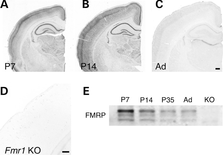
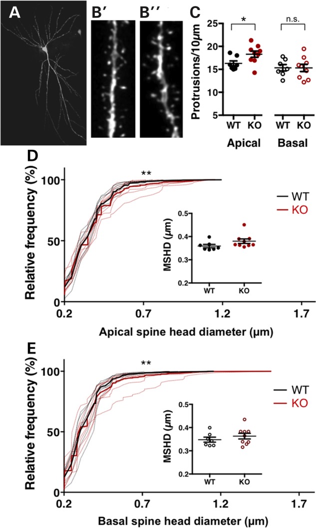
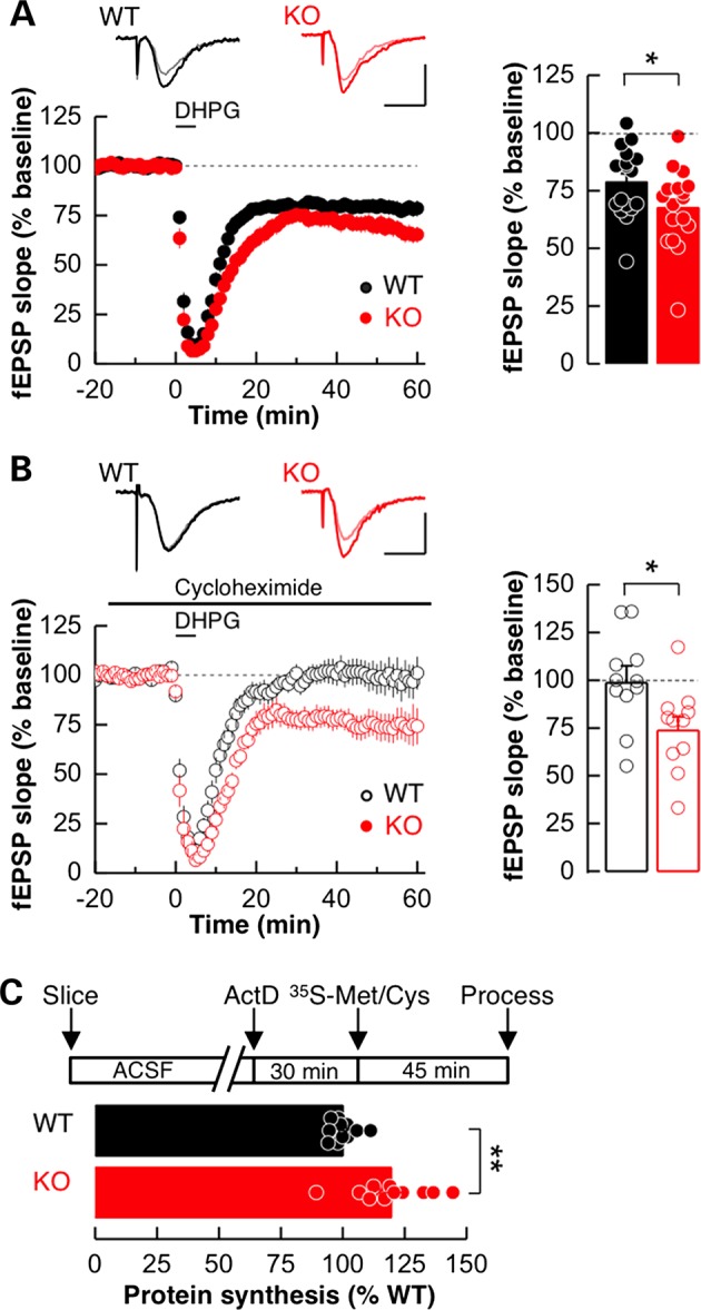
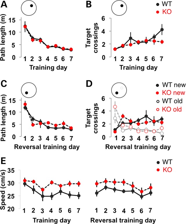
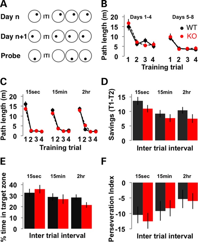
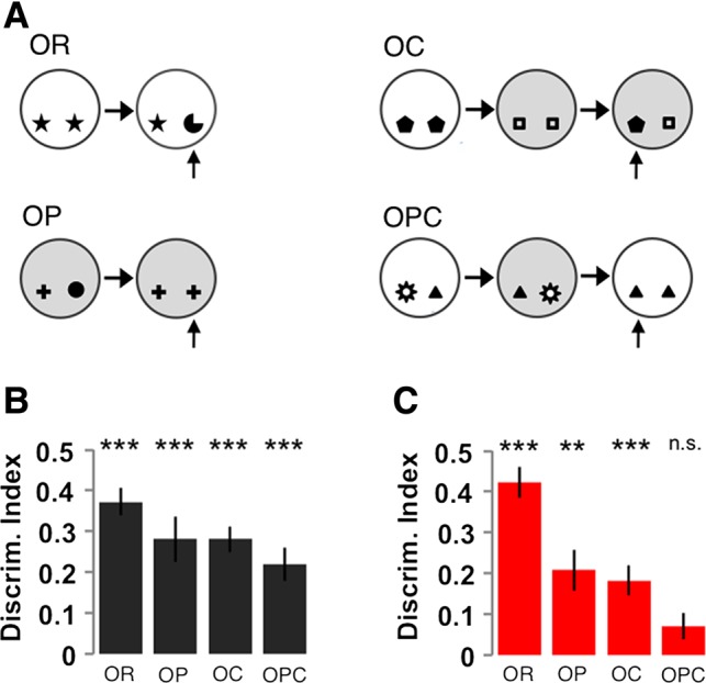
Recent advances in techniques for manipulating genomes have allowed the generation of transgenic animals other than mice. These new models enable cross-mammalian comparison of neurological disease from core cellular pathophysiology to circuit and behavioural endophenotypes. Moreover they will enable us to directly test whether common cellular dysfunction or behavioural outcomes of a genetic mutation are more conserved across species. Using a new rat model of Fragile X Syndrome, we report that Fmr1 knockout (KO) rats exhibit elevated basal protein synthesis and an increase in mGluR-dependent long-term depression in CA1 of the hippocampus that is independent of new protein synthesis. These defects in plasticity are accompanied by an increase in dendritic spine density selectively in apical dendrites and subtle changes in dendritic spine morphology of CA1 pyramidal neurons. Behaviourally, Fmr1 KO rats show deficits in hippocampal-dependent, but not hippocampal-independent, forms of associative recognition memory indicating that the loss of fragile X mental retardation protein (FMRP) causes defects in episodic-like memory. In contrast to previous reports from mice, Fmr1 KO rats show no deficits in spatial reference memory reversal learning. One-trial spatial learning in a delayed matching to place water maze task was also not affected by the loss of FMRP in rats. This is the first evidence for conservation across mammalian species of cellular and physiological hippocampal phenotypes associated with the loss of FMRP. Furthermore, while key cellular phenotypes are conserved they manifest in distinct behavioural dysfunction. Finally, our data reveal novel information about the selective role of FMRP in hippocampus-dependent associative memory.
Overview
The claims and statistics of this paper haven’t been checked yet.
Run the check — free, no account needed. It reads the open-access text and checks the sources it cites.
Integrity signals
Registry
Cites
Statistics
Figures
Verification
Claims the paper makes
The claims and statistics of this paper haven’t been checked yet.
Run the check — free, no account needed. It reads the open-access text and checks the sources it cites.
Claims other papers make about this one
Each is checked against this paper’s own text: does it say what the citing paper says it does?
The cited source says what the claim attributes to it.
In the citing paper
“Such deletion is sufficient to cause FXS-like traits, such as deficits in sustained visuospatial attention [34], altered patterns of social interaction, and cognitive impairment [28], together with abnormal synaptic plasticity [32,35,36].”
From Brain Cholesterol Biosynthetic Pathway Is Altered in a Preclinical Model of Fragile X Syndrome (2022) · cites this paper as [35]
Self-check
Percentages, sums and reported differences re-computed from the paper’s own numbers.
The self-check needs the paper’s full text.
Citations
Cites
Genome sequence of the Brown Norway rat yields insights into mammalian evolution
Gibbs, R. A., Weinstock, G. M., Metzker, M. L., Muzny, D. M., Sodergren, E. J., Scherer, S. E., Scott, G., Steffen, D. L., Worley, K. C., Burch, P. E., et al. (2004) · Nature · doi:10.1038/nature02426 · Cited by 2,170
Self-citationAltered synaptic plasticity in a mouse model of fragile X mental retardation
Huber, K. M., Gallagher, S. M., Warren, S. T., & Bear, M. F. (2002) · Proceedings of the National Academy of Sciences · doi:10.1073/pnas.122205699 · Cited by 1,349
Self-citationCorrection of Fragile X Syndrome in Mice
Dölen, G., Osterweil, E., Rao, B. S. S., Smith, G. B., Auerbach, B. D., Chattarji, S., & Bear, M. F. (2007) · Neuron · doi:10.1016/j.neuron.2007.12.001 · Cited by 979
Self-citationEvidence that fragile X mental retardation protein is a negative regulator of translation
Laggerbauer, B. (2001) · Human Molecular Genetics · doi:10.1093/hmg/10.4.329 · Cited by 571
The translation of translational control by FMRP: therapeutic targets for FXS
Darnell, J. C., & Klann, E. (2013) · Nature Neuroscience · doi:10.1038/nn.3379 · Cited by 491
Chronic Pharmacological mGlu5 Inhibition Corrects Fragile X in Adult Mice
Michalon, A., Sidorov, M., Ballard, T. M., Ozmen, L., Spooren, W., Wettstein, J. G., Jaeschke, G., Bear, M. F., & Lindemann, L. (2012) · Neuron · doi:10.1016/j.neuron.2012.03.009 · Cited by 489
Self-citationThe fragile X mental retardation protein inhibits translation via interacting with mRNA
Li, Z. (2001) · Nucleic Acids Research · doi:10.1093/nar/29.11.2276 · Cited by 448
Osterweil, E. K., Krueger, D. D., Reinhold, K., & Bear, M. F. (2010) · The Journal of Neuroscience · doi:10.1523/jneurosci.3888-10.2010 · Cited by 384
Self-citationBhattacharya, A., Kaphzan, H., Alvarez-Dieppa, A. C., Murphy, J. P., Pierre, P., & Klann, E. (2012) · Neuron · doi:10.1016/j.neuron.2012.07.022 · Cited by 316
Paradee, W., Melikian, H. E., Rasmussen, D. L., Kenneson, A., Conn, P. J., & Warren, S. T. (1999) · Neuroscience · doi:10.1016/s0306-4522(99)00285-7 · Cited by 305
Cited by
Sorted by citations
Synaptopathology Involved in Autism Spectrum Disorder
Guang, S., Pang, N., Deng, X., Yang, L., He, F. P., Wu, L., Chen, C., Yin, F., & Peng, J. (2018) · Frontiers in Cellular Neuroscience · doi:10.3389/fncel.2018.00470 · Cited by 298
Lessons learned from studying syndromic autism spectrum disorders
Sztainberg, Y., & Zoghbi, H. Y. (2016) · Nature Neuroscience · doi:10.1038/nn.4420 · Cited by 305
Sensory Processing Phenotypes in Fragile X Syndrome
Rais, M., Binder, D. K., Razak, K. A., & Ethell, I. M. (2018) · ASN NEURO · doi:10.1177/1759091418801092 · Cited by 143
Tang, X., Drotar, J., Li, K., Clairmont, C. D., Brumm, A. S., Sullins, A., Wu, H., Liu, X. S., Wang, J., Gray, N. S., et al. (2019) · Science Translational Medicine · doi:10.1126/scitranslmed.aau0164 · Cited by 120
Of Men and Mice: Modeling the Fragile X Syndrome
Dahlhaus, R. (2018) · Frontiers in Molecular Neuroscience · doi:10.3389/fnmol.2018.00041 · Cited by 133
Metadata
- Authors
- Till, S. M., Asiminas, A., Jackson, A. D., Katsanevaki, D., Barnes, S. A., Osterweil, E. K., Bear, M. F., Chattarji, S., Wood, E. R., Wyllie, D. J. A., & Kind, P. C.
- Affiliations
- University of Edinburgh (GB)
- Institute for Stem Cell Biology and Regenerative Medicine (IN)
- Howard Hughes Medical Institute (US)
- Tata Institute of Fundamental Research (IN)
- Corresponding author
- Kind, Peter C.
- ORCID
- Venue
- Human Molecular Genetics
- Publisher
- Oxford University Press (OUP)
- Published
- 2015-08-04
- Volume
- 24
- Issue
- 21
- Pages
- 5977-5984
- ISSN
- 0964-6906 (ISSN-L)
- 1460-2083
- PMC
- PMC4599667
- OpenAlex
- W1893201598
- Cited by
- 109
- References
- 28
- Citations by year
- 2026: 6 · 2025: 8 · 2024: 7 · 2023: 3 · 2022: 16 · 2021: 9 · 2020: 16 · 2019: 9 · 2018: 13 · 2017: 11 · 2016: 10 · 2015: 1
- FWCI
- 8.02
- Type
- journal article
- Language
- en
- Open access
- hybrid
- doi.org
- Licence
- cc-by-nc
- Locations
- Human Molecular Genetics (publishedVersion, cc-by-nc)
- Europe PMC (PubMed Central) (publishedVersion, cc-by-nc)
- PubMed Central (publishedVersion, cc-by-nc)
- PubMed (publishedVersion)
- Edinburgh Research Explorer (University of Edinburgh) (submittedVersion, cc-by-nc)
- DSpace@MIT (Massachusetts Institute of Technology) (submittedVersion, cc-by-nc)
- Oxford University Press eBooks
- Funders
- Wellcome Trust (104116, 104116/Z/14/Z)
- Research Councils UK
- University of Edinburgh
- Medical Research Council (MR/K014137/1, MR/M006336/1)
- Topics
- Life Sciences › Biochemistry, Genetics and Molecular Biology › Genetics › Genetics and Neurodevelopmental Disorders
- Life Sciences › Neuroscience › Cellular and Molecular Neuroscience › Neuroscience and Neuropharmacology Research
- Life Sciences › Neuroscience › Cognitive Neuroscience › Memory and Neural Mechanisms
- Keywords
- fragile X syndrome, FMRP, hippocampus, CA1 pyramidal neurons, dendritic spines, protein synthesis, synaptic plasticity, associative recognition memory, episodic-like memory, spatial memory, cross-species conservation, transgenic rats
- MeSH terms
- Animals, Disease Models, Animal, Female, Fragile X Syndrome, Hippocampus, Male, Memory Disorders, Neuronal Plasticity, Species Specificity, Rats, Sprague-Dawley, Pyramidal Cells, Maze Learning, Rats, Fragile X Mental Retardation Protein, Fragile X Messenger Ribonucleoprotein 1, Gene Knockout Techniques
About these checks
Registry, retraction, Cites and statistics results are computed, not judged, with one exception: a printed reference that no index lists is read once by a model before it counts as a citation error. Claims are extracted by a model and kept only when their quote appears verbatim in the text; each cited source is then checked, with a model’s help, for whether it says what the claim relies on. Those verdicts can be wrong — read the claim’s quote and the cited source before relying on one.
Statistics and arithmetic are recomputed, not judged.
Confidence bands are shown only for fields calibrated on our annotated test set. None is calibrated yet, so no band is shown.
What counts as a claim ›How we measure confidence ›How we check this ›
Are you an author, or do you see a problem with this page? Request a review
