

The DOI matches its Crossref record; 5 of the 5 references it cites were found in a registry or an index.
Profiling Small RNA From Brain Extracellular Vesicles in Individuals With Depression
Abstract
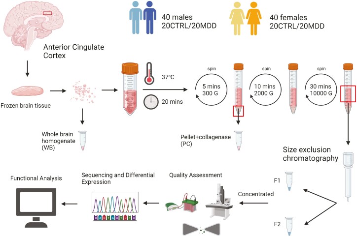
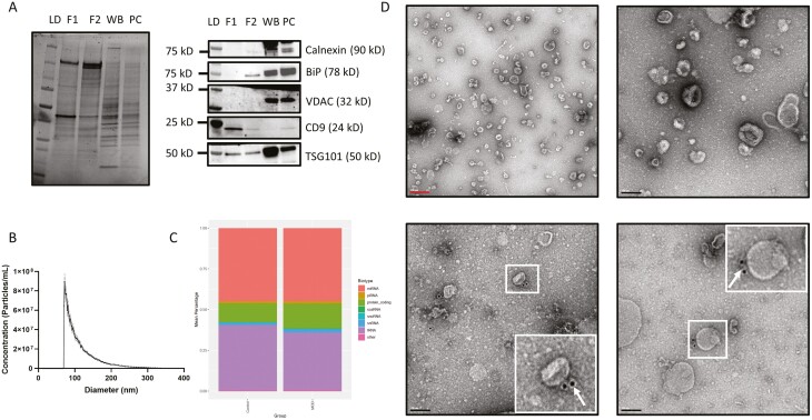
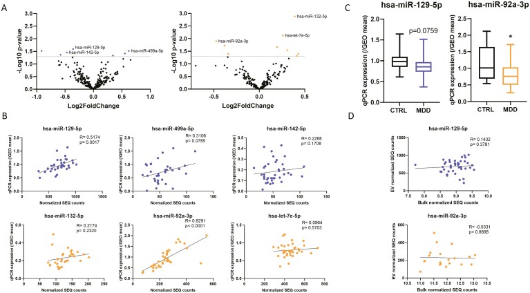
Background: Major depressive disorder (MDD) is a leading cause of disability with significant mortality risk. Despite progress in our understanding of the etiology of MDD, the underlying molecular changes in the brain remain poorly understood. Extracellular vesicles (EVs) are lipid-bound particles that can reflect the molecular signatures of the tissue of origin. We aimed to optimize a streamlined EV isolation protocol from postmortem brain tissue and determine whether EV RNA cargo, particularly microRNAs (miRNAs), have an MDD-specific profile. Methods: EVs were isolated from postmortem human brain tissue. Quality was assessed using western blots, transmission electron microscopy, and microfluidic resistive pulse sensing. EV RNA was extracted and sequenced on Illumina platforms. Functional follow-up was performed in silico. Results: Quality assessment showed an enrichment of EV markers, as well as a size distribution of 30 to 200 nm in diameter, and no contamination with cellular debris. Small RNA profiling indicated the presence of several RNA biotypes, with miRNAs and transfer RNAs being the most prominent. Exploring miRNA levels between groups revealed decreased expression of miR-92a-3p and miR-129-5p, which was validated by qPCR and was specific to EVs and not seen in bulk tissue. Finally, in silico functional analyses indicate potential roles for these 2 miRNAs in neurotransmission and synaptic plasticity. Conclusion: We provide a streamlined isolation protocol that yields EVs of high quality that are suitable for molecular follow-up. Our findings warrant future investigations into brain EV miRNA dysregulation in MDD.
Overview
The claims and statistics of this paper haven’t been checked yet.
Run the check — free, no account needed. It reads the open-access text and checks the sources it cites.
Integrity signals
Registry
Cites
Statistics
Figures
Verification
Claims the paper makes
The claims and statistics of this paper haven’t been checked yet.
Run the check — free, no account needed. It reads the open-access text and checks the sources it cites.
Claims other papers make about this one
Each is checked against this paper’s own text: does it say what the citing paper says it does?
In the citing paper
“However, it should be mentioned that recent study reported that miR-129-5p is decreased in exosomes isolated from the postmortem brain of males but not females that suffered from depression [77].”
From A role for astrocytic miR-129-5p in frontotemporal dementia (2025) · cites this paper as [77]
Self-check
Percentages, sums and reported differences re-computed from the paper’s own numbers.
The self-check needs the paper’s full text.
Citations
Cites
Otte, C., Gold, S. M., Penninx, B. W., Pariante, C. M., Etkin, A., Fava, M., Mohr, D. C., & Schatzberg, A. F. (2016) · Nature Reviews Disease Primers · doi:10.1038/nrdp.2016.65 · Cited by 2,269
Sex-specific transcriptional signatures in human depression
Labonté, B., Engmann, O., Purushothaman, I., Menard, C., Wang, J., Tan, C., Scarpa, J. R., Moy, G., Loh, Y.-H. E., Cahill, M., et al. (2017) · Nature Medicine · doi:10.1038/nm.4386 · Cited by 748
Self-citationWei, Z.-X., Xie, G.-J., Mao, X., Zou, X.-P., Liao, Y.-J., Liu, Q.-S., Wang, H., & Cheng, Y. (2020) · Neuropsychopharmacology · doi:10.1038/s41386-020-0622-2 · Cited by 227
miR-323a regulates ERBB4 and is involved in depression
Fiori, L. M., Kos, A., Lin, R., Théroux, J.-F., Lopez, J. P., Kühne, C., Eggert, C., Holzapfel, M., Huettl, R.-E., Mechawar, N., et al. (2021) · Molecular Psychiatry · doi:10.1038/s41380-020-00953-7 · Cited by 88 · cited 16×
“To determine whether these effects are specific to EVs, we correlated the expression of miRNAs from EVs to miRNA expression levels observed in bulk ACC tissue in the subjects that overlap with those from a previous study of our group (Fiori et al., 2021).”
BackgroundMethodResultInfluentialSelf-citationRapamycin reveals an mTOR-independent repression of Kv1.1 expression during epileptogenesis
Sosanya, N. M., Brager, D. H., Wolfe, S., Niere, F., & Raab-Graham, K. F. (2015) · Neurobiology of Disease · doi:10.1016/j.nbd.2014.09.011 · Cited by 49
Research works printed but not linked: 88
- Andersen, 2004, Normalization of real-time quantitative reverse transcription-PCR data: a model-based variance estimation approach to identify genes suited for normalization, applied to bladder and colon cancer data sets, Cancer Res, 64, 5245, 10.1158/0008-5472.CAN-04-0496 · DOI · Paper page
- Bahrini, 2015, Neuronal exosomes facilitate synaptic pruning by up-regulating complement factors in microglia, Sci Rep, 5, 7989, 10.1038/srep07989 · DOI
- Bakhti, 2011, Inhibition of myelin membrane sheath formation by oligodendrocyte-derived exosome-like vesicles, J Biol Chem, 286, 787, 10.1074/jbc.M110.190009 · DOI
- Banigan, 2013, Differential expression of exosomal microRNAs in prefrontal cortices of schizophrenia and bipolar disorder patients, PLoS One, 8, e48814, 10.1371/journal.pone.0048814 · DOI
- Basso, 2013, Mutant copper-zinc superoxide dismutase (SOD1) induces protein secretion pathway alterations and exosome release in astrocytes: implications for disease spreading and motor neuron pathology in amyotrophic lateral sclerosis, J Biol Chem, 288, 15699, 10.1074/jbc.M112.425066 · DOI
- Batagov, 2013, Exosomes secreted by human cells transport largely mRNA fragments that are enriched in the 3’-untranslated regions, Biol Direct, 8, 12, 10.1186/1745-6150-8-12 · DOI
- Beurel, 2020, The bidirectional relationship of depression and inflammation: double trouble, Neuron, 107, 234, 10.1016/j.neuron.2020.06.002 · DOI · Paper page
- Blair, 2012, PTEN: a new player controlling structural and functional synaptic plasticity, J Physiol, 590, 1017, 10.1113/jphysiol.2012.227868 · DOI
- Bressler, 2021, Depletion of eukaryotic initiation factor 5B (eIF5B) reprograms the cellular transcriptome and leads to activation of endoplasmic reticulum (ER) stress and c-Jun N-terminal kinase (JNK), Cell Stress Chaperones, 26, 253, 10.1007/s12192-020-01174-1 · DOI
- Buran, 2017, Treatment with TREK1 and TRPC3/6 ion channel inhibitors upregulates microRNA expression in a mouse model of chronic mild stress, Neurosci Lett, 656, 51, 10.1016/j.neulet.2017.07.017 · DOI
- Chang, 2013, Exosomes of BV-2 cells induced by alpha-synuclein: important mediator of neurodegeneration in PD, Neurosci Lett, 548, 190, 10.1016/j.neulet.2013.06.009 · DOI
- Chang, 2021, MiR-129-5p prevents depressive-like behaviors by targeting MAPK1 to suppress inflammation, Exp Brain Res, 239, 3359, 10.1007/s00221-021-06203-8 · DOI
- Chen, 2021, Prognostic plasma exosomal microRNA biomarkers in patients with substance use disorders presenting comorbid with anxiety and depression, Sci Rep, 11, 6271, 10.1038/s41598-021-84501-5 · DOI
- Chen, 2021, Mechanism of cargo sorting into small extracellular vesicles, Bioengineered, 12, 8186, 10.1080/21655979.2021.1977767 · DOI
- Colombo, 2014, Biogenesis, secretion, and intercellular interactions of exosomes and other extracellular vesicles, Annu Rev Cell Dev Biol, 30, 255, 10.1146/annurev-cellbio-101512-122326 · DOI · Paper page
- Crescitelli, 2021, Isolation and characterization of extracellular vesicle subpopulations from tissues, Nat Protoc, 16, 1548, 10.1038/s41596-020-00466-1 · DOI
- Dobricic, 2022, Common signatures of differential microRNA expression in Parkinson’s and Alzheimer’s disease brains, Brain Commun, 4, fcac274, 10.1093/braincomms/fcac274 · DOI · Paper page
- Du, 2019, Genome-wide, integrative analysis implicates exosome-derived microrna dysregulation in schizophrenia, Schizophr Bull, 45, 1257, 10.1093/schbul/sby191 · DOI
- Dumais, 2005, Is violent method of suicide a behavioral marker of lifetime aggression, Am J Psychiatry, 162, 1375, 10.1176/appi.ajp.162.7.1375 · DOI
- Fujiwara, 2022, MicroRNA-92a promotes CNS autoimmunity by modulating the regulatory and inflammatory T cell balance, J Clin Invest, 132, e155693, 10.1172/JCI155693 · DOI
- Gabrielli, 2015, Active endocannabinoids are secreted on extracellular membrane vesicles, EMBO Rep, 16, 213, 10.15252/embr.201439668 · DOI · Paper page
- Gardiner, 2016, Techniques used for the isolation and characterization of extracellular vesicles: results of a worldwide survey, J Extracell Vesicles, 5, 32945, 10.3402/jev.v5.32945 · DOI
- Giraldez, 2018, Comprehensive multi-center assessment of small RNA-seq methods for quantitative miRNA profiling, Nat Biotechnol, 36, 746, 10.1038/nbt.4183 · DOI · Paper page
- Glaesel, 2020, MiR-129-5p and miR-130a-3p regulate VEGFR-2 expression in sensory and motor neurons during development, Int J Mol Sci, 21, 3839, 10.3390/ijms21113839 · DOI
- Goetzl, 2021, Abnormal levels of mitochondrial proteins in plasma neuronal extracellular vesicles in major depressive disorder, Mol Psychiatry, 26, 7355, 10.1038/s41380-021-01268-x · DOI
- Gross, 2014, Neuron-specific regulation of class I PI3K catalytic subunits and their dysfunction in brain disorders, Front Mol Neurosci, 7, 12, 10.3389/fnmol.2014.00012 · DOI · Paper page
- Gupta, 2022, Integrative analysis of OIP5-AS1/miR-129-5p/CREBBP axis as a potential therapeutic candidate in the pathogenesis of metal toxicity-induced Alzheimer’s disease, Gene Rep, 26, 101442, 10.1016/j.genrep.2021.101442 · DOI
- Hawley, 2020, MiR-129-5p: a novel therapeutic target for amyotrophic lateral sclerosis, Noncod RNA Investig, 4, 7, 10.21037/ncri-20-5 · DOI
- He, 2017, Age-related frontal periventricular white matter hyperintensities and miR-92a-3p are associated with early-onset post-stroke depression, Front Aging Neurosci, 9, 328, 10.3389/fnagi.2017.00328 · DOI
- He, 2020, MicroRNA-92a-3p enhances functional recovery and suppresses apoptosis after spinal cord injury via targeting phosphatase and tensin homolog, Biosci Rep, 40, 10.1042/BSR20192743 · DOI
- Hessvik, 2018, Current knowledge on exosome biogenesis and release, Cell Mol Life Sci, 75, 193, 10.1007/s00018-017-2595-9 · DOI · Paper page
- Hodes, 2017, Understanding the epigenetic basis of sex differences in depression, J Neurosci Res, 95, 692, 10.1002/jnr.23876 · DOI
- Huang, 2017, The link between long noncoding RNAs and depression, Prog Neuropsychopharmacol Biol Psychiatry, 73, 73, 10.1016/j.pnpbp.2016.06.004 · DOI
- Huang, 2006, CPEB3 and CPEB4 in neurons: analysis of RNA-binding specificity and translational control of AMPA receptor GluR2 mRNA, EMBO J, 25, 4865, 10.1038/sj.emboj.7601322 · DOI
- Iguchi, 2016, Exosome secretion is a key pathway for clearance of pathological TDP-43, Brain, 139, 3187, 10.1093/brain/aww237 · DOI · Paper page
- Ji, 2023, Exposure to enriched environment ameliorated chronic unpredictable mild stress-induced depression-like symptoms in rats via regulating the miR-92a-3p/kruppel-like factor 2 (KLF2) pathway, Brain Res Bull, 195, 14, 10.1016/j.brainresbull.2023.01.002 · DOI
- Jin, 2016, MiR-17-92 cluster regulates adult hippocampal neurogenesis, anxiety, and depression, Cell Rep, 16, 1653, 10.1016/j.celrep.2016.06.101 · DOI
- Kodama, 2020, Microglial microRNAs mediate sex-specific responses to tau pathology, Nat Neurosci, 23, 167, 10.1038/s41593-019-0560-7 · DOI · Paper page
- Konoshenko, 2018, Isolation of extracellular vesicles: general methodologies and latest trends, Biomed Res Int, 2018, 8545347, 10.1155/2018/8545347 · DOI
- Krispil-Alon, 2022, Sex-specific roles of hippocampal microRNAs in stress vulnerability and resilience, Transl Psychiatry, 12, 503, 10.1038/s41398-022-02267-4 · DOI
- Lai, 2022, exoRBase 2.0: an atlas of mRNA, lncRNA and circRNA in extracellular vesicles from human biofluids, Nucleic Acids Res, 50, D118, 10.1093/nar/gkab1085 · DOI
- Letellier, 2014, MiR-92a regulates expression of synaptic GluA1-containing AMPA receptors during homeostatic scaling, Nat Neurosci, 17, 1040, 10.1038/nn.3762 · DOI · Paper page
- Li, 2017, Correcting miR92a-vGAT-mediated GABAergic dysfunctions rescues human Tau-induced anxiety in mice, Mol Ther, 25, 140, 10.1016/j.ymthe.2016.10.010 · DOI
- Li, 2020, Circular RNA in schizophrenia and depression, Front Psychiatry, 11, 392, 10.3389/fpsyt.2020.00392 · DOI
- Lin, 2021, Circular RNA circCCNT2 is upregulated in the anterior cingulate cortex of individuals with bipolar disorder, Transl Psychiatry, 11, 629, 10.1038/s41398-021-01746-4 · DOI
- Linares, 2015, High-speed centrifugation induces aggregation of extracellular vesicles, J Extracell Vesicles, 4, 29509, 10.3402/jev.v4.29509 · DOI
- Liu, 2017, MicroRNA-129-5p inhibits the development of autoimmune encephalomyelitis-related epilepsy by targeting HMGB1 through the TLR4/NF-kB signaling pathway, Brain Res Bull, 132, 139, 10.1016/j.brainresbull.2017.05.004 · DOI
- Loffreda, 2020, MiR-129-5p: a key factor and therapeutic target in amyotrophic lateral sclerosis, Prog Neurobiol, 190, 101803, 10.1016/j.pneurobio.2020.101803 · DOI
- Lutz, 2017, Association of a history of child abuse with impaired myelination in the anterior cingulate cortex: convergent epigenetic, transcriptional, and morphological evidence, Am J Psychiatry, 174, 1185, 10.1176/appi.ajp.2017.16111286 · DOI · Paper page
- Ma, 2018, Identification of miR-22-3p, miR-92a-3p, and miR-137 in peripheral blood as biomarker for schizophrenia, Psychiatry Res, 265, 70, 10.1016/j.psychres.2018.03.080 · DOI
- Meyers, 2017, TGF-beta family signaling in neural and neuronal differentiation, development, and function, Cold Spring Harb Perspect Biol, 9, a022244, 10.1101/cshperspect.a022244 · DOI
- Morgan, 2010, CPEB2, CPEB3 and CPEB4 are coordinately regulated by miRNAs recognizing conserved binding sites in paralog positions of their 3’-UTRs, Nucleic Acids Res, 38, 7698, 10.1093/nar/gkq635 · DOI · Paper page
- Morhayim, 2017, Molecular characterization of human osteoblast-derived extracellular vesicle mRNA using next-generation sequencing, Biochim Biophys Acta Mol Cell Res, 1864, 1133, 10.1016/j.bbamcr.2017.03.011 · DOI
- Mulcahy, 2014, Routes and mechanisms of extracellular vesicle uptake, J Extracell Vesicles, 4, 3
- Munoz-Llanos, 2018, MicroRNA profiling and bioinformatics target analysis in dorsal hippocampus of chronically stressed rats: relevance to depression pathophysiology, Front Mol Neurosci, 11, 251, 10.3389/fnmol.2018.00251 · DOI
- Pascua-Maestro, 2018, Extracellular vesicles secreted by astroglial cells transport apolipoprotein D to neurons and mediate neuronal survival upon oxidative stress, Front Cell Neurosci, 12, 526, 10.3389/fncel.2018.00526 · DOI
- Pena-Bautista, 2022, Plasma microRNAs as potential biomarkers in early Alzheimer disease expression, Sci Rep, 12, 15589, 10.1038/s41598-022-19862-6 · DOI
- Perez-Boza, 2018, Exploring the RNA landscape of endothelial exosomes, RNA, 24, 423, 10.1261/rna.064352.117 · DOI
- Qinlin, 2022, MiR-129-5p targets FEZ1/SCOC/ULK1/NBR1 complex to restore neuronal function in mice with post-stroke depression, Bioengineered, 13, 9708, 10.1080/21655979.2022.2059910 · DOI
- Qinlin, 2022, Differential expression analysis of microRNAs and mRNAs in the mouse hippocampus of post-stroke depression (PSD) based on transcriptome sequencing, Bioengineered, 13, 3582, 10.1080/21655979.2022.2027061 · DOI
- Ragan, 2017, Small non-coding RNA expression from anterior cingulate cortex in schizophrenia shows sex specific regulation, Schizophr Res, 183, 82, 10.1016/j.schres.2016.11.024 · DOI
- Rajendran, 2006, Alzheimer’s disease beta-amyloid peptides are released in association with exosomes, Proc Natl Acad Sci U S A, 103, 11172, 10.1073/pnas.0603838103 · DOI · Paper page
- Rajman, 2017, A microRNA-129-5p/Rbfox crosstalk coordinates homeostatic downscaling of excitatory synapses, EMBO J, 36, 1770, 10.15252/embj.201695748 · DOI · Paper page
- Sadik, 2018, Extracellular RNAs: A new awareness of old perspectives, Methods Mol Biol, 1740, 1, 10.1007/978-1-4939-7652-2_1 · DOI
- Saeedi, 2021, Neuron-derived extracellular vesicles enriched from plasma show altered size and miRNA cargo as a function of antidepressant drug response, Mol Psychiatry, 26, 7417, 10.1038/s41380-021-01255-2 · DOI
- Schumann, 2017, Possible sexually dimorphic role of miRNA and other sncRNA in ASD brain, Mol Autism, 8, 4, 10.1186/s13229-017-0117-0 · DOI
- Siedlecki-Wullich, 2019, Altered microRNAs related to synaptic function as potential plasma biomarkers for Alzheimer’s disease, Alzheimers Res Ther, 11, 46, 10.1186/s13195-019-0501-4 · DOI
- Srinivasan, 2019, Small RNA sequencing across diverse biofluids identifies optimal methods for exRNA isolation, Cell, 177, 446, 10.1016/j.cell.2019.03.024 · DOI
- Stuendl, 2016, Induction of alpha-synuclein aggregate formation by CSF exosomes from patients with Parkinson’s disease and dementia with Lewy bodies, Brain, 139, 481, 10.1093/brain/awv346 · DOI · Paper page
- Taguchi, 2018, Exploring microRNA biomarkers for Parkinson’s disease from mRNA expression profiles, Cells, 7, 245, 10.3390/cells7120245 · DOI
- Trivedi, 2022, Identification of Hsa-miR-92a-3p as a novel biomarker and PIK3R1 as a key regulatory gene to combat with the diagnostic patterns of autism spectrum disorders, Mater Today Proc, 10.1016/j.matpr.2022.12.042 · DOI
- Trujillo-Gonzalez, 2019, MicroRNA-129-5p is regulated by choline availability and controls EGF receptor synthesis and neurogenesis in the cerebral cortex, FASEB J, 33, 3601, 10.1096/fj.201801094RR · DOI
- Tsilioni, 2018, Extracellular vesicles are increased in the serum of children with autism spectrum disorder, contain mitochondrial DNA, and stimulate human microglia to secrete IL-1beta, J Neuroinflammation, 15, 239, 10.1186/s12974-018-1275-5 · DOI
- Valadi, 2007, Exosome-mediated transfer of mRNAs and microRNAs is a novel mechanism of genetic exchange between cells, Nat Cell Biol, 9, 654, 10.1038/ncb1596 · DOI · Paper page
- Vella, 2017, A rigorous method to enrich for exosomes from brain tissue, J Extracell Vesicles, 6, 1348885, 10.1080/20013078.2017.1348885 · DOI
- Videtic Paska, 2022, Suicide and changes in expression of neuronal miRNA predicted by an algorithm search through miRNA databases, Genes (Basel), 13, 562, 10.3390/genes13040562 · DOI
- Wan, 2020, LncRNA NEAT1 affects inflammatory response by targeting miR-129-5p and regulating Notch signaling pathway in epilepsy, Cell Cycle, 19, 419, 10.1080/15384101.2020.1711578 · DOI
- Wang, 2012, Astrocytes secrete exosomes enriched with proapoptotic ceramide and prostate apoptosis response 4 (PAR-4): Potential mechanism of apoptosis induction in Alzheimer disease (AD), J Biol Chem, 287, 21384, 10.1074/jbc.M112.340513 · DOI
- Wang, 2021, Plasma exosomes from depression ameliorate inflammation-induced depressive-like behaviors via sigma-1 receptor delivery, Brain Behav Immun, 94, 225, 10.1016/j.bbi.2021.02.004 · DOI
- World Health Organization, 2021, Institute of Health Metrics and Evaluation
- Wu, 2019, MicroRNA-129 modulates neuronal migration by targeting Fmr1 in the developing mouse cortex, Cell Death Dis, 10, 287, 10.1038/s41419-019-1517-1 · DOI
- Yasir, 2022, Evaluation the protective effect of zinc supplementation on depression and Mir 129-GPR39 gene expression in the hippocampus of diabetic rat, Tob Regul Sci, 8, 573
- Yeri, 2018, Evaluation of commercially available small RNASeq library preparation kits using low input RNA, BMC Genomics, 19, 331, 10.1186/s12864-018-4726-6 · DOI
- Yoshino, 2020, Non-coding RNAs in psychiatric disorders and suicidal behavior, Front Psychiatry, 11, 543893, 10.3389/fpsyt.2020.543893 · DOI
- Zhang, 2016, Clinical predictor and circulating microRNA profile expression in patients with early onset post-stroke depression, J Affect Disord, 193, 51, 10.1016/j.jad.2015.12.061 · DOI
- Zhou, 2022, A new perspective on depression and neuroinflammation: non-coding RNA, J Psychiatr Res, 148, 293, 10.1016/j.jpsychires.2022.02.007 · DOI
- Zongaro, 2013, The 3’ UTR of FMR1 mRNA is a target of miR-101, miR-129-5p and miR-221: implications for the molecular pathology of FXTAS at the synapse, Hum Mol Genet, 22, 1971, 10.1093/hmg/ddt044 · DOI
- Żurawek, 2021, The miRNome of depression, Int J Mol Sci, 22, 11312, 10.3390/ijms222111312 · DOI
Cited by
Sorted by citations
A role for astrocytic miR-129-5p in frontotemporal dementia
Kaurani, L., Pradhan, R., Schröder, S., Burkhardt, S., Schuetz, A.-L., Krüger, D. M., Pena, T., Heutink, P., Sananbenesi, F., & Fischer, A. (2025) · Translational Psychiatry · doi:10.1038/s41398-025-03338-y · Cited by 10
Metadata
- Authors
- Ibrahim, P., Denniston, R., Mitsuhashi, H., Yang, J., Fiori, L. M., Żurawek, D., Mechawar, N., Nagy, C., & Turecki, G.
- Affiliations
- Integrated Program in Neuroscience, McGill University , Montreal, Quebec , Canada
- McGill Group for Suicide Studies, Douglas Mental Health University Institute , Verdun, Quebec , Canada
- Venue
- International Journal of Neuropsychopharmacology
- Publisher
- Oxford University Press (OUP)
- Published
- 2024-03-01
- Volume
- 27
- Issue
- 3
- ISSN
- 1461-1457
- 1469-5111
- PMC
- PMC10946232
- Cited by
- 29
- References
- 5
- Language
- en
- Funders
- Canadian Institute of Health Research (FDN148374, ENP161427)
About these checks
Registry, retraction, Cites and statistics results are computed, not judged, with one exception: a printed reference that no index lists is read once by a model before it counts as a citation error. Claims are extracted by a model and kept only when their quote appears verbatim in the text; each cited source is then checked, with a model’s help, for whether it says what the claim relies on. Those verdicts can be wrong — read the claim’s quote and the cited source before relying on one.
Statistics and arithmetic are recomputed, not judged.
Confidence bands are shown only for fields calibrated on our annotated test set. None is calibrated yet, so no band is shown.
What counts as a claim ›How we measure confidence ›How we check this ›
Are you an author, or do you see a problem with this page? Request a review
How to cite
Cite CoChat for the checks on this page, and the paper for its own findings.
- This page
- CoChat. (2026). Citation check: Profiling Small RNA From Brain Extracellular Vesicles in Individuals With Depression. https://app.cochat.ai/papers/10.1093/ijnp/pyae013
- The paper
- Ibrahim, P., Denniston, R., Mitsuhashi, H., Yang, J., Fiori, L. M., Żurawek, D., Mechawar, N., Nagy, C., & Turecki, G. (2024). Profiling Small RNA From Brain Extracellular Vesicles in Individuals With Depression. International Journal of Neuropsychopharmacology. https://doi.org/10.1093/ijnp/pyae013
