

The DOI matches its Crossref record; 55 of the 58 references it cites were found in a registry or an index.
Gender Differences in Alzheimer Disease: Brain Atrophy, Histopathology Burden, and Cognition
Open accessAbstract
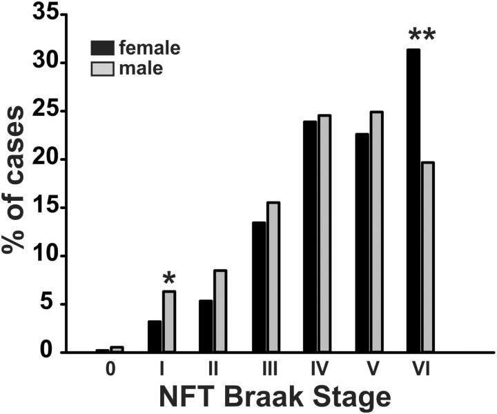
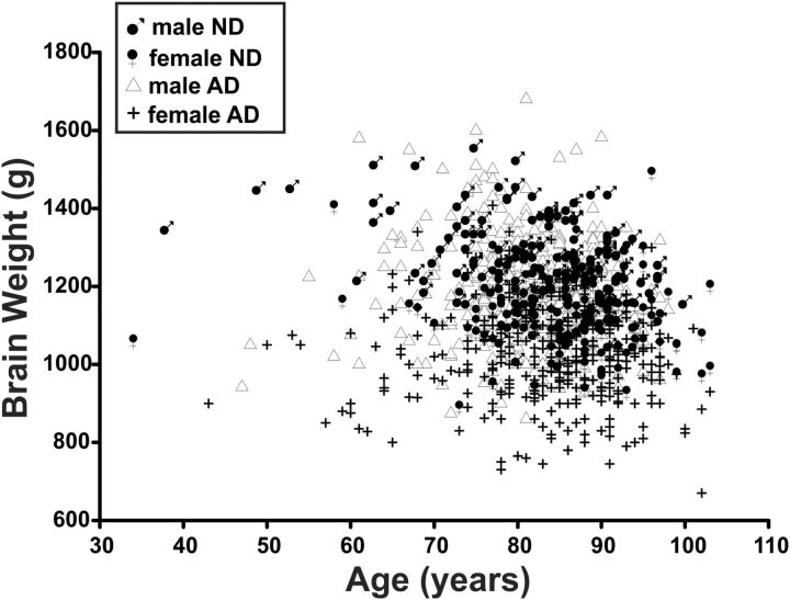
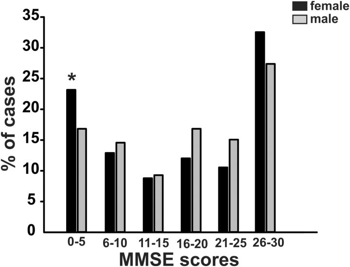
Multiple studies suggest that females are affected by Alzheimer disease (AD) more severely and more frequently than males. Other studies have failed to confirm this and the issue remains controversial. Difficulties include differences in study methods and male versus female life expectancy. Another element of uncertainty is that the majority of studies have lacked neuropathological confirmation of the AD diagnosis. We compared clinical and pathological AD severity in 1028 deceased subjects with full neuropathological examinations. The age of dementia onset did not differ by gender but females were more likely to proceed to very severe clinical and pathological disease, with significantly higher proportions having a Mini-Mental State Examination score of 5 or less and Braak stage VI neurofibrillary degeneration. Median neuritic plaque densities were similar in females and males with AD but females had significantly greater tangle density scores. In addition, we found that AD-control brain weight differences were significantly greater for females, even after adjustment for age, disease duration, and comorbid conditions. These findings suggest that when they are affected by AD, females progress more often to severe cognitive dysfunction, due to more severe neurofibrillary degeneration, and greater loss of brain parenchyma.
Overview
The claims and statistics of this paper haven’t been checked yet.
Run the check — free, no account needed. It reads the open-access text and checks the sources it cites.
Integrity signals
Registry
Cites
Statistics
Figures
Verification
Claims the paper makes
The claims and statistics of this paper haven’t been checked yet.
Run the check — free, no account needed. It reads the open-access text and checks the sources it cites.
Claims other papers make about this one
Each is checked against this paper’s own text: does it say what the citing paper says it does?
The cited source says what the claim attributes to it.
In the citing paper
“Recent studies suggest that women develop greater neurofibrillary degeneration, brain parenchymal loss, and cognitive decline.52,53,54”
From Sex differences in cognitive decline among US adults (2021) · cites this paper as [53]
Self-check
Percentages, sums and reported differences re-computed from the paper’s own numbers.
The self-check needs the paper’s full text.
Citations
Cites
McKhann, G. M., Knopman, D. S., Chertkow, H., Hyman, B. T., Jack, C. R., Kawas, C. H., Klunk, W. E., Koroshetz, W. J., Manly, J. J., Mayeux, R., et al. (2011) · Alzheimer's & Dementia · doi:10.1016/j.jalz.2011.03.005 · Cited by 18,657 · cited 1×
“Clinically defined measures used in this study included age at cognitive symptom onset and cognitive symptom duration, the latter defined as the number of years between cognitive symptom onset and death (14).”
MethodNeuropathological stageing of Alzheimer-related changes.
Braak, H., & Braak, E. (1991) · Acta Neuropathologica · doi:10.1007/bf00308809 · Cited by 16,123 · cited 13×
“Factors Affecting Alzheimer Disease-Control Brain Weight Difference, Multivariable Analysis Variable Complete Equation OR; 95% CI; Gender Values OR; 95% CI; p Value p Value Gender 1.3; 1.1–1.6; 0.01 0.6; 0.5–0.8; 0.001 Gender þ Braak stage 0.2; 0.4–0.4; <0.001 0.”
MethodInfluentialMirra, S. S., Heyman, A., McKeel, D., Sumi, S. M., Crain, B. J., Brownlee, L. M., Vogel, F. S., Hughes, J. P., Belle, G. V., Berg, L., et al. (1991) · Neurology · doi:10.1212/wnl.41.4.479 · Cited by 4,961 · cited 1×
“Neuritic plaque and neurofibrillary tangle densities were graded blindly as recommended by The Consortium to Establish a Registry for Alzheimer’s Disease (CERAD) with separate semiquantitative density estimates of none, sparse, moderate, or frequent (24).”
MethodMontine, T. J., Phelps, C. H., Beach, T. G., Bigio, E. H., Cairns, N. J., Dickson, D. W., Duyckaerts, C., Frosch, M. P., Masliah, E., Mirra, S. S., et al. (2012) · Acta Neuropathologica · doi:10.1007/s00401-011-0910-3 · Cited by 2,894
Self-citationα-Synuclein is phosphorylated in synucleinopathy lesions
Fujiwara, H., Hasegawa, M., Dohmae, N., Kawashima, A., Masliah, E., Goldberg, M. S., Shen, J., Takio, K., & Iwatsubo, T. (2002) · Nature Cell Biology · doi:10.1038/ncb748 · Cited by 2,174
Policy: NIH to balance sex in cell and animal studies
Clayton, J. A., & Collins, F. S. (2014) · Nature · doi:10.1038/509282a · Cited by 1,816 · cited 1×
“In response to this, the United States Congress approved the Revitalization Act in the early 1990s, requiring the inclusion of women in National Institutes of Health-funded clinical research (1).”
BackgroundBeach, T. G., Monsell, S. E., Phillips, L. E., & Kukull, W. (2012) · Journal of Neuropathology & Experimental Neurology · doi:10.1097/nen.0b013e31824b211b · Cited by 1,103 · cited 1×
“The accuracy of the clinical diagnosis of AD is low enough, with sensitivity ranging from 71% to 87% and specificity ranging from 44% to 71% (37), to increase the likelihood of false negative findings in studies lacking neuropathological diagnosis confirmation.”
BackgroundSelf-citationConsensus Recommendations for the Postmortem Diagnosis of Alzheimer’s Disease
(1997) · Neurobiology of Aging · doi:10.1016/s0197-4580(97)00057-2 · Cited by 981
Dementia and Alzheimer Disease Incidence: A Prospective Cohort Study
Kukull, W. A., Higdon, R., Bowen, J. D., McCormick, W. C., Teri, L., Schellenberg, G. D., van Belle, G., Jolley, L., & Larson, E. B. (2002) · Archives of Neurology · doi:10.1001/archneur.59.11.1737 · Cited by 806 · cited 1×
“Furthermore, Kukull et al (8), Rocca et al (9), and Knopman et al (33) did not find any incidence differences between males and females.”
BackgroundPhosphorylated TDP‐43 in frontotemporal lobar degeneration and amyotrophic lateral sclerosis
Hasegawa, M., Arai, T., Nonaka, T., Kametani, F., Yoshida, M., Hashizume, Y., Beach, T. G., Buratti, E., Baralle, F., Morita, M., et al. (2008) · Annals of Neurology · doi:10.1002/ana.21425 · Cited by 777
Self-citation
Research works printed but not linked: 2
- Gallyas, 1981, An argyrophil III method for the demonstration of fibrous neuroglia, Acta Morphol Acad Sci Hung, 29, 185
- Hyman, 1997, Consensus recommendations for the postmortem diagnosis of Alzheimer disease from the National Institute on Aging and the Reagan Institute Working Group on diagnostic criteria for the neuropathological assessment of Alzheimer disease, J Neuropathol Exp Neurol, 56, 1095, 10.1097/00005072-199710000-00002 · DOI · Paper page
Cited by
Sorted by citations
Understanding the impact of sex and gender in Alzheimer’s disease: A call to action
Nebel, R. A., Aggarwal, N. T., Barnes, L. L., Gallagher, A., Goldstein, J. M., Kantarci, K., Mallampalli, M. P., Mormino, E. C., Scott, L., Yu, W. H., et al. (2018) · Alzheimer's & Dementia · doi:10.1016/j.jalz.2018.04.008 · Cited by 839
“[119] Filon JR, Intorcia AJ, Sue LI, Vazquez Arreola E, Wilson J, Davis KJ, et al.”
Sex differences in cognitive decline among US adults
Levine, D. A., Gross, A. L., Briceño, E. M., Tilton, N., Giordani, B. J., Sussman, J. B., Hayward, R. A., Burke, J. F., Hingtgen, S., Elkind, M. S. V., et al. (2021) · JAMA Network Open · doi:10.1001/jamanetworkopen.2021.0169 · Cited by 440
“9%) (median [IQR] age, 58 [51-67] years at first cognitive assessment; 3636 [25.”
InfluentialSuárez‐Calvet, M., Karikari, T. K., Ashton, N. J., Lantero Rodríguez, J., Milà‐Alomà, M., Gispert, J. D., Salvadó, G., Minguillon, C., Fauria, K., Shekari, M., et al. (2020) · EMBO Molecular Medicine · doi:10.15252/emmm.202012921 · Cited by 401
“An unexpected and interesting finding was that CSF N-p-tau217 was slightly higher in women than men, which is consistent with studies that show that women have higher levels of tau pathology in pathological and PET studies (Filon et al, 2016; Buckley et al, 2019).”
ResultSex differences in Alzheimer’s disease and common neuropathologies of aging
Oveisgharan, S., Arvanitakis, Z., Yu, L., Farfel, J., Schneider, J. A., & Bennett, D. A. (2018) · Acta Neuropathologica · doi:10.1007/s00401-018-1920-1 · Cited by 330
Sex differences in Alzheimer's disease
Laws, K. R., Irvine, K., & Gale, T. M. (2018) · Current Opinion in Psychiatry · doi:10.1097/yco.0000000000000401 · Cited by 216
“Furthermore, neuroimaging [5 && ] and postmortem [6] studies point to potentially different underlying Alzheimer’s disease pathology between the sexes.”
BackgroundThe accumulation rate of tau aggregates is higher in females and younger amyloid-positive subjects
Smith, R. A., Strandberg, O. T., Mattsson, N., Leuzy, A., Palmqvist, S., Pontecorvo, M. J., Devous, M. D., Ossenkoppele, R., & Hansson, O. (2020) · Brain · doi:10.1093/brain/awaa327 · Cited by 141
“, 2010), and more females show severe tau pathology at autopsy (Filon et al., 2016).”
BackgroundMRI Deep Learning-Based Solution for Alzheimer’s Disease Prediction
Saratxaga, C. L., Moya, I., Picón, A., Acosta, M., Moreno-Fernández-de-Leceta, A., Garrote, E., & Bereciartua, A. (2021) · Journal of Personalized Medicine · doi:10.3390/jpm11090902 · Cited by 145
“The reason is that previous studies state that incidence is different in women and men [39,40], and that differences in the brain volume-associated measures are observed for male subjects [41] with an impact on the prevalence of the disease.”
BackgroundResultSex differences in neuropsychiatric symptoms in Alzheimer’s disease dementia: a meta-analysis
Eikelboom, W. S., Pan, M., Ossenkoppele, R., Coesmans, M., Gatchel, J. R., Ismail, Z., Lanctôt, K. L., Fischer, C. E., Mortby, M. E., van den Berg, E., et al. (2022) · Alzheimer s Research & Therapy · doi:10.1186/s13195-022-00991-z · Cited by 154
Sex and age interact to determine clinicopathologic differences in Alzheimer’s disease
Liesinger, A. M., Graff-Radford, N. R., Duara, R., Carter, R. E., Hanna Al-Shaikh, F. S., Koga, S., Hinkle, K. M., DiLello, S. K., Johnson, M. F., Aziz, A., et al. (2018) · Acta Neuropathologica · doi:10.1007/s00401-018-1908-x · Cited by 126
Preclinical Models of Alzheimer's Disease: Relevance and Translational Validity
Mullane, K., & Williams, M. J. (2019) · Current Protocols in Pharmacology · doi:10.1002/cpph.57 · Cited by 129
“…that while the age of onset to dementia did not differ on the basis of gender, females, once they were diagnosed, were more likely to proceed to a severe pathological phenotype with a greater severity of neurofibrillary changes and changes in brain weight as compared to males (Filon et al., 2016).”
BackgroundSex differences in Alzheimer’s disease: Understanding the molecular impact
Toro, C. A., Zhang, L., Cao, J., & Cai, D. (2019) · Brain Research · doi:10.1016/j.brainres.2019.05.031 · Cited by 105
Al‐Shaikh, F. S. H., Duara, R., Crook, J. E., Lesser, E. R., Schaeverbeke, J. M., Hinkle, K. M., Ross, O. A., Ertekin‐Taner, N., Pedraza, O., Dickson, D. W., et al. (2019) · JAMA Neurology · doi:10.1001/jamaneurol.2019.3606 · Cited by 82
Latest advances in cerebrospinal fluid and blood biomarkers of Alzheimer’s disease
Milà‐Alomà, M., Suárez‐Calvet, M., & Molinuevo, J. L. (2019) · Therapeutic Advances in Neurological Disorders · doi:10.1177/1756286419888819 · Cited by 81
Latimer, C. S., Keene, C. D., Flanagan, M. E., Hemmy, L. S., Lim, K. O., White, L. R., Montine, K. S., & Montine, T. J. (2017) · Journal of Neuropathology & Experimental Neurology · doi:10.1093/jnen/nlx030 · Cited by 73
Mielke, M. M. (2019) · The Journal of Applied Laboratory Medicine · doi:10.1373/jalm.2019.030023 · Cited by 67
“One of these studies demonstrated that hippocampal neurofibrillary tangles quantitatively differed by age at death and sex, with women showing more pronounced increases in neurofibrillary tangles in the hippocampus with age compared to men, suggesting a sex-specific neuroanatomic susceptibility (46, 47).”
ResultSex differences in mixed neuropathologies in community-dwelling older adults
Barnes, L. L., Lamar, M., & Schneider, J. A. (2019) · Brain Research · doi:10.1016/j.brainres.2019.05.028 · Cited by 62
“Relative to the large neuropathological literature on AD, few neuropathologic studies, particularly from community-dwelling participants, have systematically addressed this question (Barnes et al., 2005; Corder et al., 2004; Miller, Hicks, D'Amato, & Landis, 1984; Sandberg, Stewart, Smialek, & Troncoso, 2001; Nelson e…”
BackgroundResultSex is a defining feature of neuroimaging phenotypes in major brain disorders
Salminen, L. E., Tubi, M. A., Bright, J. K., Thomopoulos, S. I., Wieand, A., & Thompson, P. M. (2021) · Human Brain Mapping · doi:10.1002/hbm.25438 · Cited by 61
Wang, Y., Therriault, J., Servaes, S., Tissot, C., Rahmouni, N., Macedo, A. C., Arias, J. F., Mathotaarachchi, S. S., Benedet, A. L., Stevenson, J., et al. (2023) · Brain · doi:10.1093/brain/awad397 · Cited by 54
“33,34 This observation motivated investigations focusing on sex differences in tau pathology.”
Oliver, M. D., Morrison, C., Kamal, F., Graham, J., & Dadar, M. (2022) · Alzheimer s Research & Therapy · doi:10.1186/s13195-022-01138-w · Cited by 43
“Despite evidence indicating biological sex is independently associated with AD prevalence [12, 45], cortical atrophy [15, 29], and clinical progression [14, 62], the relationship between sex, cognition, and SCD classification remains relatively unexplored.”
BackgroundGlycoproteomics Landscape of Asymptomatic and Symptomatic Human Alzheimer’s Disease Brain
Suttapitugsakul, S., Stavenhagen, K., Donskaya, S., Bennett, D. A., Mealer, R. G., Seyfried, N. T., & Cummings, R. D. (2022) · Molecular & Cellular Proteomics · doi:10.1016/j.mcpro.2022.100433 · Cited by 48
“The second most frequent glycan is HexNAc(5)Hex(3)”
BackgroundResultInfluentialChang, K., Ling, J. P., Redding‐Ochoa, J., An, Y., Li, L., Dean, S. A., Blanchard, T. G., Pylyukh, T., Barrett, A., Irwin, K. E., et al. (2023) · Acta Neuropathologica · doi:10.1007/s00401-023-02653-2 · Cited by 55
“Some previous studies have also shown higher ADNC pathology scores in females [24, 32], but the presence of a greater frequency of LATE-NC in females has not been previously addressed.”
Steroid hormones sulfatase inactivation extends lifespan and ameliorates age-related diseases
Pérez‐Jiménez, M. M., Monje, J. M., Brokate-Llanos, A. M., Venegas‐Calerón, M., Sánchez‐García, A., Sansigre, P., Valladares, A., Esteban-García, S., Suárez‐Pereira, I., Vitórica, J., et al. (2021) · Nature Communications · doi:10.1038/s41467-020-20269-y · Cited by 39
Alzheimer disease seen through the lens of sex and gender
Castro‐Aldrete, L., Einsiedler, M., Martínková, J., Depypere, H. T., Ang, T. F. A., Mielke, M. M., Sindi, S., Eyre, H. A., Au, R., Dimech, A. S., et al. (2025) · Nature Reviews Neurology · doi:10.1038/s41582-025-01071-0 · Cited by 46
“…AD indicate that the subgroup of female individuals had higher Braak stages ( P < 0.05) and lower brain weights ( P < 0.001) than those of the male subgroup; the female subgroup also included significantly more individuals with severe (Braak stage VI) neurofibrillary degeneration ( P < 0.001) 57 .”
InfluentialSex differences in the progression of glucose metabolism dysfunction in Alzheimer’s disease
Park, J., Lim, H., Byun, M. S., Yi, D., Byeon, G., Jung, G., Kim, Y. K., Lee, D. Y., Han, S., & Mook‐Jung, I. (2023) · Experimental & Molecular Medicine · doi:10.1038/s12276-023-00993-3 · Cited by 44
“Although the average age at onset does not differ between sexes, women are susceptible to more severe clinical and pathological processes, including denser neuro fi brillary tangles, greater changes in brain weight, and more severe cognitive dysfunction, than men 2 – 4 .”
Marongiu, R. (2019) · Frontiers in Aging Neuroscience · doi:10.3389/fnagi.2019.00242 · Cited by 47
Goodwill, A., Campbell, S., Simpson, S., Bisignano, M., Chiang, C., Dennerstein, L., & Szoeke, C. (2018) · Maturitas · doi:10.1016/j.maturitas.2017.10.005 · Cited by 33
Neurochemical and cognitive changes precede structural abnormalities in the TgF344-AD rat model
Fowler, C., Goerzen, D., Devenyi, G. A., Madularu, D., Chakravarty, M. M., & Near, J. (2022) · Brain Communications · doi:10.1093/braincomms/fcac072 · Cited by 33
Sun, L., Yang, S., Sun, H., Li, W., & Duan, S. (2019) · Journal of Cellular and Molecular Medicine · doi:10.1111/jcmm.13852 · Cited by 32
“Sexual dimorphism in brain structure and function also contributes to sex differences in AD.(5,7) Genetic studies on the association between single‐nucleotide polymorphisms and AD have suggested that the risk of AD is greater in female than in male carriers of the apolipoprotein (Apo)E4 allele.”
Neuropathology of supercentenarians - four autopsy case studies
Takao, M., Hirose, N., Arai, Y., Mihara, B., & Mimura, M. (2016) · Acta Neuropathologica Communications · doi:10.1186/s40478-016-0368-6 · Cited by 33
“As stated earlier, age and female sex are generally considered to be risk factors for developing AD [16, 17, 40, 43].”
Hu, Y., Boonstra, J. T., McGurran, H., Stormmesand, J., Sluiter, A. A., Balesar, R. A., Verwer, R. W. H., Swaab, D. F., & Bao, A. (2021) · Neuropathology and Applied Neurobiology · doi:10.1111/nan.12729 · Cited by 28
Puris, E., Auriola, S. O. K., Petralla, S., Hartman, R., Gynther, M., de Lange, E. C. M., & Fricker, G. (2022) · Neurobiology of Disease · doi:10.1016/j.nbd.2022.105741 · Cited by 30
Libard, S., & Alafuzoff, I. I. (2019) · Acta Neuropathologica Communications · doi:10.1186/s40478-019-0748-9 · Cited by 29
Vascular Lesions, APOE ε4, and Tau Pathology in Alzheimer Disease
Nichols, J. B., Malek-Ahmadi, M., Tariot, P. N., Serrano, G. E., Sue, L. I., & Beach, T. G. (2021) · Journal of Neuropathology & Experimental Neurology · doi:10.1093/jnen/nlaa160 · Cited by 22
Self-citationSex and Sleep Disruption as Contributing Factors in Alzheimer’s Disease
Johnson, C. E., Duncan, M. J., & Murphy, M. P. (2023) · Journal of Alzheimer s Disease · doi:10.3233/jad-230527 · Cited by 34
Berezuk, C., Khan, M. Z., Callahan, B. L., Ramirez, J., Black, S. E., Zakzanis, K. K., & Initiative, A. D. N. (2022) · Journal of the International Neuropsychological Society · doi:10.1017/s1355617722000297 · Cited by 25
“For example, several studies have found that women appear to be more vulnerable to the effects of APOE4 (Altmann et al., 2014; Farrer et al., 1997; Kim et al., 2015) and AD pathology (Filon et al., 2016; Koran et al., 2017).”
Sexual Dimorphism in Neurodegenerative Diseases and in Brain Ischemia
Zalewska, T., Pawelec, P., Ziąbska, K., & Ziemka‐Nałęcz, M. (2022) · Biomolecules · doi:10.3390/biom13010026 · Cited by 23
“The datasets present a higher incidence of Alzheimer’s disease, at a ratio of approximately two to one [46,47], and women experience more progressive cognitive and physical decline [48–51].”
BackgroundSanfilippo, C., Giuliano, L., Castrogiovanni, P., Imbesi, R., Ulivieri, M., Fazio, F., Blennow, K., Zetterberg, H., & Rosa, M. D. (2022) · Current Neuropharmacology · doi:10.2174/1570159x21666221207091209 · Cited by 24
“When affected by AD, females progress more often to severe cognitive dysfunction due to more severe neurofibrillary degeneration and greater loss of brain parenchyma [110].”
BackgroundCastor, K., Shenoi, S., Edminster, S. P., Tran, T. T., King, K. S., Chui, H. C., Pogoda, J. M., Fonteh, A. N., & Harrington, M. G. (2020) · PLoS ONE · doi:10.1371/journal.pone.0231765 · Cited by 22
“Thus, dysfunctional brain mitochondria reported in AD [31, 32] may account for the reduction of some DCAs, while oxidative damage of brain lipids resulting in the loss of brain tissue in AD [33] would increase urinary excretion of oxidized DCAs [34].”
BackgroundBeach, T. G., Sue, L. I., Scott, S., Intorcia, A. J., Walker, J. E., Arce, R. A., Glass, M. J., Borja, C. I., Cline, M. P., Hemmingsen, S. J., et al. (2023) · Journal of Neuropathology & Experimental Neurology · doi:10.1093/jnen/nlad026 · Cited by 20
Self-citationChromosomal and gonadal factors regulate microglial sex effects in the aging brain
Ocañas, S. R., Ansere, V. A., Kellogg, C. M., Isola, J. V. V., Chucair‐Elliott, A. J., & Freeman, W. M. (2023) · Brain Research Bulletin · doi:10.1016/j.brainresbull.2023.02.008 · Cited by 25
Hosseininasab, S. S. M., Ebrahimi, R., Yaghoobpoor, S., Kazemi, K., Khakpour, Y., Hajibeygi, R., Mohamadkhani, A., Fathi, M., Vakili, K. G., Tavasol, A., et al. (2025) · Frontiers in Neuroscience · doi:10.3389/fnins.2024.1513095 · Cited by 20
Neubauer, A., Weissenbrunner, D., Pekrun, S., Roeber, S., Ruf, V. C., Feyen, P. L. C., Strübing, F., & Herms, J. (2025) · Acta Neuropathologica · doi:10.1007/s00401-025-02952-w · Cited by 17
“Several studies in humans found increased Aβ and tau load in women [8, 26, 57].”
Steroid hormones: risk and resilience in women’s Alzheimer disease
Calvo, N., & Einstein, G. (2023) · Frontiers in Aging Neuroscience · doi:10.3389/fnagi.2023.1159435 · Cited by 27
“In this respect, postmortem studies have shown that women have more severe neurodegeneration than men (Filon et al., 2016); and transgenic animal models of AD-like brain amyloidosis have found that female mice have increased plaque load burden and higher levels of soluble and insoluble Aβ40/Aβ42 than age-matched males…”
BackgroundSex differences in early‐onset Alzheimer's disease
Contador, J., Pérez‐Millan, A., Guillén, N., Sarto, J., Tort‐Merino, A., Balasa, M., Falgàs, N., Castellví, M., Borrego‐Écija, S., Juncà‐Parella, J., et al. (2022) · European Journal of Neurology · doi:10.1111/ene.15531 · Cited by 21
“On the other hand, women with AD have shown more accelerated longitudinal cortical thinning [32– 35] and more severe neurofibrillary degeneration and brain loss associated with faster cognitive progression [7].”
BackgroundInfluentialImpact of APOE , Klotho, and sex on cognitive decline with aging
Shibata, K., Chen, C., Tai, X. Y., Manohar, S., & Husain, M. (2025) · Proceedings of the National Academy of Sciences · doi:10.1073/pnas.2416042122 · Cited by 18
Epigenetic Mechanisms Underlying Sex Differences in Neurodegenerative Diseases
Stoccoro, A. (2025) · Biology · doi:10.3390/biology14010098 · Cited by 18
Ventricular features as reliable differentiators between bvFTD and other dementias
Manera, A. L., Dadar, M., Collins, D. L., & Ducharme, S. (2022) · NeuroImage Clinical · doi:10.1016/j.nicl.2022.102947 · Cited by 18
“MoCA 28[25-29] 19[12-23] 21[17-22] 21[10-25] 26[25-28] 23[2126] 19[14-21] <0.”
Gender Differences of NLRP1 Inflammasome in Mouse Model of Alzheimer's Disease
Zhang, J.-H., Pei, L.-H., Zang, D., Xue, Y., Wang, X.-H., Chen, Y.-Y., Li, J., Yu, J., Gao, Q.-Z., Di, W., et al. (2020) · Frontiers in Aging Neuroscience · doi:10.3389/fnagi.2020.512097 · Cited by 15
“Clinical studies have shown that women have a higher risk of suffering AD than men, and the clinical symptoms of female AD patients are more serious than males (Hebert et al., 2010; Filon et al., 2016; Canevelli et al., 2017).”
BackgroundPancani, S., Sofi, F., Cecchi, F., & Macchi, C. (2019) · The journal of nutrition health & aging · doi:10.1007/s12603-019-1203-0 · Cited by 18
“Gender differences in brain pathophysiology and dementia development have been reported in the literature (20), and in all population studies that include cohorts of very elderly people, females represent the large part of the population.”
Sex‐ and region‐dependent neuroinflammation in Alzheimer's disease
Acosta‐Martínez, M., Carter, V., Nessim, A., Panchal, R., John, D., Dhawan, J., Beach, T. G., Serrano, G. E., Sundermann, E. E., & Biegon, A. R. (2025) · Alzheimer s & Dementia · doi:10.1002/alz.14603 · Cited by 15
“8,12,13 High sensitivity of aged females to neuroinflammation is also strongly supported by the preclinical and clinical literature, 56–58 reporting higher levels of inflammatory markers and higher pro-inflammatoryresponsetolipopolysaccharide(LPS)inagedfemalemice relative to male mice of the same…”
Self-citationTian, J., Jia, K., Wang, T., Guo, L., Xuan, Z., Michaelis, E. K., Swerdlow, R. H., Initiative, A. D. N., & Du, H. (2024) · Translational Psychiatry · doi:10.1038/s41398-024-02958-0 · Cited by 17
“In addition to the tested genes, aging, sex, and ApoE4 status, which are strong risk factors associated with brain atrophy [60 – 62], were also included as critical co-variables in the analysis.”
BackgroundMethodCorrelation of Presynaptic and Postsynaptic Proteins with Pathology in Alzheimer’s Disease
Serrano, G. E., Walker, J. E., Nelson, C. M., Glass, M. J., Arce, R. A., Intorcia, A. J., Cline, M. P., Nabaty, N. L., Acuña, A. M., Steed, A. H., et al. (2024) · International Journal of Molecular Sciences · doi:10.3390/ijms25063130 · Cited by 15
“Therefore, the greater brain weight loss that we and others have observed in ADD [10] is most likely due to axonal loss and/or synaptic loss.”
Self-citationNeuropathology and cholinesterase expression in the brains of octogenarians and older
Maxwell, S. P., Cash, M. K., & Darvesh, S. (2022) · Chemico-Biological Interactions · doi:10.1016/j.cbi.2022.110065 · Cited by 15
Cognition and Diabetes: Examining Sex Differences Using a Longitudinal Sample of Older Adults
Palarino, J. V., Boardman, J. D., & Rogers, R. G. (2022) · Research on Aging · doi:10.1177/01640275221084282 · Cited by 12
“Outside the sphere of age-based cognition declines, several studies demonstrate that females experience a greater prevalence of dementia and Alzheimer’s disease than do males (Andersen et al., 1999; Filon et al., 2016; Levine et al., 2021; Li & Singh, 2014).”
BackgroundSakurai, K., Iwase, T., Kaneda, D., Uchida, Y., Inui, S., Morimoto, S., Kimura, Y., Kato, T., Nihashi, T., Ito, K., et al. (2021) · Journal of Alzheimer's Disease · doi:10.3233/jad-210638 · Cited by 12
“…of demographical variables, especially female sex which could cause more severe neurofibrillary degeneration and brain atrophy in AD patients, additional investigation with adjustment for demographical variables will be nec-essarytoclarifytheeffectofsexforthedifferentiation between AD and AGD [32].”
Boon, B. D. C., Labuzan, S. A., Peng, Z., Matchett, B. J., Kouri, N., Hinkle, K. M., Lachner, C., Ross, O. A., Ertekin‐Taner, N., Carter, R. E., et al. (2023) · Neurology · doi:10.1212/wnl.0000000000207685 · Cited by 11
Cicero, C. E., Angelini, L., Abbadessa, G., Bruno, M., Fiume, G., Nucera, B., Ornello, R., Arabia, G., Giuliano, L., Guarnieri, B., et al. (2026) · Neurological Sciences · doi:10.1007/s10072-025-08623-8 · Cited by 11
“In a recent study, there was no significant sex difference in baseline Aβ load in AD patients, but females displayed a more widespread tau pathology at follow-up, in line with previous data [21, 22 ].”
Investigation of sex differences in mutation carriers of the Dominantly Inherited Alzheimer Network
Wagemann, O., Li, Y., Hassenstab, J. J., Aschenbrenner, A. J., McKay, N. S., Gordon, B. A., Benzinger, T. L. S., Xiong, C., Cruchaga, C., Renton, A. E., et al. (2023) · Alzheimer s & Dementia · doi:10.1002/alz.13460 · Cited by 9
“…of pathology have been associated with a greater cognitive impairment in females with MCI 6,9,11,18 and studies along the spectrum of sADhavefoundfemalestograduallyperformworseonverbalmemory, verbal fluency, and domains relating to…”
Lansdell, T. A., Xu, H., Galligan, J., & Dorrance, A. (2023) · Journal of Alzheimer's Disease · doi:10.3233/jad-220905 · Cited by 10
Stites, S. D., Cao, H., James, R., Harkins, K. A., Coykendall, C., & Flatt, J. D. (2023) · Alzheimer s & Dementia Diagnosis Assessment & Disease Monitoring · doi:10.1002/dad2.12359 · Cited by 8
Giorgio, J., Jonson, C., Wang, Y.-L., Yokoyama, J. S., Wang, J.-S., & Jagust, W. (2024) · Research Square · doi:10.21203/rs.3.rs-4804430/v1 · Cited by 9
“Furthermore, females have consistently been shown to have higher tau tangle load at autopsy then men 27–29 .”
BackgroundRetinal and Choroidal Changes in Men Compared with Women with Alzheimer’s Disease
Mirzania, D., Thompson, A., Robbins, C. B., Soundararajan, S., Lee, J., Agrawal, R. V., Liu, A. J., Johnson, K. G., Grewal, D., & Fekrat, S. (2021) · Ophthalmology Science · doi:10.1016/j.xops.2021.100098 · Cited by 9
Kindler, D., Maschio, C., Ni, R., Zerbi, V., Razansky, D., & Klohs, J. (2021) · Journal of Cerebral Blood Flow & Metabolism · doi:10.1177/0271678x211062274 · Cited by 11
“…as compared to men,47,48 but it is also associated with a higher tau cerebrospinal fluid level, cortical tau load and degree of neurodegeneration (Filon et al., 2016; Hohman et al., 2018).49–51 Sexual dimorphism of the behavioral and neuropathological phenotype in animal models of tauopathy have…”
BackgroundReyes-Pablo, A. E., Campa-Córdoba, B. B., Luna‐Viramontes, N. I., Ontiveros‐Torres, M. Á., Villanueva‐Fierro, I., Bravo-Muñoz, M., Sáenz‐Ibarra, B., Barbosa, O., Guadarrama-Ortíz, P., Garcés‐Ramírez, L., et al. (2020) · Journal of Alzheimer s Disease · doi:10.3233/jad-191015 · Cited by 8
“Macro-scopically,abrainaffectedbyADpresentssymmetric atrophywithalllobesaffected,increaseddepthofthe grooves(Fig.1,arrow),dilatedventricles(Fig.1A, V),decreasedweightandcerebralvolume(Fig.1A) [18].”
Zhou, F., & Chen, S.-R. (2018) · Journal of Alzheimer's Disease · doi:10.3233/jad-170983 · Cited by 9
“Epidemiological studies suggested that, due to more severe neurodegeneration and greater loss of brain parenchyma [4], females were more severely and more frequently affected by AD, with greater risk of developing Alzheimer’s dementia, than males with greater risk of developing vascular dementia…”
Raya, M. A., Suemoto, C. K., Paes, V. R., Leite, R. E. P., Pasqualucci, C. A., Naslavsky, M. S., Rodriguez, R. D., Nitríni, R., Ferriolli, E., Allen, I. E., et al. (2026) · JAMA Neurology · doi:10.1001/jamaneurol.2026.0054 · Cited by 10
Seto, M., Clifton, M. J., Gomez, M. L., Coughlan, G., Gifford, K. A., Jefferson, A. L., Jager, P. L. D., Bennett, D. A., Wang, Y., Barnes, L. L., et al. (2025) · Nature Communications · doi:10.1038/s41467-025-64525-5 · Cited by 15
““ Anatomical structure formation involved in morphogenesis ” for males and “ orga-nonitrogen compound biosynthetic process ” in females were most signi fi cant in relation to longitudinal cognition (Supplementary Data 6).”
InfluentialGiorgio, J., Jonson, C., Wang, Y., Yokoyama, J. S., Wang, J., Jagust, W. J., & Initiative, O. B. O. T. A. D. N. (2025) · Nature Communications · doi:10.1038/s41467-025-60370-8 · Cited by 14
Whole-brain functional connectivity predicts regional tau PET in preclinical Alzheimer’s disease
Abuwarda, H., Trainer, A. E., Horien, C. L., Shen, X., Moret, S., Ju, S., Constable, R. T., & Fredericks, C. A. (2025) · Brain Communications · doi:10.1093/braincomms/fcaf274 · Cited by 10
“On the other hand, tau appears to travel along paths of FC 18-20 .”
Pan, F., Wang, Y., Wang, Y., Wang, X., Guan, Y., Xie, F., & Guo, Q. (2023) · Neurobiology of Aging · doi:10.1016/j.neurobiolaging.2023.06.013 · Cited by 9
The role of sex differences in depression in pathologically defined Alzheimer’s disease
Tremblay, C., Choudhury, P., Belden, C. M., Goldfarb, D., Lorenzini, I., Beach, T. G., & Serrano, G. E. (2023) · Frontiers in Aging Neuroscience · doi:10.3389/fnagi.2023.1156764 · Cited by 9
“Women were also shown to demonstrate a faster cognitive decline and greater clinical and pathological severity (Filon et al., 2016; Koran et al., 2017; Barnes et al., 2019; Zhu et al., 2021).”
BackgroundSelf-citationHamza, E. A., Moustafa, A. A., Tindle, R., Karki, R., Nalla, S., Hamid, M. S., & Haj, M. E. (2022) · Current Alzheimer Research · doi:10.2174/1567205020666230309113749 · Cited by 7
“For example, one study found that females progress faster to AD than males [100].”
Lee, H. I., Kang, M. K., Hwang, K., Kim, C.-Y., Kim, Y. J., Suh, K. J., Choi, B. S., Choe, G., Kim, I. A., & Jang, B. (2022) · Radiotherapy and Oncology · doi:10.1016/j.radonc.2022.09.022 · Cited by 8
Bedada, F. B., Ntekim, O. E., Nwulia, E. A., Fungwe, T. V., Nadarajah, S., & Obisesan, T. O. (2021) · Frontiers in Aging Neuroscience · doi:10.3389/fnagi.2021.641758 · Cited by 10
“For example, by eliminating Aβ deposits and preventing plaque formation, the anti-inflammatory subset of peripheral monocytes can enhance neurogenesis and produce reparative growth factors like insulin-like growth factor I (IGF-1; Gate et al., 2010; Malm et al., 2010; Schwartz and Shechter, 2010).”
BackgroundBarnes, C. A., Permenter, M. R., Vogt, J. A., Chen, K., & Beach, T. G. (2024) · The Journal of Comparative Neurology · doi:10.1002/cne.25670 · Cited by 8
Self-citationStoisavljevic, S., Ždraljević, M., Radojičić, A., Pavlovic, A. M., & Mijajlovic, M. D. (2024) · Heliyon · doi:10.1016/j.heliyon.2024.e36052 · Cited by 6
“In addition, physiologically, women exhibit greater loss of brain volume over midlife which could be a confounding factor in our cohort [40 – 43].”
BackgroundHosseinzadeh, S., Afshari, S., Molaei, S., Rezaei, N., & Dadkhah, M. (2023) · Journal of Neuroimmunology · doi:10.1016/j.jneuroim.2023.578206 · Cited by 10
Gender Difference and Dietary Supplemental Vitamin B6: Impact on Colon Luminal Environment.
Nirmagustina, D. E., Yang, Y.-S., Kumrungsee, T., Yanaka, N., & Kato, N. (2018) · Journal of Nutritional Science and Vitaminology · doi:10.3177/jnsv.64.116 · Cited by 7
Wang, Z., Ding, D., Wang, J., Ling, C., Dong, Q., Khamrai, M., Zhou, Y., Ishii, A., Sakata, K., Li, W., et al. (2025) · Cells · doi:10.3390/cells14110843 · Cited by 6
“Furthermore, female patients also tend to develop more severe clinical symptoms even with the same degree of AD pathology as male patients [68,69].”
Sex differences in vulnerability to tau pathology: Impact on cognitive decline
Zeighami, Y., Tremblay, C., & Dadar, M. (2025) · Alzheimer s & Dementia · doi:10.1002/alz.70634 · Cited by 10
“TheindirecteffectofAonrate of CDR-SB increase was significant in both sexes, but larger in females (0.437, 95% CI: 0.336–0.544, p 0.001) than in males (0.322, 95% CI: 0.222–0.422, p 0.001), with the proportion of the total effect mediated by T being 47.0% in females (95% CI: 37.2–56.5”
InfluentialAli, M., Garcia, P., Lunkes, L. P., Sciortino, A., Thomas, M. H., Heurtaux, T., Grzyb, K., Halder, R., Skupin, A., Buée, L., et al. (2025) · Acta Neuropathologica Communications · doi:10.1186/s40478-025-02013-z · Cited by 6
“Sex differences extend beyond prevalence; women with AD often experience faster cognitive decline and more severe neuropathological changes, while males tend to die faster with AD [3, 4] and have lower brain resilience to tau pathology [5].”
Alb’ool, B., & Khait, A. F. A. (2023) · Cognitive Neuropsychiatry · doi:10.1080/13546805.2023.2255342 · Cited by 4
“Females were more likely to be emotionally a ff ected by the burden of NPS. Previous research (Colombo et al., 2018; Eikelboom et al., 2021; Filon et al., 2016) has found that the amyloid burden, tau pathology, and brain volume loss seen in females with Alzheimer ’ s disease are due to sex di ff…”
BackgroundShaaban, C. E., Tudorascu, D., Glymour, M. M., Cohen, A. D., Thurston, R. C., Snyder, H. M., Hohman, T. J., Mukherjee, S., Yu, L., & Snitz, B. E. (2022) · Frontiers in Neuroimaging · doi:10.3389/fnimg.2022.978350 · Cited by 9
“Women are consistently found to have more tau accumulation in the brain than men after accounting for age (Filon et al., 2016; Hohman et al., 2018; Oveisgharan et al., 2018; Buckley et al., 2019a,b, 2020; Luchsinger et al., 2020; Edwards et al., 2021; Palta et al., 2021), with few studies reporting no differences or r…”
BackgroundAlterations in Retinal Signaling Across Age and Sex in 3xTg Alzheimer’s Disease Mice
Frame, G., Schuller, A. J., Smith, M. A., Crish, S. D., & Crish, S. D. (2022) · Journal of Alzheimer s Disease · doi:10.3233/jad-220016 · Cited by 8
Direct Consequences of Cranioplasty to the Brain: Intracranial Pressure Study
Tsianaka, E., Singh, A., Drosos, E., & Fountas, K. (2021) · The Journal of craniofacial surgery (Print) · doi:10.1097/scs.0000000000007945 · Cited by 6
Decoding alzheimer’s: The role of EEG rhythms and aperiodic components in cognitive decline
Zhang, M., Liu, X., Ren, Y., Li, L., Wang, R., Sun, L., & Ma, Q. (2025) · Clinical Neurophysiology · doi:10.1016/j.clinph.2025.2111437 · Cited by 5
Karim, F., Delaney, B. A., Mondal, R., Liang, C., Serrano, G., Beach, T., & Mukherjee, J. (2025) · Synapse · doi:10.1002/syn.70024 · Cited by 5
Self-citationNeuropathological comorbidity, genetics and cognition in a Chinese community-based autopsy cohort
Wu, W., Wang, X. L., Xu, Y., Ma, C., Qian, X., & Qiu, W. (2025) · Brain · doi:10.1093/brain/awaf039 · Cited by 6
X‐chromosome-wide association study for Alzheimer’s disease
Borgne, J. L., Gomez, L., Heikkinen, S., Amin, N., Ahmad, S., Choi, S. H., Bis, J. C., Grenier‐Boley, B., Rodriguez, O. G., Kleineidam, L., et al. (2024) · Molecular Psychiatry · doi:10.1038/s41380-024-02838-5 · Cited by 14
“For example, women have a greater tau burden than men [29 – 36] and the impact of APOE variants on the disease risk or on Tau concentration differs between males and females [37, 38].”
Self-citationPuris, E., Saveleva, L., Auriola, S., Gynther, M., Kanninen, K., & Fricker, G. (2024) · Frontiers in Pharmacology · doi:10.3389/fphar.2024.1365051 · Cited by 5
Knowledge About Alzheimer’s Disease in Saudi Arabia
El-Gamal, F., Alali, A., Mashaabi, M. H., Alsaghah, A. A., Shukr, F. A., & Kadri, S. Y. (2023) · Cureus · doi:10.7759/cureus.50188 · Cited by 5
“This increased risk is related to the depletion of endogenous estrogen in the menopausal period [14].”
BackgroundStructural signature of plasma proteins classifies the status of Alzheimer’s disease
Son, A., Kim, H., Diedrich, J. K., Bamberger, C., Wilkins, H., Burns, J. M., Morris, J. K., Rissman, R. A., Swerdlow, R. H., & Yates, J. R. (2026) · Nature Aging · doi:10.1038/s43587-026-01078-2 · Cited by 4
“Almost all individuals with AD develop neuropsychiatric symptoms (NPSs), but women reportedly develop severe cognitive impairment more rapidly than men 15–18 .”
Plasma neurofilament light reflects more severe manifestation of Alzheimer’s disease in men
Cheng, X., Wang, Z., He, K., Xia, Y., Wang, Y., Guo, Q., Xie, F., & Yuan, P. (2025) · Molecular Psychiatry · doi:10.1038/s41380-025-03149-z · Cited by 5
Neurofibrillary tangles predict dementia in patients with carotid stenosis
Zahra, S., Khakwani, K. Z. R., Acosta, D., Arias, J. C., French, S. R., Bedrick, E. J., Vitali, F., Beach, T. G., Serrano, G. E., & Weinkauf, C. C. (2025) · Journal of Vascular Surgery · doi:10.1016/j.jvs.2025.02.007 · Cited by 6
Self-citationAlzheimer’s Disease and Sex-Dependent Alterations in the Striatum: A Lesson from a Mouse Model
Barbera, L. L., & D'Amelio, M. (2023) · Journal of Alzheimer's Disease · doi:10.3233/jad-230681 · Cited by 4
“In addition, neuroimaging studies showed a tendency to increased A (cid:2) plaque density, as well as greater neurodegeneration and faster brain atrophy rate in females compared to males [3, 4].”
Sartoretti, T., Ganley, R. P., Ni, R., Freund, P., Zeilhofer, H. U., & Klohs, J. (2022) · Frontiers in Aging Neuroscience · doi:10.3389/fnagi.2022.825996 · Cited by 4
Blazer, D. (n.d.) · doi:10.4135/9781412956208.n161 · Cited by 4
Uncertainty-Aware Multimodal Learning via Conformal Shapley Intervals
Chandy, M., Johnson, M., Shen, J., Mehrotra, D., Zhou, H., Zhou, J., & Dai, X.-W. (2026) · arXiv.org · doi:10.48550/arxiv.2602.00171 · Cited by 3
“These patterns align with evidence that Alzheimer’s disease progression differ by sex (Mei et al., 2025; Arenaza-Urquijo et al., 2024; Filon et al., 2016; Moutinho, 2025).”
Kalumbilo, L., Mpolya, E., & Vianney, J.-M. (2023) · The Pan African Medical Journal · doi:10.11604/pamj.2023.45.1.36831 · Cited by 3
“Additionally, our results are consistent with other CT-scan-based published studies that report that there is more brain atrophy with aging in men than in women [20-24].”
ResultPotential Explanatory Models of the Female Preponderance in Very Late Onset Schizophrenia
Johnstone, S., Cruz, G. A. D., Girard, T. A., Rajji, T. K., & Castle, D. J. (2022) · Women · doi:10.3390/women2040033 · Cited by 3
Metadata
- Authors
- Filon, J. R., Intorcia, A. J., Sue, L. I., Vazquez Arreola, E., Wilson, J., Davis, K. J., Sabbagh, M. N., Belden, C. M., Caselli, R. J., Adler, C. H., Woodruff, B. K., Rapscak, S. Z., Ahern, G. L., Burke, A. D., Jacobson, S., Shill, H. A., Driver-Dunckley, E., Chen, K., Reiman, E. M., Beach, T. G., & Serrano, G. E.
- Affiliations
- University of Arizona (US)
- Corresponding author
- Serrano, Geidy E.
- ORCID
- Venue
- Journal of Neuropathology & Experimental Neurology
- Publisher
- Oxford University Press (OUP)
- Published
- 2016-06-11
- Volume
- 75
- Issue
- 8
- Pages
- 748-754
- ISSN
- 0022-3069 (ISSN-L)
- 1554-6578
- PMC
- PMC7299435
- OpenAlex
- W2418237732
- Cited by
- 143
- References
- 58
- Citations by year
- 2026: 20 · 2025: 26 · 2024: 13 · 2023: 20 · 2022: 21 · 2021: 19 · 2020: 6 · 2019: 9 · 2018: 6 · 2017: 2 · 2016: 1
- FWCI
- 5.33
- Type
- journal article
- Language
- en
- Open access
- hybrid
- doi.org
- Licence
- Locations
- Journal of Neuropathology & Experimental Neurology (publishedVersion, cc-by-nc)
- PubMed Central (publishedVersion, cc-by-nc)
- PubMed (publishedVersion)
- HighWire Press Open Archive (submittedVersion)
- Funders
- NIH
- National Institute of Neurological Disorders and Stroke (U24 NS072026)
- National Institute on Aging (P30 AG19610)
- Arizona Department of Health Services (211002)
- Arizona Biomedical Research Commission (4001, 0011, 05-901, 1001)
- Michael J. Fox Foundation for Parkinson's Research
- Topics
- Health Sciences › Medicine › Psychiatry and Mental health › Dementia and Cognitive Impairment Research
- Life Sciences › Neuroscience › Cellular and Molecular Neuroscience › Alzheimer's disease research and treatments
- Life Sciences › Neuroscience › Neurology › Neuroinflammation and Neurodegeneration Mechanisms
- Keywords
- Alzheimer's disease, sex differences, brain atrophy, neurofibrillary tangles, Braak stages, neuritic plaques, cognitive impairment, dementia severity, neuropathology, brain weight, Mini-Mental State Examination
About these checks
Registry, retraction, Cites and statistics results are computed, not judged, with one exception: a printed reference that no index lists is read once by a model before it counts as a citation error. Claims are extracted by a model and kept only when their quote appears verbatim in the text; each cited source is then checked, with a model’s help, for whether it says what the claim relies on. Those verdicts can be wrong — read the claim’s quote and the cited source before relying on one.
Statistics and arithmetic are recomputed, not judged.
Confidence bands are shown only for fields calibrated on our annotated test set. None is calibrated yet, so no band is shown.
What counts as a claim ›How we measure confidence ›How we check this ›
Are you an author, or do you see a problem with this page? Request a review
