

Decellularized extracellular matrix-based composite scaffolds for tissue engineering and regenerative medicine
Open accessThe DOI matches its Crossref record; 223 of the 226 references it cites were found in a registry or an index.
Abstract
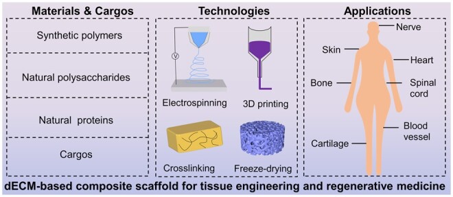
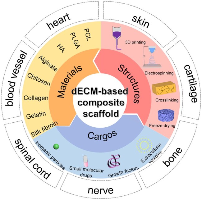
Despite the considerable advancements in fabricating polymeric-based scaffolds for tissue engineering, the clinical transformation of these scaffolds remained a big challenge because of the difficulty of simulating native organs/tissues’ microenvironment. As a kind of natural tissue-derived biomaterials, decellularized extracellular matrix (dECM)-based scaffolds have gained attention due to their unique biomimetic properties, providing a specific microenvironment suitable for promoting cell proliferation, migration, attachment and regulating differentiation. The medical applications of dECM-based scaffolds have addressed critical challenges, including poor mechanical strength and insufficient stability. For promoting the reconstruction of damaged tissues or organs, different types of dECM-based composite platforms have been designed to mimic tissue microenvironment, including by integrating with natural polymer or/and syntenic polymer or adding bioactive factors. In this review, we summarized the research progress of dECM-based composite scaffolds in regenerative medicine, highlighting the critical challenges and future perspectives related to the medical application of these composite materials.
Overview
The claims and statistics of this paper haven’t been checked yet.
Free: it reads the open-access text and checks the first 5 claims that cite a source we can check. Create a free account to check every claim.
Integrity signals
Registry
Cites
Statistics
Figures


Verification
Claims the paper makes
The claims and statistics of this paper haven’t been checked yet.
Free: it reads the open-access text and checks the first 5 claims that cite a source we can check. Create a free account to check every claim.
Claims other papers make about this one
Each is checked against this paper’s own text: does it say what the citing paper says it does?
The source backs part of the claim, not all of it — or backs it for a different population, condition or comparison.
In the citing paper
“Inorganic materials such as hydroxyapatite(HA) can be added to PLCL to design a scaffold with higher hardness and mechanical strength, which can improve the mechanical properties of the osteochondral repair interface [195].”
From Application of PLCL as a biodegradable polymer in biomedical engineering (2026) · cites this paper as [195]
Experimental — accuracy not yet measuredThe cited source says what the claim attributes to it.
In the citing paper
“Poor mechanical properties; rapid degradation rate.”
From Constructing biomimetic microenvironments for liver regeneration (2025) · doi:10.1186/s12951-025-03729-9 · cites this paper as [134]
In the citing paper
“Imitates the liver’s complexity and exhibits excellent biocompatibility.”
From Constructing biomimetic microenvironments for liver regeneration (2025) · doi:10.1186/s12951-025-03729-9 · cites this paper as [134]
In the citing paper
“These materials are produced through specialized physical, chemical, or biological processing methods that remove all cellular components from native tissues or organs [149].”
From Constructing biomimetic microenvironments for liver regeneration (2025) · doi:10.1186/s12951-025-03729-9 · cites this paper as [149]
In the citing paper
“Chen et al. further advanced the field by fabricating a bioactive dECM platform embedding dihydrolipoic acid–stabilised Pt nanoclusters doped with Cu nanozymes within cell-laden microgels [149].”
From Constructing biomimetic microenvironments for liver regeneration (2025) · doi:10.1186/s12951-025-03729-9 · cites this paper as [149]
Self-check
Percentages, sums and reported differences re-computed from the paper’s own numbers.
The self-check needs the paper’s full text.
Citations
Cites
1 retracted
Gholipourmalekabadi, M., Seifalian, A. M., Urbanska, A. M., Omrani, M. D., Hardy, J. G., Madjd, Z., Hashemi, S. M., Ghanbarian, H., Brouki Milan, P., Mozafari, M., et al. (2018) · Biomacromolecules · doi:10.1021/acs.biomac.7b01807 · Cited by 89 · cited 1×
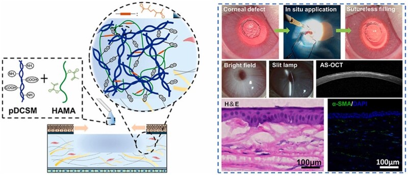
“[143] designed electrospun nanofibrous silk fibroin that coated with human amniotic dECM membrane to seeded adipose-tissue-derived MSC, which could accelerate wound healing process regeneration and significantly reduces scars formation in burn wound.”
BackgroundRetractedMaterials design for bone-tissue engineering
Koons, G. L., Diba, M., & Mikos, A. G. (2020) · Nature Reviews Materials · doi:10.1038/s41578-020-0204-2 · Cited by 1,699
Injectable and biodegradable hydrogels: gelation, biodegradation and biomedical applications
Li, Y., Rodrigues, J., & Tomás, H. (2012) · Chem. Soc. Rev. · doi:10.1039/c1cs15203c · Cited by 1,445
Biomimetic materials for tissue engineering
Ma, P. X. (2008) · Advanced Drug Delivery Reviews · doi:10.1016/j.addr.2007.08.041 · Cited by 1,310
Alginate-based composite materials for wound dressing application:A mini review
Varaprasad, K., Jayaramudu, T., Kanikireddy, V., Toro, C., & Sadiku, E. R. (2020) · Carbohydrate Polymers · doi:10.1016/j.carbpol.2020.116025 · Cited by 691
Synthetic biodegradable functional polymers for tissue engineering: a brief review
Guo, B., & Ma, P. X. (2014) · Science China Chemistry · doi:10.1007/s11426-014-5086-y · Cited by 592
Bone tissue engineering via growth factor delivery: from scaffolds to complex matrices
De Witte, T.-M., Fratila-Apachitei, L. E., Zadpoor, A. A., & Peppas, N. A. (2018) · Regenerative Biomaterials · doi:10.1093/rb/rby013 · Cited by 513
Multifunctional GelMA platforms with nanomaterials for advanced tissue therapeutics
Kurian, A. G., Singh, R. K., Patel, K. D., Lee, J.-H., & Kim, H.-W. (2022) · Bioactive Materials · doi:10.1016/j.bioactmat.2021.06.027 · Cited by 444 · cited 1×
“GelMA maintained functional sequences of gelatin (Arg–Gly–Asp and the matrix metalloproteinase) and could form hydrogel with tunable physicochemical properties by photo-crosslinking [125].”
BackgroundExtracellular vesicle-loaded hydrogels for tissue repair and regeneration
Ju, Y., Hu, Y., Yang, P., Xie, X., & Fang, B. (2023) · Materials Today Bio · doi:10.1016/j.mtbio.2022.100522 · Cited by 440
Gelatin carriers for drug and cell delivery in tissue engineering
Santoro, M., Tatara, A. M., & Mikos, A. G. (2014) · Journal of Controlled Release · doi:10.1016/j.jconrel.2014.04.014 · Cited by 395 · cited 1×
“Gelatin is an essential protein obtained from native collagen by hydrolysis, which can be obtained by disrupting the intermolecular bonds and breaking the a -helix conformation of collagen [109, 110].”
Background
Research works printed but not linked: 1
- -pyrimidinone unit modified with gelatin; Thiol-Ene PEG -Thiol terminated 3- mercaptopropionic acid functioned poly(ethylene glycol); 3D -Three-dimensional; 4-arm-PEG-NHS -4-arm poly(ethylene glycol) succinimidyl glutarate. Page 33 of 68 https://mc.manuscriptcentral.com/rb Manuscripts submitted to Regenerative Biomaterials Downloaded from https://academic.oup.com/rb/advance-article/doi/10.1093/rb/rbad107/7457501 by guest on 02 December 2023 References · doi:10.1093/rb/rbad107/7457501
Cited by
Liu, Y., & Ge, L. (2025) · Bioengineering · doi:10.3390/bioengineering12111178 · Cited by 84
“Conductive polymers may contain residual monomers or dopants that could be inflammatory [87].”
Conductive hydrogels: intelligent dressings for monitoring and healing chronic wounds
Fang, Y. Z., Han, Y., Lu, Y., Kankala, R. K., Wang, S., Chen, A., & Fu, C. (2024) · Regenerative Biomaterials · doi:10.1093/rb/rbae127 · Cited by 73
Self-citationDesign and Applications of Extracellular Matrix Scaffolds in Tissue Engineering and Regeneration
Mangani, S., Vetoulas, M., Mineschou, K., Spanopoulos, K., Vivanco, M. D., Piperigkou, Z., & Karamanos, N. K. (2025) · Cells · doi:10.3390/cells14141076 · Cited by 78
“More-over, unresolved issues in appropriate sterilization and disinfection methods, cell sourcing, regulatory classification, cost-efficiency, and the lack of robust preclinical data—especially in large animal models—pose further barriers [115,117–120].”
Advances in Electrospun Poly(ε-caprolactone)-Based Nanofibrous Scaffolds for Tissue Engineering
Robles, K. N., Zahra, F. T., Mu, R. R., & Giorgio, T. D. (2024) · Polymers · doi:10.3390/polym16202853 · Cited by 67
“Decellularized ECM (dECM) is biocompatible and can form 3D hydrogels; however, it lacks mechanical Decellularized ECM-based composite scaffolds have been fabricated with synthetic polymers, natural polysaccharides, and natural proteins for tissue engineering [2]. strength.”
BackgroundLiu, B., Alimi, O. A., Wang, Y., Kong, Y., Kuss, M. A., Krishnan, M. A., Hu, G., Xiao, Y., Dong, J., DiMaio, D. J., et al. (2024) · Journal of Controlled Release · doi:10.1016/j.jconrel.2024.02.019 · Cited by 48
Milan, P. B., Masoumi, F., Biazar, E., Jalise, S. Z., & Mehrabi, A. (2024) · Macromolecular Bioscience · doi:10.1002/mabi.202400322 · Cited by 46
Decellularized Extracellular Matrices for Skin Wound Treatment
Liang, R., Pan, R. L., He, L., Dai, Y., Jiang, Y., He, S., Li, B., & Li, Y. (2025) · Materials · doi:10.3390/ma18122752 · Cited by 49
van Hengel, E. V. A., van der Laan, L. J. W., de Jonge, J., & Verstegen, M. M. A. (2025) · Bioengineering · doi:10.3390/bioengineering12020136 · Cited by 48
Recent advances in fabrication of dECM-based composite materials for skin tissue engineering
Xu, P., Cao, J., Duan, Y., Kankala, R. K., & Chen, A. (2024) · Frontiers in Bioengineering and Biotechnology · doi:10.3389/fbioe.2024.1348856 · Cited by 44
Self-citationAdvanced biomaterials in immune modulation: The future of regenerative therapies
Biglari, N., Razzaghi, M., Afkham, Y., Azimi, G., Gross, J. D., & Samadi, A. (2025) · International Journal of Pharmaceutics · doi:10.1016/j.ijpharm.2025.125972 · Cited by 39
Yücer, S., Sarac, B., & Çiftçi, F. (2025) · Bioengineering & Translational Medicine · doi:10.1002/btm2.70080 · Cited by 34
Xia, H., Ye, Y., Li, J., Gong, Z.-Z., Meng, R.-Y., Chen, A., Wang, S., & Kankala, R. K. (2025) · ACS Applied Materials & Interfaces · doi:10.1021/acsami.5c15412 · Cited by 29
Self-citationβ-Cyclodextrin-based nanoassemblies for the treatment of atherosclerosis
Ji, W., Zhang, Y., Shao, W., Kankala, R. K., & Chen, A. (2024) · Regenerative Biomaterials · doi:10.1093/rb/rbae071 · Cited by 29
Self-citationLin, X., Zhang, Y., Li, J., Oliver, B. G. G., Wang, B., Li, H., Yong, K., & Li, J. J. (2024) · Bioactive Materials · doi:10.1016/j.bioactmat.2024.10.001 · Cited by 60
Li, Y., Wu, J., Ye, P., Cai, Y., Shao, M., Zhang, T., Guo, Y., Zeng, S., & Pathak, J. L. (2024) · ACS Biomaterials Science & Engineering · doi:10.1021/acsbiomaterials.4c01764 · Cited by 30
Biomaterials for neuroengineering: applications and challenges
Wu, H., Feng, E., Yin, H., Zhang, Y., Chen, G., Zhu, B., Yue, X., Zhang, H., Liu, Q., & Xiong, L. (2025) · Regenerative Biomaterials · doi:10.1093/rb/rbae137 · Cited by 29
Liu, H., Xing, F., Yu, P., Lu, R., Ma, S., Shakya, S., Xiang, Z., Peng, K., Zhang, D., & Liu, M. (2024) · Materials & Design · doi:10.1016/j.matdes.2024.113072 · Cited by 37
Luo, S., Wang, Q., Li, M., Xu, P., Wang, Y., Wang, Y., Kankala, R. K., Wang, S., & Chen, A. (2024) · Regenerative Biomaterials · doi:10.1093/rb/rbae113 · Cited by 23
“In an increasing number of studies, researchers have turned to dECM as a biomaterial to establish tissue-specific bioactive platforms, aiming to retain the bioactive composition of the orga-notypic ECM to the greatest extent possible [14–16].”
BackgroundSelf-citationRational design matrix materials for organoid development and application in biomedicine
Huang, Y. F., Zhang, X., Zhang, W., Tang, J., & Liu, J. (2025) · Regenerative Biomaterials · doi:10.1093/rb/rbaf038 · Cited by 23
Bhattacharya, T., Kumari, M., Kaur, K., Kaity, S., Arumugam, S., Ravichandiran, V., & Roy, S. (2024) · Journal of Materials Chemistry B · doi:10.1039/d4tb00680a · Cited by 21
Decellularized extracellular matrix-based disease models for drug screening
Chen, Z., Wang, J., Kankala, R. K., Jiang, M., Long, L., Li, W., Zou, L., Chen, A., & Liu, Y. (2024) · Materials Today Bio · doi:10.1016/j.mtbio.2024.101280 · Cited by 26
“These innovative strategies find broad utility in tissue engineering and regenerative medicine endeavors [79].”
BackgroundSelf-citationWang, Z., Lin, X., Shi, Y., Yan, H., Shang, Y., & Ren, H. (2026) · Biomedical Technology · doi:10.1016/j.bmt.2025.100131 · Cited by 22
Chen, Z., Long, L., Wang, J., Li, W., Wang, A., Kankala, R. K., Zou, L., & Liu, Y. (2024) · ACS Omega · doi:10.1021/acsomega.3c09265 · Cited by 17
Self-citationAdvances in Adhesive Materials for Oral and Maxillofacial Soft Tissue Diseases
Cheng, Q., Xu, P., Luo, S., & Chen, A. (2024) · Macromolecular Bioscience · doi:10.1002/mabi.202400494 · Cited by 19
Self-citationZhu, J., Ma, H., Du, J., Fang, H., Cheng, Y. Y., Xu, J., Pan, B., & Song, K. (2024) · International Journal of Biological Macromolecules · doi:10.1016/j.ijbiomac.2024.139056 · Cited by 18
Nature‐Inspired Macromolecular Biocomposites Based on Decellularized Extracellular Matrix
Lin, Y., Lin, Y.-T., Hu, H., Ma, P., Luo, Z., Tan, G., & Wu, Y. (2025) · Macromolecular Rapid Communications · doi:10.1002/marc.202401049 · Cited by 15
Chen, Z., Long, L., Wang, J., Jiang, M., Li, W., Cui, W., & Zou, L. (2024) · Small · doi:10.1002/smll.202402040 · Cited by 15
Ghadirian, S., Shariati, L., & Karbasi, S. (2025) · Biomaterials Advances · doi:10.1016/j.bioadv.2025.214249 · Cited by 16
Zhou, X., Wu, S., Liu, P., Wang, L., & Xie, F. (2025) · Carbohydrate Polymers · doi:10.1016/j.carbpol.2025.124023 · Cited by 15
Soltanmohammadi, F., Gharehbaba, A. M., & Javadzadeh, Y. (2025) · Biomedicine & Pharmacotherapy · doi:10.1016/j.biopha.2025.118200 · Cited by 16
Constructing biomimetic microenvironments for liver regeneration
(2025) · doi:10.1186/s12951-025-03729-9 · Cited by 11
“These materials are produced through specialized physical, chemical, or biological processing methods that remove all cellular components from native tissues or organs [149].”
Advancement in Scaffold-Based 3D Cell Culture Models for Osteosarcoma Drug Screening
Madaiah, P. M., Ghosh, R. N., Namboothiri, P. K., & Peter, M. (2025) · ACS Biomaterials Science & Engineering · doi:10.1021/acsbiomaterials.5c01174 · Cited by 12
Liang, L., Lin, Z., Duan, Z., Agbedor, S.-O., Li, N., Baker, I. A., Wang, B., Tang, L., & Wu, H. P. (2024) · Regenerative Biomaterials · doi:10.1093/rb/rbae104 · Cited by 10
“It is urgently needed to develop advanced biomaterials promoting bone regeneration for the treatment of large bone defects [3].”
BackgroundZhou, L., Zhang, Y., Yi, X., Chen, Y., & Li, Y. (2024) · International Journal of Biological Macromolecules · doi:10.1016/j.ijbiomac.2024.136788 · Cited by 15
Sundaram, M. K., Balavigneswaran, C. K., Saravanakumar, I., Jayaraman, G., & Muthuvijayan, V. (2025) · Journal of Materials Chemistry B · doi:10.1039/d5tb00248f · Cited by 9
Fang, Y. Z., Xu, K., Lu, X., Feng, X., Wang, Y., Lin, J., Chen, B., Kankala, R. K., Chen, A., Wang, S., et al. (2025) · ACS Sensors · doi:10.1021/acssensors.5c02154 · Cited by 10
Self-citationZhang, R., Xu, Z., Xu, S., Li, R., & Qian, H. (2025) · International Journal of Nanomedicine · doi:10.2147/ijn.s550390 · Cited by 11
Tang, Y., Su, T., Huang, B., Xu, Q., Chen, Q., Zhang, S., & Li, W. (2026) · Frontiers in Cell and Developmental Biology · doi:10.3389/fcell.2025.1730032 · Cited by 13
“Regulation of lineage transitions seems to occur via some combination of growth factors and cytokines and their interactions with the ECM, resulting in local organization of the wound microenvironment-to varying degrees (Xu et al., 2024).”
Domingues, M. F., Carvalho, M. S., Sanjuan‐Alberte, P., & Silva, J. C. (2025) · RSC Advances · doi:10.1039/d5ra02106e · Cited by 9
Cao, J., Luo, S., Huang, W., Lu, X., Kankala, R. K., Wang, S., Xu, P., & Chen, A. (2025) · Materials Today Bio · doi:10.1016/j.mtbio.2025.102141 · Cited by 7
“Nanofiber membranes fabricated by electrospinning are commonly used as wound dressings because of their biomimetic topological characteristics that approximate the microstructure of natural dermal fibers and their potential for sustained and controlled release of bioactive components [6].”
Self-citation3D In Vitro Models of the Bone Marrow Niche
Scala, P., Serio, B., & Giudice, V. (2025) · ACS Biomaterials Science & Engineering · doi:10.1021/acsbiomaterials.5c01421 · Cited by 14
Application of PLCL as a biodegradable polymer in biomedical engineering
Zhao, Y., & Liu, H. (2026) · Journal of Materials Science Materials in Medicine · doi:10.1007/s10856-026-07016-3 · Cited by 7
Yoon, J., Lee, Y., Kim, M., Park, J. Y., & Jang, J. (2025) · Advanced Healthcare Materials · doi:10.1002/adhm.202403118 · Cited by 7
Abdulkadhar, M. J., Jayakodi, S., Purushothaman, R., Ali, B. M. S., Vinayagam, S., Gnanasekaran, L., Ramakrishnan, P., & Sundaram, T. (2025) · Chemistry - An Asian Journal · doi:10.1002/asia.202401879 · Cited by 9
Engineering of tissue in microphysiological systems demonstrated by modelling skeletal muscle
Gao, Y., Zhang, Z., Yao, Y., Zhang, J., Li, X., Yang, K., Si, N., Chen, Z., Gu, Z., & Yin, N. (2025) · Regenerative Biomaterials · doi:10.1093/rb/rbaf059 · Cited by 8
Yuan, H., Li, B., Shao, X., Xi, Y., Cheng, S., Liu, A., & Zheng, B. (2025) · Nano Research · doi:10.26599/nr.2025.94907777 · Cited by 7
Historical evolution, hotspots, and trends in tendon tissue engineering: A bibliometric analysis
Hou, W., Liu, W., Tan, M., Fang, J., Cao, K., Du, X., & Zhang, H. (2025) · Regenerative Therapy · doi:10.1016/j.reth.2025.04.009 · Cited by 5
“It is important to note that when preparing composite scaffolds, the proportions of various materials need to be carefully regulated to create scaffolds that meet the desired requirements [56,57].”
Fabrication of collagen-based biomaterials for sports medicine application
Jiang, B., Zhou, Z., Yu, C., Li, H., Li, S., Hua, C., Huang, R., Xia, Z., Kong, B., & Chen, J. (2025) · Regenerative Biomaterials · doi:10.1093/rb/rbaf116 · Cited by 6
Farag, A. A. G., Hendawy, H. A., Emam, M. H., Hasegawa, M., Mandour, A. S., & Tanaka, R. (2025) · Biomolecules · doi:10.3390/biom15030371 · Cited by 6
“By providing an environment conducive to cell growth, scaffold technology can support the regeneration of damaged heart tissues, offering a promising route to improve cardiac function post-injury [191–193].”
Designed Biomaterial‐Enhanced Cell Transplantation for Neural Tissue Engineering
Tang, Y., Friec, A. L., Chen, M., & Sun, D. (2025) · Aggregate · doi:10.1002/agt2.70022 · Cited by 5
Zhao, Z., Xu, M., Zhang, Z., Yin, X., Pu, X., Wang, J., Liao, X., Huang, Z., Cao, S., & Yin, G. (2025) · Biomaterials Science · doi:10.1039/d5bm00469a · Cited by 5
Duan, Y., Liao, B.-H., Huang, W., Wang, W.-H., Kankala, R. K., Wang, S., Xu, P., & Chen, A. (2025) · Chemical Engineering Journal · doi:10.1016/j.cej.2025.168082 · Cited by 8
Self-citationTissue-Informed Biomaterial Innovations Advance Pulmonary Regenerative Engineering
Kary, A. D., Noelle, H., & Magin, C. M. (2025) · ACS Macro Letters · doi:10.1021/acsmacrolett.5c00075 · Cited by 5
Irastorza, A., Vázquez‐Aristizabal, P., Olaskoaga, L. Z., Mateo‐Abad, M., Guerrero, P., de la Caba, K., & Izeta, A. (2024) · bioRxiv (Cold Spring Harbor Laboratory) · doi:10.1101/2024.09.23.614437 · Cited by 5
Decellularized Scaffolds for Tissue Regeneration: Techniques and Applications
Biazar, E. (2024) · Biomedical engineering · doi:10.5772/intechopen.1007748 · Cited by 5
Bio-fabrication of fibrotic liver microtissues as a scalable and drug-responsive in vitro model
Majidi, F., Choshali, M. A., Izadi, A., Hajari, M. A., Saheli, M., Ziai, S. A., Vosough, M., & Piryaei, A. (2025) · International Journal of Pharmaceutics · doi:10.1016/j.ijpharm.2025.126242 · Cited by 5
Luo, S., Li, M.-T., Wang, Y.-C., Kankala, R. K., Wang, S.-B., Xu, P., & Chen, A. (2026) · ACS Applied Bio Materials · doi:10.1021/acsabm.5c01821 · Cited by 7
Self-citationXiao, L., Tao, M., Quan, L., Zhang, H., Xin, Y., Wu, X., Fang, X., Fan, J., Tian, X., Wang, X., et al. (2024) · Regenerative Biomaterials · doi:10.1093/rb/rbae054 · Cited by 6
“This has led to an increasing shift toward natural materials like chitosan [10–12] and collagen [13, 14].”
BackgroundPromotion of cardiac microtissue assembly within G-CSF-enriched collagen I-cardiogel hybrid hydrogel
Khodayari, H., Khodayari, S., Rezaee, M., Rezaeiani, S., Choshali, M. A., Erfanian, S., Muhammadnejad, A., Nili, F., Pourmehran, Y., Pirjani, R., et al. (2024) · Regenerative Biomaterials · doi:10.1093/rb/rbae072 · Cited by 7
“The composition of hydrogels play a determining role in physical stiffness and is one the most important issues in hydrogel fabrication to construct an engineered tissue [42–44].”
BackgroundVeettil, J. P., Lolitha, D. S., & Umashankar, P. R. (2025) · International Journal of Biological Macromolecules · doi:10.1016/j.ijbiomac.2025.141804 · Cited by 4
Natural Biomaterial Composites for Tissue Engineering: Challenges and Opportunities
Shirbacheh, A., Shirbacheh, K., Shirbache, K., & Karbalaee, M. (2024) · Journal of Composites and Compounds · doi:10.61186/jcc.6.3.4 · Cited by 7
Kadam, O., Gumathannavar, R., Basu, K., Saini, N., Sapre, N., Wankar, S., Bennur, T., & Kulkarni, A. (2025) · Discover Materials · doi:10.1007/s43939-025-00321-8 · Cited by 4
Jia, L., Cheng, J., Wen, X., Zhang, Z., Bao, H., Fei, J., Ding, Z., Li, J., & Li, Z. (2026) · Journal of Materials Chemistry B · doi:10.1039/d6tb00068a · Cited by 3
Cui, X., Liu, H., Liu, Y., Yu, Z., Wang, D., Wei, W., & Wang, S. (2025) · International Journal of Biological Macromolecules · doi:10.1016/j.ijbiomac.2025.144469 · Cited by 7
Self-citationThe Combination of Natural Products with Biomaterials for Regenerative Medicine: Current Trends
Janani, B. M., Srinivasan, S., Vijayalekha, A., & Pandurangan, A. K. (2025) · Current Pharmacology Reports · doi:10.1007/s40495-025-00421-2 · Cited by 2
Hsu, P.-S., Hsieh, M., Shie, M., Hsieh, W.-C., Lung, C.-F., Lin, Y.-H., Yao, C., Tsai, C.-S., & Hsu, Y. (2025) · Journal of the Taiwan Institute of Chemical Engineers · doi:10.1016/j.jtice.2025.106325 · Cited by 2
Chen, S., Li, Q., Ye, C., Pan, J., Chen, S., Zhou, J., Liu, L., Zhang, J., Xu, Z., Li, B., et al. (2025) · Regenerative Biomaterials · doi:10.1093/rb/rbaf122 · Cited by 3
Physical decellularization of fish skin utilizing electrical fields
Chen, M., Chen, S., Song, Y., Wang, S., Qiaoyin, Z., Zhifeng, Y., Peng, W., Li, H., & Wen, F. (2026) · Regenerative Biomaterials · doi:10.1093/rb/rbag005 · Cited by 4
Self-citationLu, X., Chen, B., Fang, Y., Cao, J., Huang, R., Kankala, R. K., Wang, S., & Chen, A. (2026) · Regenerative Biomaterials · doi:10.1093/rb/rbag010 · Cited by 4
Self-citationZhou, Y., Xu, T., Han, P., Wang, C., Bartold, P. M., & Ivanovski, S. (2026) · Journal of Periodontal Research · doi:10.1111/jre.70078 · Cited by 5
Toward 4D printed functional soft tissues
Shabazz, A., Fitlin, J., Orozco-Contreras, H., Guo, S., Zhang, L. G., & Fisher, J. P. (2026) · Acta Biomaterialia · doi:10.1016/j.actbio.2026.02.035 · Cited by 5
Kuppa, S. S., Sathiyamoorthy, P., Kim, H. K., Kang, J. Y., Sankaranarayanan, J., Park, I.-K., & Seon, J. K. (2026) · ACS Biomaterials Science & Engineering · doi:10.1021/acsbiomaterials.5c02109 · Cited by 4
Decellularized matrix grafts and peripheral nerve regeneration
Zhang, Q., Liu, X., Zhu, Y., Qian, T., Wang, S., & Li, M. (2025) · Neural Regeneration Research · doi:10.4103/nrr.nrr-d-25-00526 · Cited by 2
Self-citationNatural Biomaterials for Osteochondral Repair: From Source to Strategy
Liu, H., Zhang, H., Yang, W., Chen, H., Wang, J., & Chang, F. (2026) · Advanced Healthcare Materials · doi:10.1002/adhm.71344 · Cited by 3
Impact of Diabetic Condition on the Remodeling of In Situ Tissue-Engineered Heart Valves
Yang, F., Du, X., Zhao, Z., Guo, G., & Wang, Y. (2024) · ACS Biomaterials Science & Engineering · doi:10.1021/acsbiomaterials.4c01273 · Cited by 2
Negishi, J., Yamaguchi, A., Tanaka, D., Hashimoto, Y., Zhang, Y., & Funamoto, S. (2024) · Advanced Biology · doi:10.1002/adbi.202400398 · Cited by 2
Sarabia‐Vallejos, M. A., la Fuente, S. R. D., Cohn-Inostroza, N. A., Terraza, C. A., Rodríguez‐Hernández, J., & González-Henríquez, C. M. (2024) · Gels · doi:10.3390/gels10120761 · Cited by 1
Decellularized Extracellular Matrices: A Future for Musculoskeletal Regeneration
Jeevannavar, S. S., Goni, M., Keshav Shenoy, S., Daragad, M., Baindoor, P., & Fernandes, S. (2025) · Springer eBooks · doi:10.1007/978-981-96-3017-2_10 · Cited by 2
Chierici, A., D’Atri, G., Manferdini, C., Lambertini, E., Lisignoli, G., Piva, R., Nastruzzi, C., & Penolazzi, L. (2026) · ACS Biomaterials Science & Engineering · doi:10.1021/acsbiomaterials.5c02006 · Cited by 1
Liu, H., He, L., Liu, X., Tang, Y., Cheng, Y. Y., Li, X., Wang, H., Gao, H., & Song, K. (2026) · Biotechnology Journal · doi:10.1002/biot.70218 · Cited by 1
“Additionally, omentum-derived dECM combined with silk fibroin has exhibited significant angiogenesis-promoting and tissue remodeling capabilities in diabetic wound models [134].”
He, Y., Hua, C., Lin, L., Huang, C., Li, Y., Huang, J., Luo, C., & Zhang, Z. (2026) · Biomaterials Research · doi:10.34133/bmr.0383 · Cited by 1
“Notably, the decellularized MSC-matrix retains essential proteins even after the cellular components are removed [ 27 , 28 ].”
InfluentialEmerging Regenerative Medicine for Spinal Cord Injury: Spinal Cord Organoids-on-a-Chip
Khan, M., Choi, H., Ahmad, S., Lee, S., Park, J., & Han, I. (2026) · International Journal of Molecular Sciences · doi:10.3390/ijms27157060 · Cited by 1
“This loss is dependent on the parameters of the decellularization process and cannot be fully standardized amongst different laboratories [88,89].”
Investigation of anti-adhesion ability of 8-arm PEGNHS-modified porcine pericardium
Say, S., Suzuki, M., Hashimoto, Y., Kimura, T., & Kishida, A. (2024) · Biomedical Materials · doi:10.1088/1748-605x/ad2ed3
Fabrication Strategies for the Synthesis of Natural Product-Based Scaffold
Cruz, R. D. C. (2024) · Springer eBooks · doi:10.1007/978-981-97-3111-4_2
Scaffold for Tissue Engineering Design Fabrication and Applications
Iqbal, N., Ullah, M. A., Khalid, M. Z., & Hussain, S. T. (2025) · IGI Global eBooks · doi:10.4018/979-8-3373-1305-4.ch010
Xie, L., Zhou, Y., Luo, W., Zhang, J., & Wei, D. (2025) · Journal of Polymer Science · doi:10.1002/pol.20250625
HSIAO, M., Kimura, T., Suzuki, M., Hashimoto, Y., Yamada, M., Kim, Y., HARAZONO, Y., YOSHITAKE, H., Terauchi, M., Kishida, A., et al. (2025) · Dental Materials Journal · doi:10.4012/dmj.2025-168
Khorasani, E., Garazhian, S., & Vahidi, B. (2025) · National and International Iranian Conference on Biomedical Engineering (ICBME) · doi:10.1109/icbme68496.2025.11392517
“This strategy includes the use of scaffold as the temporary structure, which hopefully resembles the mechanical and bio-chemical conditions to the natural bone [4].”
Advances in thin film development for applications in regenerative medicine
Parveen, K. S. S., Jayachandran, K., & Radhakrishnan, E. K. (2026) · Elsevier eBooks · doi:10.1016/b978-0-443-18929-6.00012-8
Artificial ovaries and the future of women's health: Shaping novel functions
Parra, J. P. R. L. L., Abuna, R. P. F., Miglino, M. A., Arantes, L. B., Laurindo, L. F., Damous, L. L., Garcia, M. H. H., Barbalho, S. M., Meirelles, F. V., Direito, R., et al. (2026) · Translational research · doi:10.1016/j.trsl.2026.03.003
Decellularized extracellular matrix for gastrointestinal organoid culture: a comprehensive review
He, Y., Wong, C.-S., Chen, J., Leong, C.-N., Lan, J., Lei, L.-H., Wei, S., Yang, D., & Yan, L. (2026) · In vitro models · doi:10.1007/s44164-025-00103-9
“Several dECM products have already received regulatory approval for wound healing and soft tissue reconstruction, demonstrating a clear pathway for organoid-based therapies [131, 132].”
Heidarian, E., Naji, M., Setareyi, R., Shirinsokhan, A., Rahimpour, M., Torbati, P. M., Ansari, E., Kianirad, S., Feizollahi, N., & Nekoonam, S. (2026) · Journal of Biomedical Materials Research Part A · doi:10.1002/jbma.70015 · Cited by 1
Bruck, H., Sofer, S., Lion, A., Ornoy, A., & Sarig, U. (2026) · Frontiers in Bioengineering and Biotechnology · doi:10.3389/fbioe.2026.1774476
Sun, F., Wang, J., Zheng, T., Wu, L., Cao, W., Sun, Q., Li, M., Dai, X., Qian, T., Meng, C., et al. (2026) · Results in Engineering · doi:10.1016/j.rineng.2026.110624
Chen, S., Wu, Q., Chen, Z., Xie, C., Liang, Y., Deng, C., Jiang, W., Lu, T., Huang, C., Liu, Y., et al. (2026) · Genes & Diseases · doi:10.1016/j.gendis.2026.102209
Extracellular Vesicles-Loaded Silk Fibroin Scaffolds for Targeted Therapeutic Delivery
Linu-Mithran, K., Gounder, S. S., Masarudin, M. J., & Thilakavathy, K. (2026) · Regenerative Engineering and Translational Medicine · doi:10.1007/s40883-026-00590-6
Adams, C., Gómez, I., Rodríguez, O., Vigil, A., Hernández, C., Ceballos, J., Cisterna, B. A., & Reginensi, D. (2026) · Materials · doi:10.3390/ma19091803
“Additionally, dSIS shows more uniform thickness and mechanical consistency, facilitating reproducibility [19,23–25].”
Natural biopolymeric scaffolds for tissue engineering applications
Puri, A. V., Mohite, P. B., Ramole, A., Nagare, S., & Singh, S. (2026) · Elsevier eBooks · doi:10.1016/b978-0-443-33391-0.00017-0
Novel Biomaterials for Diagnosis and Therapeutics
Harun‐Ur‐Rashid, M., & Jahan, I. (2026) · Engineering materials · doi:10.1007/978-981-95-4216-1_9
Drzyzga, Ł. R., Śpiewak, D., Dorecka, M., & Wyględowska‐Promieńska, D. (2026) · Bioengineering · doi:10.3390/bioengineering13050548 · Cited by 1
“Combined with 3D bioprinting technologies, these approaches may enable fabrication of patient-specific corneal substitutes with customized curvature, thickness, and biomechanical properties [114,115].”
Metadata
- Authors
- Xu, P., Kankala, R. K., Wang, S., & Chen, A.
- Affiliations
- Institute of Biomaterials and Tissue Engineering, Huaqiao University , Xiamen, Fujian 361021, PR China
- Huaqiao University (CN)
- Venue
- Regenerative Biomaterials
- Publisher
- Oxford University Press (OUP)
- Published
- 2023-12-01
- Volume
- 11
- Pages
- rbad107-rbad107
- ISSN
- 2056-3418 (ISSN-L)
- 2056-3426
- PMC
- PMC10761212
- OpenAlex
- W4389243078
- Cited by
- 129
- References
- 226
- Citations by year
- 2026: 56 · 2025: 45 · 2024: 28
- FWCI
- 8.33
- Type
- journal article
- Language
- en
- Open access
- gold
- doi.org
- Licence
- Locations
- Regenerative Biomaterials (publishedVersion, cc-by)
- PubMed Central (publishedVersion, cc-by)
- PubMed (publishedVersion)
- Funders
- National Natural Science Foundation of China (NSFC 32271410, 32071323, 81971734)
- Science and Technology Projects in Fujian Province (2022FX1, 2023Y4008)
- Scientific Research Funds of Huaqiao University (21BS113)
- Open Research Fund of Academy of Advanced Carbon Conversion Technology, Huaqiao University (AACCT0004)
- Topics
- Health Sciences › Medicine › Surgery › Tissue Engineering and Regenerative Medicine
- Physical Sciences › Materials Science › Biomaterials › Hydrogels: synthesis, properties, applications
- Physical Sciences › Engineering › Biomedical Engineering › 3D Printing in Biomedical Research
- Keywords
- decellularized extracellular matrix, composite scaffolds, tissue engineering, regenerative medicine, biomimetic microenvironment, natural polymers, bioactive agents, mechanical strength, tissue reconstruction
About these checks
Registry, retraction, Cites and statistics results are computed, not judged, with one exception: a printed reference that no index lists is read once by a model before it counts as a citation error. Claims are extracted by a model and kept only when their quote appears verbatim in the text; each cited source is then checked, with a model’s help, for whether it says what the claim relies on. Those verdicts can be wrong — read the claim’s quote and the cited source before relying on one.
Statistics and arithmetic are recomputed, not judged.
Confidence bands are shown only for fields calibrated on our annotated test set. None is calibrated yet, so no band is shown.
What counts as a claim ›How we measure confidence ›How we check this ›
Are you an author, or do you see a problem with this page? Request a review
Data sources
Bibliographic data: Semantic Scholar, OpenAlex, Crossref.
Contains information from the Semantic Scholar Academic Graph Datasets, made available under the ODC Attribution License (ODC-BY 1.0).
Semantic Scholar’s data is described in: Kinney, R., et al. (2023). The Semantic Scholar Open Data Platform. arXiv:2301.10140.
