

Decellularized extracellular matrix-based composite scaffolds for tissue engineering and regenerative medicine
Open accessThe DOI matches its Crossref record; 223 of the 226 references it cites were found in a registry or an index.
Abstract
Despite the considerable advancements in fabricating polymeric-based scaffolds for tissue engineering, the clinical transformation of these scaffolds remained a big challenge because of the difficulty of simulating native organs/tissues’ microenvironment. As a kind of natural tissue-derived biomaterials, decellularized extracellular matrix (dECM)-based scaffolds have gained attention due to their unique biomimetic properties, providing a specific microenvironment suitable for promoting cell proliferation, migration, attachment and regulating differentiation. The medical applications of dECM-based scaffolds have addressed critical challenges, including poor mechanical strength and insufficient stability. For promoting the reconstruction of damaged tissues or organs, different types of dECM-based composite platforms have been designed to mimic tissue microenvironment, including by integrating with natural polymer or/and syntenic polymer or adding bioactive factors. In this review, we summarized the research progress of dECM-based composite scaffolds in regenerative medicine, highlighting the critical challenges and future perspectives related to the medical application of these composite materials.
Overview
The claims and statistics of this paper haven’t been checked yet.
Free: it reads the open-access text and checks the first 5 claims that cite a source we can check. Create a free account to check every claim.
Integrity signals
Registry
Cites
Statistics
Figures


Verification
Claims the paper makes
The claims and statistics of this paper haven’t been checked yet.
Free: it reads the open-access text and checks the first 5 claims that cite a source we can check. Create a free account to check every claim.
Claims other papers make about this one
Each is checked against this paper’s own text: does it say what the citing paper says it does?
The source backs part of the claim, not all of it — or backs it for a different population, condition or comparison.
In the citing paper
“Inorganic materials such as hydroxyapatite(HA) can be added to PLCL to design a scaffold with higher hardness and mechanical strength, which can improve the mechanical properties of the osteochondral repair interface [195].”
From Application of PLCL as a biodegradable polymer in biomedical engineering (2026) · cites this paper as [195]
Experimental — accuracy not yet measuredThe cited source says what the claim attributes to it.
In the citing paper
“Poor mechanical properties; rapid degradation rate.”
From Constructing biomimetic microenvironments for liver regeneration (2025) · doi:10.1186/s12951-025-03729-9 · cites this paper as [134]
In the citing paper
“Imitates the liver’s complexity and exhibits excellent biocompatibility.”
From Constructing biomimetic microenvironments for liver regeneration (2025) · doi:10.1186/s12951-025-03729-9 · cites this paper as [134]
In the citing paper
“These materials are produced through specialized physical, chemical, or biological processing methods that remove all cellular components from native tissues or organs [149].”
From Constructing biomimetic microenvironments for liver regeneration (2025) · doi:10.1186/s12951-025-03729-9 · cites this paper as [149]
In the citing paper
“Chen et al. further advanced the field by fabricating a bioactive dECM platform embedding dihydrolipoic acid–stabilised Pt nanoclusters doped with Cu nanozymes within cell-laden microgels [149].”
From Constructing biomimetic microenvironments for liver regeneration (2025) · doi:10.1186/s12951-025-03729-9 · cites this paper as [149]
Self-check
Percentages, sums and reported differences re-computed from the paper’s own numbers.
The self-check needs the paper’s full text.
Citations
Cites
1 retracted
Gholipourmalekabadi, M., Seifalian, A. M., Urbanska, A. M., Omrani, M. D., Hardy, J. G., Madjd, Z., Hashemi, S. M., Ghanbarian, H., Brouki Milan, P., Mozafari, M., et al. (2018) · Biomacromolecules · doi:10.1021/acs.biomac.7b01807 · Cited by 89 · cited 1×
“[143] designed electrospun nanofibrous silk fibroin that coated with human amniotic dECM membrane to seeded adipose-tissue-derived MSC, which could accelerate wound healing process regeneration and significantly reduces scars formation in burn wound.”
BackgroundRetractedMaterials design for bone-tissue engineering
Koons, G. L., Diba, M., & Mikos, A. G. (2020) · Nature Reviews Materials · doi:10.1038/s41578-020-0204-2 · Cited by 1,699
Injectable and biodegradable hydrogels: gelation, biodegradation and biomedical applications
Li, Y., Rodrigues, J., & Tomás, H. (2012) · Chem. Soc. Rev. · doi:10.1039/c1cs15203c · Cited by 1,445
Biomimetic materials for tissue engineering
Ma, P. X. (2008) · Advanced Drug Delivery Reviews · doi:10.1016/j.addr.2007.08.041 · Cited by 1,310
Alginate-based composite materials for wound dressing application:A mini review
Varaprasad, K., Jayaramudu, T., Kanikireddy, V., Toro, C., & Sadiku, E. R. (2020) · Carbohydrate Polymers · doi:10.1016/j.carbpol.2020.116025 · Cited by 691
Synthetic biodegradable functional polymers for tissue engineering: a brief review
Guo, B., & Ma, P. X. (2014) · Science China Chemistry · doi:10.1007/s11426-014-5086-y · Cited by 592
Bone tissue engineering via growth factor delivery: from scaffolds to complex matrices
De Witte, T.-M., Fratila-Apachitei, L. E., Zadpoor, A. A., & Peppas, N. A. (2018) · Regenerative Biomaterials · doi:10.1093/rb/rby013 · Cited by 513
Multifunctional GelMA platforms with nanomaterials for advanced tissue therapeutics
Kurian, A. G., Singh, R. K., Patel, K. D., Lee, J.-H., & Kim, H.-W. (2022) · Bioactive Materials · doi:10.1016/j.bioactmat.2021.06.027 · Cited by 444 · cited 1×
“GelMA maintained functional sequences of gelatin (Arg–Gly–Asp and the matrix metalloproteinase) and could form hydrogel with tunable physicochemical properties by photo-crosslinking [125].”
BackgroundExtracellular vesicle-loaded hydrogels for tissue repair and regeneration
Ju, Y., Hu, Y., Yang, P., Xie, X., & Fang, B. (2023) · Materials Today Bio · doi:10.1016/j.mtbio.2022.100522 · Cited by 440
Gelatin carriers for drug and cell delivery in tissue engineering
Santoro, M., Tatara, A. M., & Mikos, A. G. (2014) · Journal of Controlled Release · doi:10.1016/j.jconrel.2014.04.014 · Cited by 395 · cited 1×
“Gelatin is an essential protein obtained from native collagen by hydrolysis, which can be obtained by disrupting the intermolecular bonds and breaking the a -helix conformation of collagen [109, 110].”
BackgroundThe role of natural polymers in bone tissue engineering
Guo, L., Liang, Z., Yang, L., Du, W., Yu, T., Tang, H., Li, C., & Qiu, H. (2021) · Journal of Controlled Release · doi:10.1016/j.jconrel.2021.08.055 · Cited by 352
Anisotropic Materials for Skeletal‐Muscle‐Tissue Engineering
Jana, S., Levengood, S. K. L., & Zhang, M. (2016) · Advanced Materials · doi:10.1002/adma.201600240 · Cited by 345
Mehrali, M., Thakur, A., Pennisi, C. P., Talebian, S., Arpanaei, A., Nikkhah, M., & Dolatshahi‐Pirouz, A. (2017) · Advanced Materials · doi:10.1002/adma.201603612 · Cited by 345 · cited 1×
“Adding the inorganic particles into biomaterials could remedy the mechanical shortcomings and increase their bio-activity for tissue regeneration [191].”
BackgroundApplications of chitin and chitosan nanofibers in bone regenerative engineering
Tao, F., Cheng, Y., Shi, X., Zheng, H., Du, Y., Xiang, W., & Deng, H. (2020) · Carbohydrate Polymers · doi:10.1016/j.carbpol.2019.115658 · Cited by 341
Gao, G., Lee, J. H., Jang, J., Lee, D. H., Kong, J., Kim, B. S., Choi, Y., Jang, W. B., Hong, Y. J., Kwon, S., et al. (2017) · Advanced Functional Materials · doi:10.1002/adfm.201700798 · Cited by 336 · cited 1×
“[187] produced atorvastatin-loaded PLGA microspheres for delivering endothelial progenitor cells and mixed with dECM and alginate-based bioink to construct bio-blood-vessel.”
MethodGelatin-Based Materials in Ocular Tissue Engineering
Rose, J., Pacelli, S., Haj, A., Dua, H., Hopkinson, A., White, L., & Rose, F. (2014) · Materials · doi:10.3390/ma7043106 · Cited by 327 · cited 1×
“Compared with collagen, gelatin maintains the RGD (Arg– Gly–Asp) sequence to promote cell adhesion and also shows lower immunogenicity [111].”
BackgroundSarrigiannidis, S. O., Rey, J. M., Dobre, O., González-García, C., Dalby, M. J., & Salmeron-Sanchez, M. (2021) · Materials Today Bio · doi:10.1016/j.mtbio.2021.100098 · Cited by 309 · cited 1×
“Collagen is the most abundant protein in vertebrate animals and a prevalent component found in ECM [102].”
BackgroundXing, H., Zhang, Z., Mao, Q., Wang, C., Zhou, Y., Zhou, X., Ying, L., Xu, H., Hu, S., & Zhang, N. (2021) · Journal of Nanobiotechnology · doi:10.1186/s12951-021-00991-5 · Cited by 296
Silk fibroin for skin injury repair: Where do things stand?
Gholipourmalekabadi, M., Sapru, S., Samadikuchaksaraei, A., Reis, R. L., Kaplan, D. L., & Kundu, S. C. (2020) · Advanced Drug Delivery Reviews · doi:10.1016/j.addr.2019.09.003 · Cited by 288 · cited 1×
“In recent years, owing to the excellent biocompatibility, tunable degradability and abundant sources of silk fibroin, silk fibroin-based bio-scaffolds have received considerable attention for tissue regeneration [138, 139].”
BackgroundCurrent advances in electrospun gelatin-based scaffolds for tissue engineering applications
Aldana, A. A., & Abraham, G. A. (2017) · International Journal of Pharmaceutics · doi:10.1016/j.ijpharm.2016.09.044 · Cited by 286 · cited 1×
“Gelatin is an essential protein obtained from native collagen by hydrolysis, which can be obtained by disrupting the intermolecular bonds and breaking the a -helix conformation of collagen [109, 110].”
BackgroundBrown, M., Li, J., Moraes, C., Tabrizian, M., & Li-Jessen, N. Y. K. (2022) · Biomaterials · doi:10.1016/j.biomaterials.2022.121786 · Cited by 282
Progress in Gelatin as Biomaterial for Tissue Engineering
Lukin, I., Erezuma, I., Maeso, L., Zarate, J., Desimone, M. F., Al-Tel, T. H., Dolatshahi-Pirouz, A., & Orive, G. (2022) · Pharmaceutics · doi:10.3390/pharmaceutics14061177 · Cited by 278 · cited 1×
“Due to their biocompatibility and acceptable toxicity, gelatin-based medical products (such as Cutanplast, Surgiflo and Floseal) have been used for hemostasis in surgeries [112].”
BackgroundExtracellular‐Matrix‐Reinforced Bioinks for 3D Bioprinting Human Tissue
De Santis, M. M., Alsafadi, H. N., Tas, S., Bölükbas, D. A., Prithiviraj, S., Da Silva, I. A. N., Mittendorfer, M., Ota, C., Stegmayr, J., Daoud, F., et al. (2021) · Advanced Materials · doi:10.1002/adma.202005476 · Cited by 233
Alven, S., Peter, S., Mbese, Z., & Aderibigbe, B. A. (2022) · Polymers · doi:10.3390/polym14040724 · Cited by 231
Mao, Q., Wang, Y., Li, Y., Juengpanich, S., Li, W., Chen, M., Yin, J., Fu, J., & Cai, X. (2020) · Materials Science and Engineering: C · doi:10.1016/j.msec.2020.110625 · Cited by 215
Gelatin-Based Hydrogels: Potential Biomaterials for Remediation
Andreazza, R., Morales, A., Pieniz, S., & Labidi, J. (2023) · Polymers · doi:10.3390/polym15041026 · Cited by 213 · cited 1×
“Moreover, gelatin can act as an excellent candidate to produce biomedical materials, such as hydrogels, scaffolds or films for promoting tissue repair [113, 114].”
BackgroundLi, Z., & Tan, B. H. (2014) · Materials Science and Engineering: C · doi:10.1016/j.msec.2014.06.003 · Cited by 212
Baiguera, S., Del Gaudio, C., Lucatelli, E., Kuevda, E., Boieri, M., Mazzanti, B., Bianco, A., & Macchiarini, P. (2014) · Biomaterials · doi:10.1016/j.biomaterials.2013.10.060 · Cited by 211 · cited 1×
“Thus, gelatin is also commonly used to fabricate hybrid scaffolds with dECM for tissue generation [115, 116].”
BackgroundExtracellular vesicles for tissue repair and regeneration: Evidence, challenges and opportunities
Nagelkerke, A., Ojansivu, M., van der Koog, L., Whittaker, T. E., Cunnane, E. M., Silva, A. M., Dekker, N., & Stevens, M. M. (2021) · Advanced Drug Delivery Reviews · doi:10.1016/j.addr.2021.04.013 · Cited by 208 · cited 1×
“Due to the biological activities, the bioactive properties of extracellular vesicles have been used to cooperate with scaffolds in tissue re-generation [211–213].”
Chen, W., Xu, Y., Li, Y., Jia, L., Mo, X., Jiang, G., & Zhou, G. (2020) · Chemical Engineering Journal · doi:10.1016/j.cej.2019.122986 · Cited by 193 · cited 2×
“…o w s c a p u l a r c a r t i l a ge E l e c t r o s p i nn i n g a n d 3 D p r i n t i n g 3 D - p r i n t e d fi b e r - r e i n f o r c e d s c a ff o l d e nh a n c e d r e p a i r a r t i c u l a r c a r t i l a ge e ff e c t i n r a bb i t s d e f e c t m o d e l [ 120 ] G e l a t i n , H A -”
MethodLi, Q., Yu, H., Zhao, F., Cao, C., Wu, T., Fan, Y., Ao, Y., & Hu, X. (2023) · Advanced Science · doi:10.1002/advs.202303650 · Cited by 185 · cited 1×
“[217] designed a 3D double-network hydrogel by using GelMA, oxidative HA, dopamine-conjugated HA, cartilage dECM and bone dECM as raw materials, encapsulating with human adipose MSC exosomes.”
MethodShin, Y. J., Shafranek, R. T., Tsui, J. H., Walcott, J., Nelson, A., & Kim, D.-H. (2021) · Acta Biomaterialia · doi:10.1016/j.actbio.2020.11.006 · Cited by 174 · cited 1×
“[208] bioprinted a PEGDA and cardiac dECM hydrogel reinforced with Laponite to model cardiac tissue.”
MethodRai, V., Dilisio, M. F., Dietz, N. E., & Agrawal, D. K. (2017) · Journal of Biomedical Materials Research Part A · doi:10.1002/jbm.a.36087 · Cited by 171
Cryogenic 3D printed hydrogel scaffolds loading exosomes accelerate diabetic wound healing
Hu, Y., Wu, B., Xiong, Y., Tao, R., Panayi, A. C., Chen, L., Tian, W., Xue, H., Shi, L., Zhang, X., et al. (2021) · Chemical Engineering Journal · doi:10.1016/j.cej.2021.130634 · Cited by 164 · cited 1×
“[219] designed a 3D scaffold based on dECM, mesoporous bioactive glass and exosomes by cryogenic printing technology.”
MethodKim, M. K., Jeong, W., Lee, S. M., Kim, J. B., Jin, S., & Kang, H.-W. (2020) · Biofabrication · doi:10.1088/1758-5090/ab5d80 · Cited by 161 · cited 1×
“Thus, gelatin is also commonly used to fabricate hybrid scaffolds with dECM for tissue generation [115, 116].”
BackgroundLee, J., Hong, J., Kim, W., & Kim, G. H. (2020) · Carbohydrate Polymers · doi:10.1016/j.carbpol.2020.116914 · Cited by 157
Zamboni, F., Wong, C. K., & Collins, M. N. (2022) · Bioactive Materials · doi:10.1016/j.bioactmat.2022.04.023 · Cited by 156
Zhang, X., Liu, Y., Luo, C., Zhai, C., Li, Z., Zhang, Y., Yuan, T., Dong, S., Zhang, J., & Fan, W. (2021) · Materials Science and Engineering: C · doi:10.1016/j.msec.2020.111388 · Cited by 154
3D cell-printing of biocompatible and functional meniscus constructs using meniscus‐derived bioink
Chae, S., Lee, S.-S., Choi, Y.-J., Hong, D. H., Gao, G., Wang, J. H., & Cho, D.-W. (2021) · Biomaterials · doi:10.1016/j.biomaterials.2020.120466 · Cited by 146
Janani, G., Kumar, M., Chouhan, D., Moses, J. C., Gangrade, A., Bhattacharjee, S., & Mandal, B. B. (2019) · ACS Applied Bio Materials · doi:10.1021/acsabm.9b00576 · Cited by 145
Recent advances in decellularized biomaterials for wound healing
Xiao, H., Chen, X., Liu, X., Wen, G., & Yu, Y. (2023) · Materials Today Bio · doi:10.1016/j.mtbio.2023.100589 · Cited by 143
Lee, H., Yang, G. H., Kim, M., Lee, J., Huh, J., & Kim, G. (2017) · Materials Science and Engineering C · doi:10.1016/j.msec.2017.11.013 · Cited by 140 · cited 1×
“[150] developed a 3D porous scaffold that consisted of collagen, dECM and silk fibroin using low-temperature printing technology.”
MethodDecellularization systems and devices: State-of-the-art
Choudhury, D., Yee, M., Sheng, Z. L. J., Amirul, A., & Naing, M. W. (2020) · Acta Biomaterialia · doi:10.1016/j.actbio.2020.07.060 · Cited by 135
Hong, J. Y., Seo, Y., Davaa, G., Kim, H.-W., Kim, S. H., & Hyun, J. K. (2020) · Acta Biomaterialia · doi:10.1016/j.actbio.2019.11.012 · Cited by 134 · cited 1×
“Hong et al . produced collagen-based hydrogels that contained various concentrations of dECM (1, 5 and 8mg/ml) [106].”
BackgroundBiohybrid cardiac ECM-based hydrogels improve long term cardiac function post myocardial infarction
Efraim, Y., Sarig, H., Anavy, N. C., Sarig, U., de-Berardinis, E., Chaw, S. Y., Krishnamoorthi, M. K., Kalifa, J., Bogireddi, H., Duc, T. V., et al. (2016) · Acta Biomaterialia · doi:10.1016/j.actbio.2016.12.015 · Cited by 131
Silk fibroin/amniotic membrane 3D bi-layered artificial skin
Gholipourmalekabadi, M., Samadikuchaksaraei, A., Seifalian, A. M., Urbanska, A. M., Ghanbarian, H., Hardy, J. G., Omrani, M. D., Mozafari, M., Reis, R. L., & Kundu, S. C. (2018) · Biomedical Materials · doi:10.1088/1748-605x/aa999b · Cited by 131 · cited 1×
“Based on the unique propriety, the silk fibroin–dECM composite scaffold with different shapes was constructed by previous studies, which provided a new approach to tissue regeneration [140, 141].”
MethodGrowth Factor and Its Polymer Scaffold-Based Delivery System for Cartilage Tissue Engineering
Chen, L., Liu, J., Guan, M., Zhou, T., Duan, X., & Xiang, Z. (2020) · International Journal of Nanomedicine · doi:10.2147/ijn.s249829 · Cited by 130 · cited 1×
“TGF-b superfamily has been widely used in tissue engineering because they could stimulate multiple cell functions, including TGF-b 1/2/3 and bone morphogenetic proteins (BMPs) [159].”
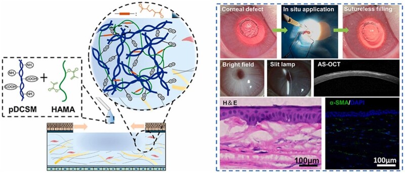
BackgroundDual-crosslinked regenerative hydrogel for sutureless long-term repair of corneal defect
Shen, X., Li, S., Zhao, X., Han, J., Chen, J., Rao, Z., Zhang, K., Quan, D., Yuan, J., & Bai, Y. (2022) · Bioactive Materials · doi:10.1016/j.bioactmat.2022.06.006 · Cited by 124
Decellularized Extracellular Matrix for Remodeling Bioengineering Organoid's Microenvironment
Zhu, L., Yuhan, J., Yu, H., Zhang, B., Huang, K., & Zhu, L. (2023) · Small · doi:10.1002/smll.202207752 · Cited by 123
Xu, J., Fang, H., Zheng, S., Li, L., Jiao, Z., Wang, H., Nie, Y., Liu, T., & Song, K. (2021) · International Journal of Biological Macromolecules · doi:10.1016/j.ijbiomac.2021.07.162 · Cited by 120 · cited 2×
“F r ee z e - d r y i n g m e t h o d T h e b i o l o g i c a l f un c t i o n a l h y b r i d s c a ff o l d s h o w e d e x - c e ll e n t a n t i b a c t e r i a l a c t i v i t y f o r w o un d h e a l i n g [ 122 ] G e l M A P o r c i n e t r a b e c u l a r”
BackgroundTsui, J. H., Leonard, A., Camp, N. D., Long, J. T., Nawas, Z. Y., Chavanachat, R., Smith, A. S. T., Choi, J. S., Dong, Z., Ahn, E. H., et al. (2021) · Biomaterials · doi:10.1016/j.biomaterials.2021.120764 · Cited by 117 · cited 1×
“[202] developed a reduced graphene oxide-contained dECM scaffold to mimic the cardiac microenvironment.”
BackgroundLi, Y., Liu, Y., Xun, X., Zhang, W., Xu, Y., & Gu, D. (2019) · ACS Applied Materials & Interfaces · doi:10.1021/acsami.9b12206 · Cited by 112 · cited 2×
“[119] indicated that the gelatin/PCL nanofiber in the dECM scaffold could increase its mechanical properties and structural stability.”
BackgroundKC, P., Hong, Y., & Zhang, G. (2019) · Regenerative Biomaterials · doi:10.1093/rb/rbz017 · Cited by 107
Liu, S., Zhao, Y., Li, M., Nie, L., Wei, Q., Okoro, O. V., Jafari, H., Wang, S., Deng, J., Chen, J., et al. (2023) · Chemical Engineering Journal · doi:10.1016/j.cej.2023.143016 · Cited by 107 · cited 1×
“[177] fabricated polydopamine nanoparticles loaded with asiaticoside and mixed with dECM and GelMA composite hydrogel, which has the potential for scarless wound healing application.”
MethodSelf-citationPhoto-crosslinkable amniotic membrane hydrogel for skin defect healing
Zhang, Q., Chang, C., Qian, C., Xiao, W., Zhu, H., Guo, J., Meng, Z., Cui, W., & Ge, Z. (2021) · Acta Biomaterialia · doi:10.1016/j.actbio.2021.02.043 · Cited by 107 · cited 2×
“T h e p h o t o - c r o ss l i n k e d h y d r o ge l w i t h o u t s t a n d i n g m e c h a n i c a l p r o p e r t i e s c o u l d a c t a s a p r o m i s i n g m a t e r i a l f o r s k i n d e f e c t h e a l i n g [ 132 ] G e l M A P o r c i n e d e n t a l f o ll i c l e s”
BackgroundChen, S., Zhang, Q., Nakamoto, T., Kawazoe, N., & Chen, G. (2015) · Tissue Engineering Part C Methods · doi:10.1089/ten.tec.2015.0281 · Cited by 106 · cited 1×
“Moreover, gelatin can act as an excellent candidate to produce biomedical materials, such as hydrogels, scaffolds or films for promoting tissue repair [113, 114].”
BackgroundRecent Advancements in Polyurethane-based Tissue Engineering
Singh, S., Paswan, K. K., Kumar, A., Gupta, V., Sonker, M., Khan, M. A., Kumar, A., & Shreyash, N. (2023) · ACS Applied Bio Materials · doi:10.1021/acsabm.2c00788 · Cited by 104
Kang, Y., Xu, J., Meng, L., Su, Y., Fang, H., Liu, J., Cheng, Y. Y., Jiang, D., Nie, Y., & Song, K. (2023) · Biofabrication · doi:10.1088/1758-5090/acb6b8 · Cited by 104 · cited 1×
“In addition, extracellular vesicles were combined with other bio-active factors in dECM-based scaffold forms, which exerted synergistic effects in tissue engineering [218].”
BackgroundChitosan-based scaffolds as drug delivery systems in bone tissue engineering
Bharathi, R., Ganesh, S. S., Harini, G., Vatsala, K., Ramprasad, A., Aravind, S., Abinaya, S., & Selvamurugan, N. (2022) · International Journal of Biological Macromolecules · doi:10.1016/j.ijbiomac.2022.09.058 · Cited by 103 · cited 1×
“By adding small molecular drugs with special functions into scaffolds, the hybrid scaffold can be used to achieve drug release in demand to improve the activities of drugs [172].”
BackgroundMultiphasic scaffolds for the repair of osteochondral defects: Outcomes of preclinical studies
Chen, R., Pye, J. S., Li, J., Little, C. B., & Li, J. J. (2023) · Bioactive Materials · doi:10.1016/j.bioactmat.2023.04.016 · Cited by 102
Zheng, C., Yang, Z., Chen, S., Zhang, F., Rao, Z., Zhao, C., Quan, D., Bai, Y., & Shen, J. (2021) · Theranostics · doi:10.7150/thno.50825 · Cited by 99
Yang, Z., Zhao, T., Gao, C., Cao, F., Li, H., Liao, Z., Fu, L., Li, P., Chen, W., Sun, Z., et al. (2021) · ACS Applied Materials & Interfaces · doi:10.1021/acsami.1c01844 · Cited by 98 · cited 1×
“To this end, several studies applied the TGF-b superfamily to design multifunctional systems combined with dECM [160].”
MethodDeng, R., Luo, Z., Rao, Z., Lin, Z., Chen, S., Zhou, J., Zhu, Q., Liu, X., Bai, Y., & Quan, D. (2022) · Advanced Fiber Materials · doi:10.1007/s42765-021-00124-5 · Cited by 96
Lih, E., Park, K. W., Chun, S. Y., Kim, H., Kwon, T. G., Joung, Y. K., & Han, D. K. (2016) · ACS Applied Materials & Interfaces · doi:10.1021/acsami.6b03771 · Cited by 92
Casali, D. M., Handleton, R. M., Shazly, T. M., & Matthews, M. (2017) · The Journal of Supercritical Fluids · doi:10.1016/j.supflu.2017.07.021 · Cited by 91
Chae, S., Sun, Y., Choi, Y.-J., Ha, D.-H., Jeon, I., & Cho, D.-W. (2021) · Biofabrication · doi:10.1088/1758-5090/abd159 · Cited by 91
Yang, X., Ma, Y., Wang, X., Yuan, S., Huo, F., Yi, G., Zhang, J., Yang, B., & Tian, W. (2023) · Advanced Science · doi:10.1002/advs.202205041 · Cited by 91 · cited 2×
“[134] prepared a 3D biomimetic periodontal module that containing GelMA, dECM and human dental follicle cells (DFUs).”
MethodPetrou, C. L., D’Ovidio, T. J., Bölükbas, D. A., Tas, S., Brown, R. D., Allawzi, A., Lindstedt, S., Nozik-Grayck, E., Stenmark, K. R., Wagner, D. E., et al. (2020) · Journal of Materials Chemistry B · doi:10.1039/d0tb00613k · Cited by 89
Yu, Y., Wang, Y., Zhang, W., Wang, H., Li, J., Pan, L., Han, F., & Li, B. (2020) · Acta Biomaterialia · doi:10.1016/j.actbio.2020.06.030 · Cited by 88 · cited 1×
“The GelMA-combined dECM scaffold with different forms has shown extraordinary behaviors in regenerating various tissues [126–128].”
BackgroundKim, H. S., Mandakhbayar, N., Kim, H.-W., Leong, K. W., & Yoo, H. S. (2021) · Biomaterials · doi:10.1016/j.biomaterials.2020.120214 · Cited by 88
Ko, K.-W., Park, S.-Y., Lee, E. H., Yoo, Y.-I., Kim, D.-S., Kim, J. Y., Kwon, T. G., & Han, D. K. (2021) · ACS Nano · doi:10.1021/acsnano.1c01098 · Cited by 88 · cited 2×
“W o u n d h e a l i n g [ 206 ] G e l M A , d E C M K i d n e y r ege n e r a t i o n [ 221 ] b F G F , b a s i c fi b r o b l a s t g r o w t h f a c t o r ; B M P - 2 , b o n e m o r p h o ge n e t i c p r o t e i n - 2 ; B M P - 7 , b o n e m o r p h o ge n e t i c p r o t e i n - 7 ; B M S C ,…”
BackgroundFeng, B., Ji, T., Wang, X., Fu, W., Ye, L., Zhang, H., & Li, F. (2020) · Materials & Design · doi:10.1016/j.matdes.2020.108773 · Cited by 86
Yang, Y., Lei, D., Zou, H., Huang, S., Yang, Q., Li, S., Qing, F.-L., Ye, X., You, Z., & Zhao, Q. (2019) · Acta Biomaterialia · doi:10.1016/j.actbio.2019.06.037 · Cited by 85 · cited 2×
“[188] fabricated vascular grafts with small diameter that consisted of rapamycin-loaded PCL nanofibrous as an outer layer to coat on the rat aorta dECM layer.”
MethodNanofibrillar Decellularized Wharton's Jelly Matrix for Segmental Tracheal Repair
Xu, Y., Duan, L., Li, Y., She, Y., Zhu, J., Zhou, G., Jiang, G., & Yang, Y. (2020) · Advanced Functional Materials · doi:10.1002/adfm.201910067 · Cited by 85
Zheng, Z., Chen, Y., Hong, H., Shen, Y., Wang, Y., Sun, J., & Wang, X. (2020) · Advanced Healthcare Materials · doi:10.1002/adhm.202000631 · Cited by 82 · cited 2×
“[203] synthesized magnesium-enriched graphene oxide nanoparticles for modulating the inflammatory responses and then combined them with bone dECM scaffold for bone regeneration.”
Deng, M., Tan, J., Hu, C., Hou, T., Peng, W., Liu, J., Yu, B., Dai, Q., Zhou, J., Yang, Y., et al. (2020) · Advanced Healthcare Materials · doi:10.1002/adhm.202000353 · Cited by 81
Glycocalyx‐Like Hydrogel Coatings for Small Diameter Vascular Grafts
Dimitrievska, S., Wang, J., Lin, T., Weyers, A. M., Bai, H., Qin, L., Li, G., Cai, C. Y. L., Kypson, A. P., Kristofik, N. J., et al. (2020) · Advanced Functional Materials · doi:10.1002/adfm.201908963 · Cited by 74
Kumar, A., Nune, K. C., & Misra, R. D. K. (2016) · Journal of Biomedical Materials Research Part A · doi:10.1002/jbm.a.35664 · Cited by 74 · cited 1×
“Researchers developed lots of dECM/hydroxyapatite composite scaffolds by different manufacturing processes, achieving a better bone repair effect than using those materials alone [192, 193].”
MethodPatel, K. H., Dunn, A. J., Talovic, M., Haas, G. J., Marcinczyk, M., Elmashhady, H., Kalaf, E. G., Sell, S. A., & Garg, K. (2019) · Biomedical Materials · doi:10.1088/1748-605x/ab0b06 · Cited by 73
Kara, A., Distler, T., Polley, C., Schneidereit, D., Seitz, H., Friedrich, O., Tıhmınlıoğlu, F., & Boccaccini, A. R. (2022) · Materials Today Bio · doi:10.1016/j.mtbio.2022.100309 · Cited by 72 · cited 2×
“T h e 3 D s c a ff o l d e nh a n c e d t h e m e c h a n i c a l s t r e n g t h , d eg r a d a t i o n r a t e a n d w a s s u i t a b l e f o r b o n e t i ss u e e n g i n ee r i n g [ 117 ] G e l a t i n H u m a n a m n i o t i c m e m b r a n e”
BackgroundKayabolen, A., Keskin, D., Aykan, A., Karslıoglu, Y., Zor, F., & Tezcaner, A. (2017) · Biomedical Materials · doi:10.1088/1748-605x/aa6a63 · Cited by 69 · cited 2×
“[146] prepared silk fibroin/dECM matrix hydrogel with different ratios via crosslinking to mimic the native tissue microenvironment.”
MethodWu, K., Wang, Y., Yang, H. Y., Chen, Y., Lu, K., Wu, Y., Liu, C., Zhang, H., Meng, H., Yu, Q., et al. (2023) · ACS Applied Materials & Interfaces · doi:10.1021/acsami.2c16682 · Cited by 69 · cited 1×
“In recent research, different kinds of growth factors, including basic fibroblast growth factor (bFGF), TGF-b superfamily, vascular endothelial growth factors (VEGF) and other types of growth factors were incorporated into dECM-based materials [152–154].”
BackgroundTan, J., Zhang, Q., Song, Y., Huang, K., Jiang, Y., Chen, J., Wang, R., Zou, C., Li, Q., Qin, B., et al. (2022) · Composites Part B Engineering · doi:10.1016/j.compositesb.2022.110149 · Cited by 68 · cited 1×
“[162] impregnated BMP-2 into dECM hydrogel, aiming to achieve bone defects repair in rat model.”
MethodBrown-Etris, M., Milne, C. T., & Hodde, J. P. (2019) · Journal of Tissue Viability · doi:10.1016/j.jtv.2018.11.001 · Cited by 66
Singh, H., Purohit, S. D., Bhaskar, R., Yadav, I., Bhushan, S., Gupta, M. K., & Mishra, N. C. (2021) · Journal of Biomedical Materials Research Part B Applied Biomaterials · doi:10.1002/jbm.b.34903 · Cited by 66 · cited 1×
“Following the previous research, antibacterial agents were loaded onto dECM to fabricate new-generation dressings [173].”
MethodApplication of chitosan with different molecular weights in cartilage tissue engineering
Zhang, R., Chang, S. J., Jing, Y., Wang, L., Chen, C.-J., & Liu, J. (2023) · Carbohydrate Polymers · doi:10.1016/j.carbpol.2023.120890 · Cited by 66
Lee, H., Kim, W., Lee, J., Yoo, J. J., Kim, G. H., & Lee, S. J. (2019) · ACS Applied Materials & Interfaces · doi:10.1021/acsami.9b12639 · Cited by 65
Guo, W., Chen, M., Wang, Z., Tian, Y., Zheng, J., Gao, S., Li, Y., Zheng, Y., Li, X., Huang, J., et al. (2021) · Bioactive Materials · doi:10.1016/j.bioactmat.2021.02.019 · Cited by 65
Ma, Y., Shi, H., Wei, Q.-S., Deng, Q., Sun, J.-H., Liu, Z., Lai, B., Li, G., Ding, Y., Niu, W., et al. (2021) · Biomaterials · doi:10.1016/j.biomaterials.2021.121192 · Cited by 65
Zheng, L., Liu, Y., Jiang, L. H., Wang, X., Chen, Y., Li, L., Song, M., Zhang, H., Zhang, Y. S., & Zhang, X. (2022) · Acta Biomaterialia · doi:10.1016/j.actbio.2022.11.047 · Cited by 65 · cited 2×
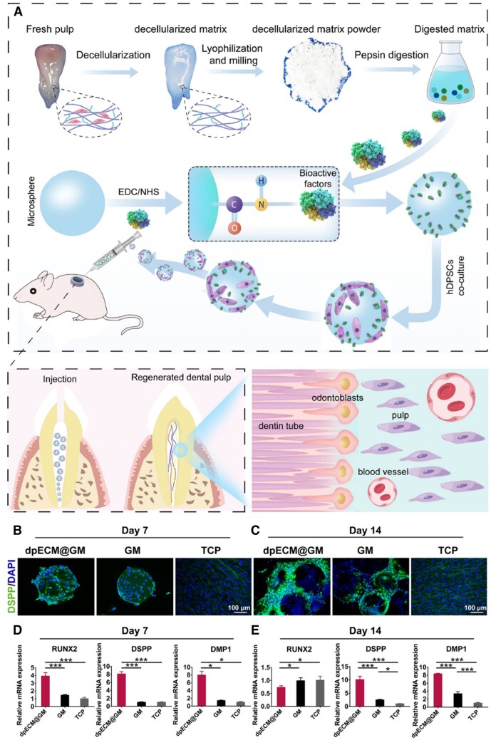
“[131] developed GelMA microspheres and conjugated dental pulp dECM to mimic pulp-specific microenvironment (Figure 6).”
BackgroundEmulsion templated scaffolds of poly(ε-caprolactone) – a review
Yadav, A. L., Ghosh, S., Samanta, A., Pal, J., & Srivastava, R. K. (2021) · Chemical Communications · doi:10.1039/d1cc04941k · Cited by 64
BMSC exosome-enriched acellular fish scale scaffolds promote bone regeneration
Wang, Y., Kong, B., Chen, X. '., Liu, R., Zhao, Y., Gu, Z., & Jiang, Q. Y. (2022) · Journal of Nanobiotechnology · doi:10.1186/s12951-022-01646-9 · Cited by 64 · cited 1×
“[214] designed fish skin dECM hydrogel to bind to BMSCs, leading to a stronger synergistic effect on promoting bone regeneration.”
BackgroundMultimodal therapy strategy based on a bioactive hydrogel for repair of spinal cord injury
Roh, E. J., Kim, D., Kim, J. H., Lim, C. S., Choi, H., Kwon, S. Y., Park, S. Y., Kim, J. Y., Kim, H., Hwang, D., et al. (2023) · Biomaterials · doi:10.1016/j.biomaterials.2023.122160 · Cited by 63 · cited 1×
“[220] incorporated MSC-derived extracellular vesicles and polydeoxyribonucleotide into HA and dECM-based scaffold, studying its spinal cord regeneration activity.”
Chen, M., Li, Y., Liu, S., Feng, Z., Wang, H., Yang, D., Guo, W., Yuan, Z., Gao, S., Zhang, Y., et al. (2021) · Bioactive Materials · doi:10.1016/j.bioactmat.2020.12.009 · Cited by 62
Lee, H., Ju, Y. M., Kim, I., Elsangeedy, E., Lee, J. H., Yoo, J. J., Atala, A., & Lee, S. J. (2020) · Methods · doi:10.1016/j.ymeth.2019.06.027 · Cited by 61
Kim, T. H., Jung, Y., & Kim, S. H. (2018) · Tissue Engineering Part A · doi:10.1089/ten.tea.2017.0318 · Cited by 59
TGF-β1-supplemented decellularized annulus fibrosus matrix hydrogels promote annulus fibrosus repair
Wei, Q., Liu, D., Chu, G., Yu, Q., Liu, Z., Li, J., Meng, Q., Wang, W., Han, F., & Li, B. (2023) · Bioactive Materials · doi:10.1016/j.bioactmat.2022.04.025 · Cited by 59 · cited 1×
“[163] fabricated a multifunctional system made by polyethylene glycol diacrylate (PEGDA) and annulus fibrosus dECM capable of continuously releasing TGF-b 1 in 7days.”
MethodJeon, E. Y., Joo, K. I., & Chi, H. J. (2020) · Acta Biomaterialia · doi:10.1016/j.actbio.2020.07.033 · Cited by 59
Xu, Y., Wang, Z., Hua, Y., Zhu, X., Wang, Y., Duan, L., Zhu, L., Jiang, G., Xia, H., She, Y., et al. (2020) · Materials Science and Engineering C · doi:10.1016/j.msec.2020.111628 · Cited by 59 · cited 2×
“[121] designed a photo-crosslinked hydrogel system consisting of HA-NB/gelatin/dECM for loading chondrocytes.”
MethodLu, Y., Wang, Y., Zhang, H., Tang, Z., Cui, X., Li, X., Liang, J., Wang, Q., Fan, Y., & Zhang, X. (2021) · ACS Applied Materials & Interfaces · doi:10.1021/acsami.1c07530 · Cited by 59 · cited 1×
“[107] fabricated collagen scaffolds that functionalized cartilage dECM in solution and particle forms.”
3D bioprinting of conductive hydrogel for enhanced myogenic differentiation
Wang, Y., Wang, Q., Luo, S., Chen, Z., Zheng, X., Kankala, R. K., Chen, A., & Wang, S. (2021) · Regenerative Biomaterials · doi:10.1093/rb/rbab035 · Cited by 59 · cited 1×
“Due to the poor mechanical strength and mismatched degradation of the gelatin-based scaffold, the chemical modification of gelatin with methacryloyl (GelMA) has been increasingly developed [123, 124].”
BackgroundSelf-citationLiu, X., Zhang, J., Cheng, X., Liu, P., Feng, Q., Wang, S., Li, Y., Gu, H., Zhong, L., Chen, M., et al. (2023) · Regenerative Biomaterials · doi:10.1093/rb/rbac085 · Cited by 58 · cited 1×
“Due to the biological activities, the bioactive properties of extracellular vesicles have been used to cooperate with scaffolds in tissue re-generation [211–213].”
Self-citationSingh, H., Bashir, S. M., Purohit, S. D., Bhaskar, R., Rather, M. A., Ali, S. I., Yadav, I., Makhdoomi, D. M., Dar, M. U. D., Gani, M. A., et al. (2022) · Biomaterials Advances · doi:10.1016/j.bioadv.2022.212806 · Cited by 58 · cited 2×
“W o u n d h e a l i n g [ 206 ] G e l M A , d E C M K i d n e y r ege n e r a t i o n [ 221 ] b F G F , b a s i c fi b r o b l a s t g r o w t h f a c t o r ; B M P - 2 , b o n e m o r p h o ge n e t i c p r o t e i n - 2 ; B M P - 7 , b o n e m o r p h o ge n e t i c p r o t e i n - 7 ; B M S C ,…”
Electrospun Decellularized Lung Matrix Scaffold for Airway Smooth Muscle Culture
Young, B. M., Shankar, K., Allen, B. P., Pouliot, R. A., Schneck, M. B., Mikhaiel, N. S., & Heise, R. L. (2017) · ACS Biomaterials Science & Engineering · doi:10.1021/acsbiomaterials.7b00384 · Cited by 56
Jin, Y., Zhang, J., Xu, Y., Yi, K., Li, F., Zhou, H., Wang, H., Chan, H. F., Lao, Y.-H., Lv, S., et al. (2023) · Bioactive Materials · doi:10.1016/j.bioactmat.2023.05.001 · Cited by 55 · cited 1×
“[210] developed a synergistic platform based on copper oxide nanoparticles-loaded PLGA nanofibers and dECM hydrogel with human adipose-derived MSC.”
BackgroundCurley, C. J., Dolan, E. B., Otten, M., Hinderer, S., Duffy, G. P., & Murphy, B. P. (2019) · Drug Delivery and Translational Research · doi:10.1007/s13346-018-00601-2 · Cited by 55
Gao, E., Gao, L., Cao, R., Xia, H., Xu, Y. F., Jiang, G., Xiao, K., Chen, J., Chen, R., & Duan, L. (2021) · Composites Part B Engineering · doi:10.1016/j.compositesb.2021.109470 · Cited by 55 · cited 2×
“…u s t o m i z e d m o l d a n d f r ee z e - d r y i n g T h e r i n g - s h a p e d p o r o u s s c a ff o l d d i s p l a y e d s u i t a b l e p o r e s i z e a n d p o r o s i t y f o r c e ll c o l o n i z a t i o n [ 147 ] S i l k fi b r o i n , P C L H u m a n u m b i l i c a l c o r d M S C”
MethodYang, Y., Zhang, Y., Yan, Y., Ji, Q., Dai, Y., Jin, S., Liu, Y., Chen, J., & Teng, L. (2020) · Polymers · doi:10.3390/polym12030535 · Cited by 54
Zhang, X., Liu, Y., Zuo, Q., Wang, Q., Li, Z., Yan, K., Yuan, T., Zhang, Y., Shen, K., Xie, R., et al. (2024) · International Journal of Bioprinting · doi:10.18063/ijb.v7i4.401 · Cited by 54 · cited 1×
“[166] designed a silk fibroin and dECM composite scaffold as an integrated 3D platform to deliver TGF-b and BMP-2 to achieve osteochondral regeneration.”
MethodIşık, M., Karakaya, E., Arslan, T. S., Atila, D., Erdoğan, Y. K., Arslan, Y. E., Eskizengin, H., Eylem, C. C., Nemutlu, E., Ercan, B., et al. (2023) · Advanced Healthcare Materials · doi:10.1002/adhm.202203044 · Cited by 54
Kim, D.-H., Kim, M.-J., Kwak, S.-Y., Jeong, J., Choi, D., Choi, S. W., Ryu, J., & Kang, K.-S. (2023) · Nature Communications · doi:10.1038/s41467-023-35941-2 · Cited by 53 · cited 2×
“[200] crosslinked the dECM scaffold with nano-graphene oxide to alleviate the enzymatic degradation activity and enhance the mechanical rigidity.”
Datta, S., Rameshbabu, A. P., Bankoti, K., Roy, M., Gupta, C., Jana, S., Das, A. K., Sen, R., & Dhara, S. (2020) · Materials Science and Engineering C · doi:10.1016/j.msec.2020.111604 · Cited by 53 · cited 1×
“[100] prepared a novel biohybrid hydrogel using fatty acid-modified chitosan and bone dECM to deliver human amnion-derived MSCs.”
Gong, M., Sun, J., Liu, G., Li, L., Wu, S., & Xiang, Z. (2020) · Materials Science and Engineering C · doi:10.1016/j.msec.2020.111603 · Cited by 53 · cited 2×
“Based on the unique mechanical and electronic properties, graphene oxide functioned scaffold has attracted increased attention [197, 198].”
BackgroundDual-Bionic Regenerative Microenvironment for Peripheral Nerve Repairing
Guan, Y., Ren, Z., Yang, B., Xu, W., Wu, W., Li, X., Zhang, T., Li, D., Chen, S., Bai, J., et al. (2022) · Bioactive Materials · doi:10.1016/j.bioactmat.2023.02.002 · Cited by 53 · cited 2×
“T h e e x t r a c e ll u l a r m a t r i x - m o d i fi e d e l e c t r o s p un fi b e r s c o u l d p r o m o t e axo n r ege n e r a t i o n o f S c h w a nn c e ll s [ 148 ] S i l k fi b r o i n , P C L”
BackgroundChoi, E., Kim, D., Kang, D., Yang, G. H., Jung, B., Yeo, M., Park, M.-J., An, S., Lee, K., Kim, J. S., et al. (2021) · Regenerative Biomaterials · doi:10.1093/rb/rbab001 · Cited by 52 · cited 1×
“Due to the poor mechanical strength and mismatched degradation of the gelatin-based scaffold, the chemical modification of gelatin with methacryloyl (GelMA) has been increasingly developed [123, 124].”
BackgroundEngineering Densely Packed Adipose Tissue via Environmentally Controlled In‐Bath 3D Bioprinting
Ahn, M., Cho, W., Kim, B. S., & Cho, D. (2022) · Advanced Functional Materials · doi:10.1002/adfm.202200203 · Cited by 52
Vascularized adipose tissue engineering: moving towards soft tissue reconstruction
Peirsman, A., Nguyen, H. T., Waeyenberge, M. V., Ceballos‐González, C. F., Bolívar‐Monsalve, E. J., Kawakita, S., Vanlauwe, F., Tirpáková, Z., Dorpe, S. V., Damme, L. V., et al. (2023) · Biofabrication · doi:10.1088/1758-5090/acd7a5 · Cited by 52
Yang, X., Li, Y., Liu, X., Huang, Q., Zhang, R., & Feng, Q. (2018) · Regenerative Biomaterials · doi:10.1093/rb/rby014 · Cited by 51
Silk fibroin protein modified acellular dermal matrix for tissue repairing and regeneration
Dhasmana, A., Singh, L., Roy, P., & Mishra, N. C. (2019) · Materials Science and Engineering: C · doi:10.1016/j.msec.2018.12.038 · Cited by 51 · cited 1×
“[144] modified silk fibroin on the acellular dermal matrix scaffold to prolong the degradation time and lower immune response in wound healing.”
BackgroundWu, R., Li, H., Yang, Y., Zheng, Q., Li, S., & Chen, Y. (2021) · ACS Applied Bio Materials · doi:10.1021/acsabm.1c00654 · Cited by 51 · cited 1×
“Compared to collagen and gelation-derived biomaterials, silk fibroin purified from silkworm cocoons has achieved superior mechanical performance [137].”
BackgroundYang, L., Jin, S., Shi, L., Ullah, I., Yu, K., Zhang, W., Bo, L., Zhang, X., & Guo, X. (2021) · Chemical Engineering Journal · doi:10.1016/j.cej.2021.133459 · Cited by 51 · cited 2×
“B o n e r ege n e r a t i o n [ 195 ] d E C M P o r c i n e s m a ll i n t e s t i n e S r 2 þ / F e 3 þ c o - d o p e d h y - d r ox y a p a t i t e B o n e r ege n e r a t i o n [ 196 ] d E C M P o r c i n e f e m o r a l c o n d y l e”
MethodBiomaterial strategies for the application of reproductive tissue engineering
Liu, X., Wu, K., Gao, L., Wang, L., & Shi, X. (2022) · Bioactive Materials · doi:10.1016/j.bioactmat.2021.11.023 · Cited by 50
Liu, Y.-K., Fan, J., Lv, M.-Q., She, K.-P., Sun, J., Lu, Q.-H., Han, C.-G., Ding, S.-T., Zhao, S., Wang, G.-X., et al. (2021) · Regenerative Biomaterials · doi:10.1093/rb/rbab048 · Cited by 50 · cited 1×
“In recent years, owing to the excellent biocompatibility, tunable degradability and abundant sources of silk fibroin, silk fibroin-based bio-scaffolds have received considerable attention for tissue regeneration [138, 139].”
BackgroundLi, S., Deng, R., Zou, X., Rong, Q., Shou, J., Rao, Z., Wu, W., Wu, G., Quan, D., Zhou, M., et al. (2022) · Composites Part B Engineering · doi:10.1016/j.compositesb.2022.109620 · Cited by 50
Decellularization Strategies for Regenerating Cardiac and Skeletal Muscle Tissues
Tan, Y. H., Helms, H. R., & Nakayama, K. H. (2022) · Frontiers in Bioengineering and Biotechnology · doi:10.3389/fbioe.2022.831300 · Cited by 50
Hewawasam, R. S., Blomberg, R., Šerbedžija, P., & Magin, C. M. (2023) · ACS Applied Materials & Interfaces · doi:10.1021/acsami.2c18330 · Cited by 48
Carbon nanodot decorated acellular dermal matrix hydrogel augments chronic wound closure
Bankoti, K., Rameshbabu, A. P., Datta, S., Roy, M., Goswami, P., Roy, S., Das, A. K., Ghosh, S. K., & Dhara, S. (2020) · Journal of Materials Chemistry B · doi:10.1039/d0tb01574a · Cited by 48 · cited 2×
“Combining the outstanding merits of other inorganic nanoparticles (nanoceria, laponite, metal or non-metal particles) with dECM, the hybrid scaffold exhibited excellent performance in tissue regeneration [204].”
BackgroundDong, C., Qiao, F., Chen, G., & Lv, Y. (2021) · Journal of Materials Chemistry B · doi:10.1039/d1tb00895a · Cited by 47
Hua, Y., Huo, Y., Bai, B., Hao, J., Hu, G., Ci, Z., Wu, X., Yu, M., Wang, X., Chen, H., et al. (2022) · Materials Today Bio · doi:10.1016/j.mtbio.2022.100489 · Cited by 47 · cited 1×
“Then, they incorporated bone dECM particles to effectively promote osteochondral regeneration [105].”
MethodNie, X., & Wang, D. (2018) · Biomaterials Science · doi:10.1039/c8bm00772a · Cited by 46
Parmaksiz, M., Elçin, A. E., & Elçin, Y. M. (2019) · Materials Science and Engineering: C · doi:10.1016/j.msec.2018.10.011 · Cited by 46 · cited 2×
“[194] prepared a multilayer composite scaffold using small intestinal submucosa dECM as layer and PCL-hydroxyapatite microparticles as a binder.”
MethodLiao, Z., Ke, W., Liu, H., Tong, B., Wang, K., Feng, X., Hua, W., Wang, B., Song, Y., Luo, R., et al. (2022) · Journal of Nanobiotechnology · doi:10.1186/s12951-022-01624-1 · Cited by 46 · cited 1×
“[216] fabricated a thermo-responsive hydrogel that composed of Pluronic F127 and nucleus pulposus dECM for human MSC-derived extracellular vesicle delivery.”
Shi, J., Teng, Y., Li, D., He, J., Midgley, A. C., Guo, X., Wang, X., Yang, X., Wang, S., Feng, Y., et al. (2023) · Materials Today Bio · doi:10.1016/j.mtbio.2023.100709 · Cited by 46 · cited 1×
“[189] developed a tri-layered vascular graft via electrospun by sandwiching media dECM powders loaded in PLCL between outer layers that loaded intima powders and inner layers that salidroside.”
BackgroundSelf-citationApplication of Graphene Oxide-Based Hydrogels in Bone Tissue Engineering
Zhihui, K., & Min, D. (2022) · ACS Biomaterials Science & Engineering · doi:10.1021/acsbiomaterials.2c00396 · Cited by 45
Yang, Z., Cao, F., Li, H., He, S., Zhao, T., Deng, H., Li, J., Sun, Z., Hao, C., Xu, J., et al. (2022) · Acta Biomaterialia · doi:10.1016/j.actbio.2022.07.029 · Cited by 45 · cited 1×
“[165] developed PLGA microspheres to control the TGF-b 3 releasement and then loaded them with a functional cartilage scaffold based on PCL, dECM and GelMA.”
MethodZhang, D., Fu, Q., Fu, H., Zeng, J., Jia, L., & Chen, M. (2023) · International Journal of Bioprinting · doi:10.18063/ijb.718 · Cited by 45
Suhaeri, M., Subbiah, R., Kim, S.-H., Kim, C.-H., Oh, S. J., Kim, S.-H., & Park, K. (2017) · ACS Applied Materials & Interfaces · doi:10.1021/acsami.6b14020 · Cited by 44
A pH-driven genipin gelator to engineer decellularized extracellular matrix-based tissue adhesives
Nishiguchi, A., & Taguchi, T. (2021) · Acta Biomaterialia · doi:10.1016/j.actbio.2021.06.033 · Cited by 43
Du, J., Hu, X., Su, Y., Wei, T., Jiao, Z., Liu, T., Wang, H., Nie, Y., Li, X., & Song, K. (2022) · International Journal of Biological Macromolecules · doi:10.1016/j.ijbiomac.2022.04.188 · Cited by 43
Preparation of Acellular Homogenates From Muscle Samples
Poel, W. E. (1948) · Science · doi:10.1126/science.108.2806.390.b · Cited by 43
Yeleswarapu, S., Dash, A., Chameettachal, S., & Pati, F. (2023) · Biomaterials Advances · doi:10.1016/j.bioadv.2023.213494 · Cited by 43
Lin, C.-Y., Liu, T.-Y., Chen, M., Sun, J., & Chen, M. (2016) · Biomedical Materials · doi:10.1088/1748-6041/11/3/035010 · Cited by 42
Lee, S., Lee, H. S., Chung, J. J., Kim, S. H., Park, J. W., Lee, K., & Jung, Y. (2021) · International Journal of Molecular Sciences · doi:10.3390/ijms22062886 · Cited by 42
Zhan, A., Chen, L., Sun, W., Tang, Y., Chen, J., Yu, D., & Zhang, W. (2022) · Materials & Design · doi:10.1016/j.matdes.2022.110660 · Cited by 41
Liu, C., Jin, Z., Ge, X., Zhang, Y., & Xu, H. (2019) · Tissue Engineering Part A · doi:10.1089/ten.tea.2018.0297 · Cited by 41 · cited 1×
“[157] fabricated a dECM and chitosan-based hydrogel with bFGF for supporting annulus fibrosus-derived stem cell growth.”
Liu, Z., Liu, J., Liu, N., Zhu, X., & Tang, R. (2021) · Materials Science and Engineering C · doi:10.1016/j.msec.2021.112235 · Cited by 41
Wang, Y., Wei, X., Wang, L., Qian, Z., Liu, H., & Fan, Y. (2022) · Composites Part B Engineering · doi:10.1016/j.compositesb.2022.110311 · Cited by 41
Modeling dECM-based inflammatory cartilage microtissues in vitro for drug screening
Chen, Y., Chen, L., Wang, Y., Duan, Y., Luo, S., Zhang, J.-T., Kankala, R. K., Wang, S., & Chen, A. (2022) · Composites Part B Engineering · doi:10.1016/j.compositesb.2022.110437 · Cited by 41
Self-citationZhu, Y., Wang, D., Yao, X., Wang, M., Zhao, Y., Lu, Y., Wang, Z., & Guo, Y. (2021) · Journal of Biomaterials Science, Polymer Edition · doi:10.1080/09205063.2020.1818018 · Cited by 40 · cited 1×
“[145] fabricated a hybrid nanofiber scaffold silk fibroin and dECM using the electrospinning method for islet transplantation.”
MethodCao, H., Wang, X., Chen, M., Liu, Y., Cui, X., Liang, J., Wang, Q., Fan, Y., & Zhang, X. (2021) · ACS Applied Materials & Interfaces · doi:10.1021/acsami.1c19447 · Cited by 40 · cited 1×
“[108] obtained rabbit articular dECM cartilage at 3, 100 and 200days (Col 3d-S, Col 100d-S and Col 200d-S) and crosslinked with collagen to investigate the chondrogenic inducibilities.”
MethodHe, W., Zhang, X., Li, X., Ju, D., Mao, T., Lu, Y., Gu, Y., Qi, L., Wang, Q., Wu, Q., et al. (2022) · Journal of Materials Chemistry B · doi:10.1039/d2tb00792d · Cited by 40 · cited 1×
“[130] prepared spinal cord dECM and GelMA hydrogel and achieved a synergistic effect with human menstrual blood-derived stem cells for spinal cord injury treatment.”
BackgroundLiu, J., Chen, D., Zhu, X., Liu, N., Zhang, H., Tang, R., & Liu, Z. (2022) · Acta Biomaterialia · doi:10.1016/j.actbio.2022.09.009 · Cited by 40 · cited 2×
“[149] constructed human amniotic membrane dECM-based grafts with tubular structures and rolled with PCL/ silk fibroin nanofiber to form the luminal surface.”
BackgroundXu, P., Kankala, R. K., Li, Y., Wang, S., & Chen, A. (2022) · Regenerative Biomaterials · doi:10.1093/rb/rbac072 · Cited by 40
Self-citationKo, E., Alberti, K., Lee, J. S., Yang, K., Jin, Y., Shin, J., Yang, H. S., Xu, Q., & Cho, S.-W. (2016) · ACS Applied Materials & Interfaces · doi:10.1021/acsami.6b05358 · Cited by 39 · cited 2×
“B o n e r ege n e r a t i o n [ 193 ] P C L , d E C M B o v i n e s m a ll i n t e s t i - n a l s u b m u c o s a”
MethodBo, Q., Yan, L., Li, H., Jia, Z., Zhan, A., Chen, J., Yuan, Z., Zhang, W., Gao, B., & Chen, R. (2020) · Materials & Design · doi:10.1016/j.matdes.2020.109152 · Cited by 39
Chen, L., Song, X., Yao, Z., Zhou, C., Yang, J., Yang, Q., Chen, J., Wu, J., Sun, Z., Gu, L., et al. (2023) · Biomaterials · doi:10.1016/j.biomaterials.2023.122207 · Cited by 39 · cited 2×
“[118] integrated the gelatin nanofiber membrane into the dECM membrane based on the interfacial covalent bonding to maintain the biological activity of dECM.”
BackgroundSun, W., Yang, Y., Wang, L., Tang, H., Zhang, L., She, Y., Xiao, X., Hu, X., Feng, Q., & Chen, C. (2022) · Advanced Functional Materials · doi:10.1002/adfm.202201257 · Cited by 38
Riley, J. A. (1948) · Science · doi:10.1126/science.108.2806.390-a · Cited by 37
Wiggenhauser, P. S., Schwarz, S., Körber, L., Hoffmann, T. K., & Rotter, N. (2019) · Journal of Materials Science Materials in Medicine · doi:10.1007/s10856-019-6323-x · Cited by 37
Gao, C., Sow, W. T., Wang, Y., Wang, Y., Yang, D., Lee, B. H., Matičić, D., Fang, L., Li, H., & Zhang, C. (2021) · Journal of Materials Chemistry B · doi:10.1039/d0tb02588g · Cited by 37 · cited 2×
“[129] incorporated bone dECM particles into GelMA hydrogel for bone defect reconstruction.”
MethodSani, M., Hosseinie, R., Latifi, M., Shadi, M., Razmkhah, M., Salmannejad, M., Parsaei, H., & Talaei‐Khozani, T. (2022) · Biomaterials Advances · doi:10.1016/j.bioadv.2022.213019 · Cited by 37 · cited 1×
“In recent research, different kinds of growth factors, including basic fibroblast growth factor (bFGF), TGF-b superfamily, vascular endothelial growth factors (VEGF) and other types of growth factors were incorporated into dECM-based materials [152–154].”
BackgroundLiu, W. Y., Sun, Y., Dong, X., Yin, Q., Zhu, H., Li, S., Zhou, J., & Wang, C. (2020) · Regenerative Biomaterials · doi:10.1093/rb/rbaa011 · Cited by 36 · cited 1×
“[142] cultured cardiac fibroblasts in silk fibroin scaffold and decellularized to obtain the cell dECM-coated scaffold to mimic the myocardial microenvironment.”
MethodDai, Y., Han, X., Hu, L., Wu, H., Huang, S., & Lu, Y. (2020) · Regenerative Biomaterials · doi:10.1093/rb/rbaa007 · Cited by 36 · cited 1×
“The supplementation of growth factors in the scaffold could promote tissue regeneration by promoting cell proliferation and differentiation [151].”
BackgroundBarthold, J. E., McCreery, K. P., Martinez, J., Bellerjeau, C., Ding, Y., Bryant, S. J., Whiting, G. L., & Neu, C. P. (2022) · Biofabrication · doi:10.1088/1758-5090/ac584c · Cited by 36
Lin, Q., Wong, H. L., Tian, F., Huang, Y., Xu, J., Yang, J., Chen, P.-P., Fan, Z.-L., Lu, C., & Zhao, Y. (2016) · International Journal of Pharmaceutics · doi:10.1016/j.ijpharm.2016.12.028 · Cited by 35
Bai, H., Wang, Z., Li, M., Liu, Y., Wang, W., Sun, P., Wei, S., Wang, Z., Li, J., & Dardik, A. (2020) · Journal of Biomedical Materials Research Part B Applied Biomaterials · doi:10.1002/jbm.b.34574 · Cited by 35 · cited 1×
“Thus, different drug agent was incorporated in dECM-based vascular grafts to achieve better performance in tissue regeneration [185, 186].”
BackgroundPeng, B., Du, L., Zhang, T., Chen, J., & Xu, B. (2023) · Biomaterials Science · doi:10.1039/d2bm01862d · Cited by 35
Datta, S., Rameshbabu, A. P., Bankoti, K., Jana, S., Roy, S., Sen, R., & Dhara, S. (2021) · Journal of Materials Chemistry B · doi:10.1039/d1tb00255d · Cited by 34 · cited 1×
“[184] designed alendronate-loaded gelatin microspheres embedded in bone dECM/oleoyl chitosan hydrogel for loading BMP-2.”
MethodCuenca, J. P., Kang, H.-J., Fahad, M. A. A., Park, M., Choi, M., Lee, H.-Y., & Lee, B. (2022) · Journal of Biomaterials Science Polymer Edition · doi:10.1080/09205063.2022.2069398 · Cited by 34 · cited 1×
“VEGF, an essential growth factor regulating neovascularization and angiogenesis, has also been processed into a dECM-based scaffold for tissue regeneration [168].”
BackgroundChandika, P., Khan, F., Heo, S.-Y., Kim, Y., Yi, M., & Jung, W. (2022) · Biomaterials Advances · doi:10.1016/j.bioadv.2022.213046 · Cited by 34 · cited 1×
“[174] mixed usnic acid with PCL and dECM to develop novel composite nanofibrous using electrospinning technology.”
MethodHwangbo, H., Lee, J., & Kim, G. (2022) · International Journal of Biological Macromolecules · doi:10.1016/j.ijbiomac.2022.07.040 · Cited by 33 · cited 2×
“B o n e r ege n e r a t i o n [ 195 ] d E C M P o r c i n e s m a ll i n t e s t i n e S r 2 þ / F e 3 þ c o - d o p e d h y - d r ox y a p a t i t e B o n e r ege n e r a t i o n [ 196 ] d E C M P o r c i n e f e m o r a l c o n d y l e”
MethodXie, X., Wang, W., Cheng, J. Q., Liang, H., Lin, Z., Zhang, T., Lu, Y., & Li, Q. (2020) · Colloids and Surfaces B Biointerfaces · doi:10.1016/j.colsurfb.2020.110903 · Cited by 32 · cited 1×
“[182] fabricated the nanofibers composed of dECM and PLGA-loaded pifithrin-a acting as bilayer bone scaffold for enhancing osteoinductivity.”
MethodSingh, H., Purohit, S. D., Bhaskar, R., Yadav, I., Bhushan, S., Gupta, M. K., Gautam, S., Showkeen, M., & Mishra, N. C. (2021) · Biomedical Materials · doi:10.1088/1748-605x/abdb74 · Cited by 31 · cited 2×
“[205] found that the activity accelerates tissue regeneration by combining nanoceria and dECM.”
BackgroundZhou, J., Wu, Y., Tang, Z., Zou, K., Chen, J., Lei, Z., Wan, X., Liu, Y., Zhang, H., Wang, Y., et al. (2022) · Regenerative Biomaterials · doi:10.1093/rb/rbac057 · Cited by 31
Jin, B., Yu, Y., Chen, X., Yang, Y., Xiong, Y., Im, Y. J., Zhao, Y., & Xiao, J. (2022) · Bioactive Materials · doi:10.1016/j.bioactmat.2022.08.027 · Cited by 31
Fish scale-derived scaffolds with MSCs loading for photothermal therapy of bone defect
Shen, S., Liu, R., Song, C., Shen, T., Zhou, Y., Guo, J., Kong, B., & Jiang, Q. Y. (2023) · Nano Research · doi:10.1007/s12274-023-5460-1 · Cited by 31 · cited 1×
“[209] designed a black phosphorus nanosheets degradation loaded photothermal scaffold based on fish scale dECM and GelMA to accelerate bone regeneration.”
Arin, A., Rahaman, M. S., Farwa, U., & Lee, B.-T. (2023) · Advanced Functional Materials · doi:10.1002/adfm.202212325 · Cited by 30 · cited 1×
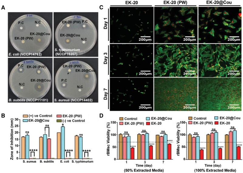
“[175] used decellularized kelp as crosslinking material to strengthen the physical structure of liver dECM and then loaded it with p-coumaric acid (EK-20).”
MethodHong, H., Kim, H., Han, S. J., Jang, J., Kim, H. K., Cho, D., & Kim, D. S. (2019) · Materials Science and Engineering C · doi:10.1016/j.msec.2019.109837 · Cited by 30 · cited 1×
“[104] mixed compressed collagen with cornea dECM to construct a dense collagenous scaffold for mimicking biomimetic corneal stroma analog.”
MethodLarochette, N., El‐Hafci, H., Potier, E., Setterblad, N., Bensidhoum, M., Petite, H., & Logeart‐Avramoglou, D. (2020) · Acta Biomaterialia · doi:10.1016/j.actbio.2020.09.003 · Cited by 30 · cited 1×
“[161] developed bone morphogenetic protein-2 (BMP-2)-incorporated osteogenic-differentiated MSC-derived dECM scaffolds for ectopic bone formation.”
BackgroundWang, F., Zhao, L., Song, F., Wu, J., Zhou, Q., & Xie, L. (2021) · Journal of Materials Chemistry B · doi:10.1039/d1tb00263e · Cited by 29 · cited 1×
“To achieve sustained drug release, voriconazole-loaded PLGA microspheres were produced by using emulsion evaporation technique before being mixed with dECM and gelatin composite hydrogel [176].”
MethodNishiguchi, A., Ito, S., Nagasaka, K., & Taguchi, T. (2023) · Biomacromolecules · doi:10.1021/acs.biomac.2c01210 · Cited by 29 · cited 2×
“[135] functionalized gelatin with the 2-ureido-4[1H]-pyrimi- dinone unit (TGUPy) and mixed it with urinary bladder dECM to obtain a composite patch.”
MethodMokhtarinia, K., & Masaeli, E. (2023) · Biomaterials Science · doi:10.1039/d2bm02111k · Cited by 28
Decellularized extracellular matrix particle-based biomaterials for cartilage repair applications
Guo, P., Jiang, N., Mini, C., Miklosic, G., Zhu, S., Vernengo, J., D’Este, M., Grad, S., Alini, M., & Li, Z. (2023) · Journal of Material Science and Technology · doi:10.1016/j.jmst.2023.03.019 · Cited by 28
Chen, Y., Lin, Y.-H., Lin, T.-L., Lee, A. K.-X., Yu, M., & Shie, M. (2023) · Biofabrication · doi:10.1088/1758-5090/ace5e1 · Cited by 28 · cited 1×
“[133] fabricated GelMA and dECM-based scaffolds to understand the influence of tensile stimulation on chondrocytes.”
Hwang, S. H., Kim, J., Heo, C., Yoon, J., Kim, H., Lee, S., Park, H. W., Heo, M. S., Moon, H. E., Kim, C., et al. (2022) · Acta Biomaterialia · doi:10.1016/j.actbio.2022.11.050 · Cited by 27 · cited 1×
“[171] designed a new generation of 3D dECM-based scaffolds to support tissue regeneration that explores the incorporation of VEGF and FGF.”
BackgroundPlacenta Powder-Infused Thiol-Ene PEG Hydrogels as Potential Tissue Engineering Scaffolds
Fan, Y., Lüchow, M., Badria, A. F., Hutchinson, D. J., & Malkoch, M. (2023) · Biomacromolecules · doi:10.1021/acs.biomac.2c01355 · Cited by 27
An adhesive gelatin-coated small intestinal submucosa composite hydrogel dressing aids wound healing
Wang, L., Liu, F., Zhai, X., Dong, W. F., Wei, W., & Hu, Z. (2023) · International Journal of Biological Macromolecules · doi:10.1016/j.ijbiomac.2023.124622 · Cited by 27 · cited 2×
“[136] designed o-nitrobenzene-modified gelatin coated with dECM hydrogel as a multifunctional wound dressing.”
Ghosh, P., Gruber, S. M. S., Lin, C.-Y. J., & Whitlock, P. W. (2018) · Biofabrication · doi:10.1088/1758-5090/aaa637 · Cited by 26
Guo, Z., Ci, H., Ma, C., Li, Z., Jiang, W., Chen, L., Wang, Z., Zhou, M., & Sun, J. (2022) · Acta Biomaterialia · doi:10.1016/j.actbio.2022.05.053 · Cited by 26 · cited 1×
“[158] designed a miniaturized porous PCL chamber by 3D printing and filled it with bFGF-loaded adipose tissue dECM hydrogel.”
MethodSong, E. S., Park, J.-H., Ha, S. S., Cha, P.-H., Kang, J.-T., Park, C. Y., & Park, K. (2022) · ACS Applied Materials & Interfaces · doi:10.1021/acsami.2c01709 · Cited by 25
Kong, Y., Wang, D., Weı, Q., & Yang, Y. (2022) · Frontiers in Bioengineering and Biotechnology · doi:10.3389/fbioe.2021.840421 · Cited by 24
Chu, T. L., Tripathi, G., Park, M., Bae, S., & Lee, B. (2022) · International Journal of Biological Macromolecules · doi:10.1016/j.ijbiomac.2022.05.085 · Cited by 24
Han, Y., Lian, M., Zhang, C., Jia, B., Wu, Q., Sun, B., Qiao, Z., Sun, B., & Dai, K. (2022) · Applied Materials Today · doi:10.1016/j.apmt.2022.101547 · Cited by 24
Gupta, V., Lyne, D. V., Laflin, A. D., Zabel, T. A., Barragan, M., Bunch, J. T., Pacicca, D. M., & Detamore, M. S. (2016) · ACS Biomaterials Science & Engineering · doi:10.1021/acsbiomaterials.6b00071 · Cited by 23
Xu, J., Fang, Q., Liu, Y., Zhou, Y., Ye, Z., & Tan, W. (2020) · Colloids and Surfaces B Biointerfaces · doi:10.1016/j.colsurfb.2020.111374 · Cited by 23
Pereira, A. T., Schneider, K. H., Henriques, P. C., Grasl, C., Melo, S. F., Fernandes, I. P., Kiss, H., Martins, M. C. L., Bergmeister, H., & Gonçalves, I. C. (2021) · ACS Applied Materials & Interfaces · doi:10.1021/acsami.1c04028 · Cited by 23 · cited 2×
“V a s c u l a r g r a f t [ 199 ] d E C M R a t l i v e r”
Liu, K., Zhao, M., Li, Y., Luo, L., & Hu, D. (2021) · Journal of Biomaterials Science Polymer Edition · doi:10.1080/09205063.2021.2002235 · Cited by 23 · cited 1×
“[169] found that the combination of dECM and VEGF hydrogel could enhance tube formation and accelerate wound healing process.”
BackgroundGuagliano, G., Volpini, C., Sardelli, L., Bloise, N., Vangosa, F. B., Cornaglia, A. I., Dotti, S., Villa, R., Visai, L., & Petrini, P. (2022) · ACS Biomaterials Science & Engineering · doi:10.1021/acsbiomaterials.2c01226 · Cited by 22
Kafili, G., Tamjid, E., Niknejad, H., & Simchi, A. (2022) · European Polymer Journal · doi:10.1016/j.eurpolymj.2022.111566 · Cited by 22 · cited 1×
“Based on the 2D structure and special composition, Laponite has been used to enhance the mechanical properties and degradation time of dECM in tissue engineering [207].”
MethodNi, R., Luo, Y. H., Jiang, L., Mao, X., Feng, Y., Tuersun, S., Hu, Z., & Zhu, Y. (2023) · International Journal of Biological Macromolecules · doi:10.1016/j.ijbiomac.2023.125556 · Cited by 21 · cited 1×
“Apart from the antibacterial drugs, other drugs, such as anti-resorptive agents and antioxidants, among others, were also combinated with dECM-based scaffolds in biomedical applications, including bone and vascular tissue engineering, among others [179–181].”
Background
Cited by
Liu, Y., & Ge, L. (2025) · Bioengineering · doi:10.3390/bioengineering12111178 · Cited by 84
“Conductive polymers may contain residual monomers or dopants that could be inflammatory [87].”
Conductive hydrogels: intelligent dressings for monitoring and healing chronic wounds
Fang, Y. Z., Han, Y., Lu, Y., Kankala, R. K., Wang, S., Chen, A., & Fu, C. (2024) · Regenerative Biomaterials · doi:10.1093/rb/rbae127 · Cited by 73
Self-citationDesign and Applications of Extracellular Matrix Scaffolds in Tissue Engineering and Regeneration
Mangani, S., Vetoulas, M., Mineschou, K., Spanopoulos, K., Vivanco, M. D., Piperigkou, Z., & Karamanos, N. K. (2025) · Cells · doi:10.3390/cells14141076 · Cited by 77
“More-over, unresolved issues in appropriate sterilization and disinfection methods, cell sourcing, regulatory classification, cost-efficiency, and the lack of robust preclinical data—especially in large animal models—pose further barriers [115,117–120].”
Advances in Electrospun Poly(ε-caprolactone)-Based Nanofibrous Scaffolds for Tissue Engineering
Robles, K. N., Zahra, F. T., Mu, R. R., & Giorgio, T. D. (2024) · Polymers · doi:10.3390/polym16202853 · Cited by 67
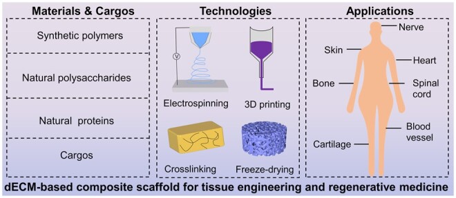
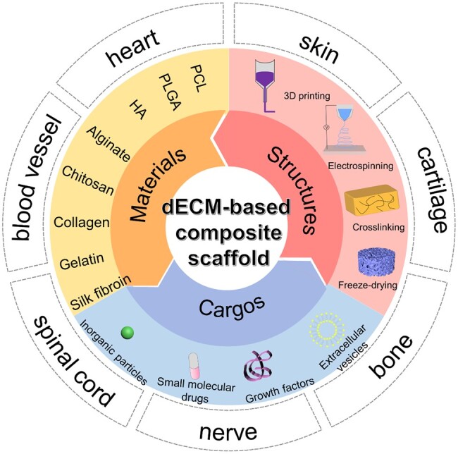
“Decellularized ECM (dECM) is biocompatible and can form 3D hydrogels; however, it lacks mechanical Decellularized ECM-based composite scaffolds have been fabricated with synthetic polymers, natural polysaccharides, and natural proteins for tissue engineering [2]. strength.”
BackgroundLiu, B., Alimi, O. A., Wang, Y., Kong, Y., Kuss, M. A., Krishnan, M. A., Hu, G., Xiao, Y., Dong, J., DiMaio, D. J., et al. (2024) · Journal of Controlled Release · doi:10.1016/j.jconrel.2024.02.019 · Cited by 48
Metadata
- Authors
- Xu, P., Kankala, R. K., Wang, S., & Chen, A.
- Affiliations
- Institute of Biomaterials and Tissue Engineering, Huaqiao University , Xiamen, Fujian 361021, PR China
- Huaqiao University (CN)
- Venue
- Regenerative Biomaterials
- Publisher
- Oxford University Press (OUP)
- Published
- 2023-12-01
- Volume
- 11
- Pages
- rbad107-rbad107
- ISSN
- 2056-3418 (ISSN-L)
- 2056-3426
- PMC
- PMC10761212
- OpenAlex
- W4389243078
- Cited by
- 129
- References
- 226
- Citations by year
- 2026: 56 · 2025: 45 · 2024: 28
- FWCI
- 8.33
- Type
- journal article
- Language
- en
- Open access
- gold
- doi.org
- Licence
- Locations
- Regenerative Biomaterials (publishedVersion, cc-by)
- PubMed Central (publishedVersion, cc-by)
- PubMed (publishedVersion)
- Funders
- National Natural Science Foundation of China (NSFC 32271410, 32071323, 81971734)
- Science and Technology Projects in Fujian Province (2022FX1, 2023Y4008)
- Scientific Research Funds of Huaqiao University (21BS113)
- Open Research Fund of Academy of Advanced Carbon Conversion Technology, Huaqiao University (AACCT0004)
- Topics
- Health Sciences › Medicine › Surgery › Tissue Engineering and Regenerative Medicine
- Physical Sciences › Materials Science › Biomaterials › Hydrogels: synthesis, properties, applications
- Physical Sciences › Engineering › Biomedical Engineering › 3D Printing in Biomedical Research
- Keywords
- decellularized extracellular matrix, composite scaffolds, tissue engineering, regenerative medicine, biomimetic microenvironment, natural polymers, bioactive agents, mechanical strength, tissue reconstruction
About these checks
Registry, retraction, Cites and statistics results are computed, not judged, with one exception: a printed reference that no index lists is read once by a model before it counts as a citation error. Claims are extracted by a model and kept only when their quote appears verbatim in the text; each cited source is then checked, with a model’s help, for whether it says what the claim relies on. Those verdicts can be wrong — read the claim’s quote and the cited source before relying on one.
Statistics and arithmetic are recomputed, not judged.
Confidence bands are shown only for fields calibrated on our annotated test set. None is calibrated yet, so no band is shown.
What counts as a claim ›How we measure confidence ›How we check this ›
Are you an author, or do you see a problem with this page? Request a review
Data sources
Bibliographic data: Semantic Scholar, OpenAlex, Crossref.
Contains information from the Semantic Scholar Academic Graph Datasets, made available under the ODC Attribution License (ODC-BY 1.0).
Semantic Scholar’s data is described in: Kinney, R., et al. (2023). The Semantic Scholar Open Data Platform. arXiv:2301.10140.
