

The DOI matches its Crossref record; 35 of the 35 references it cites were found in a registry or an index.
Robust deep learning based protein sequence design using ProteinMPNN
Open accessPreprint — published as 10.1126/science.add2187 · Paper page
Abstract
While deep learning has revolutionized protein structure prediction, almost all experimentally characterized de novo protein designs have been generated using physically based approaches such as Rosetta. Here we describe a deep learning based protein sequence design method, ProteinMPNN, with outstanding performance in both in silico and experimental tests. The amino acid sequence at different positions can be coupled between single or multiple chains, enabling application to a wide range of current protein design challenges. On native protein backbones, ProteinMPNN has a sequence recovery of 52.4%, compared to 32.9% for Rosetta. Incorporation of noise during training improves sequence recovery on protein structure models, and produces sequences which more robustly encode their structures as assessed using structure prediction algorithms. We demonstrate the broad utility and high accuracy of ProteinMPNN using X-ray crystallography, cryoEM and functional studies by rescuing previously failed designs, made using Rosetta or AlphaFold, of protein monomers, cyclic homo-oligomers, tetrahedral nanoparticles, and target binding proteins. One-sentence summary: A deep learning based protein sequence design method is described that is widely applicable to current design challenges and shows outstanding performance in both in silico and experimental tests.
Overview
The claims and statistics of this paper haven’t been checked yet.
Run the check — free, no account needed. It reads the open-access text and checks the sources it cites.
Integrity signals
Registry
Cites
Statistics
Figures
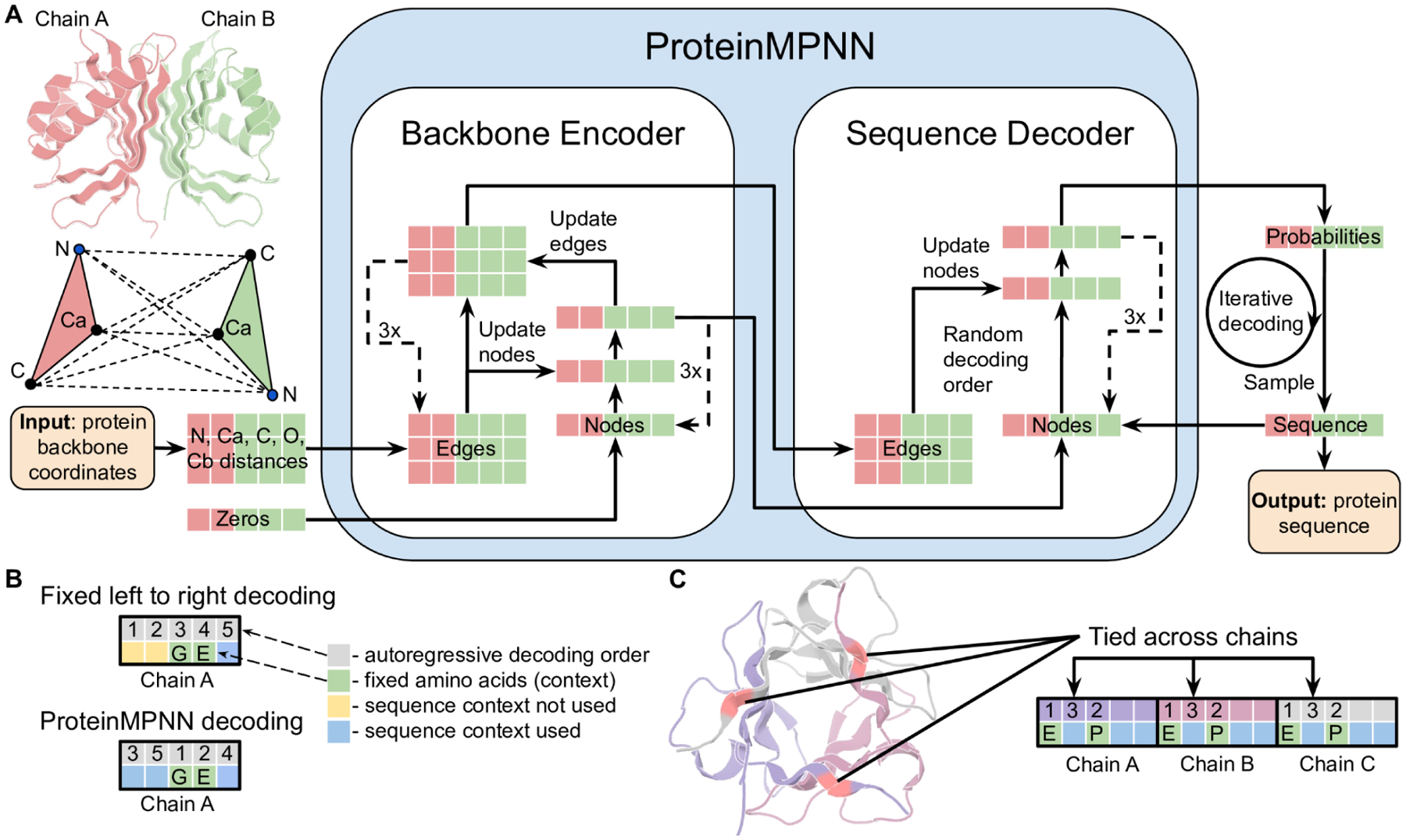
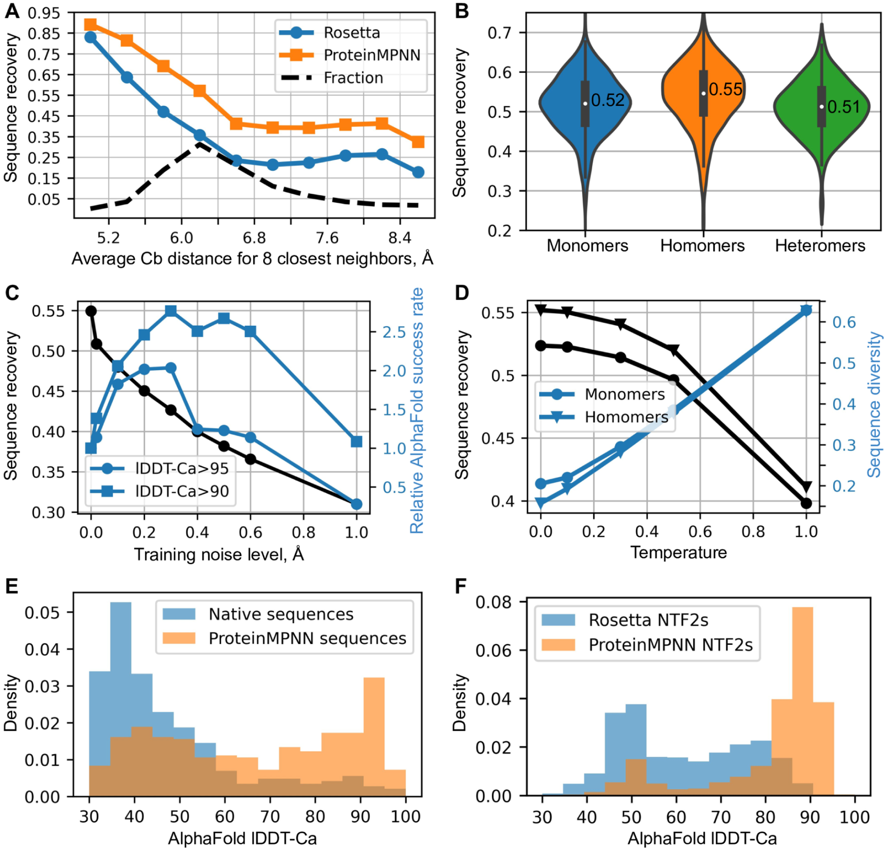
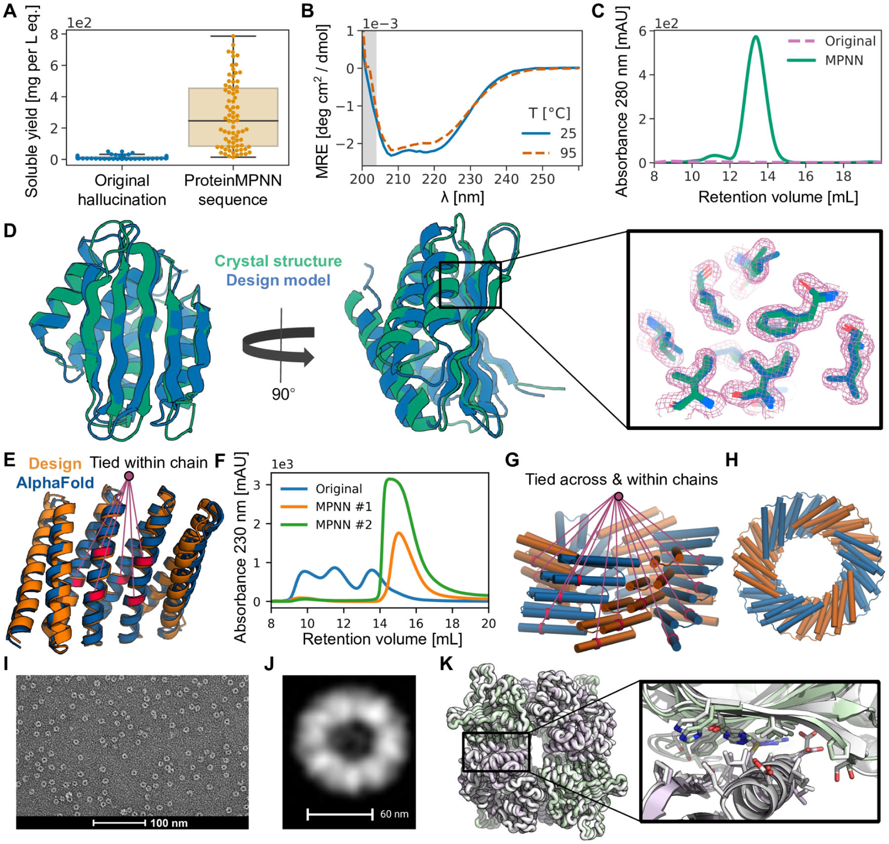
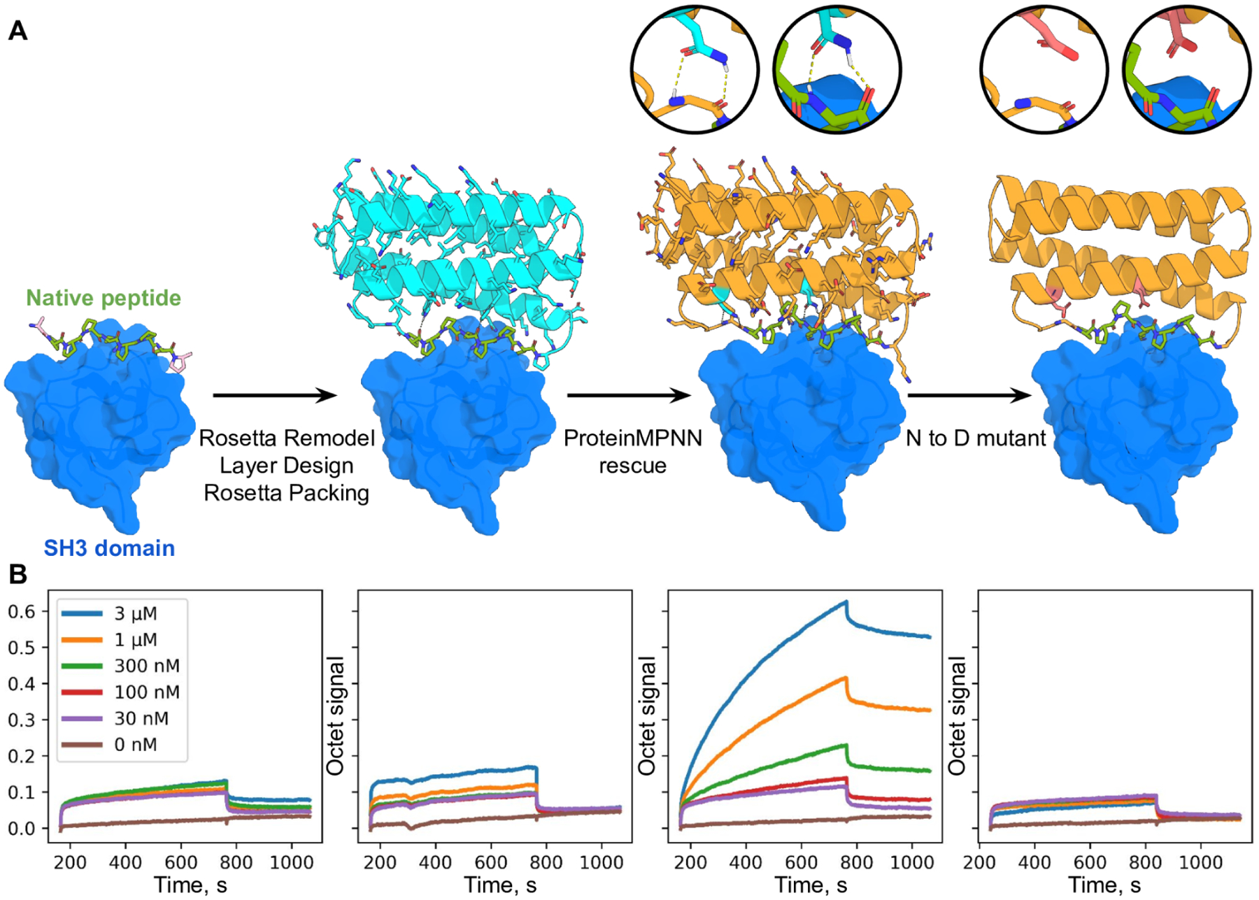
Verification
Claims the paper makes
The claims and statistics of this paper haven’t been checked yet.
Run the check — free, no account needed. It reads the open-access text and checks the sources it cites.
Self-check
Percentages, sums and reported differences re-computed from the paper’s own numbers.
The self-check needs the paper’s full text.
Citations
Cites
Vaswani, A., Shazeer, N., Parmar, N., Uszkoreit, J., Jones, L., Gomez, A. N., Kaiser, L., & Polosukhin, I. (2017) · arXiv · doi:10.48550/arxiv.1706.03762 · Cited by 120,404
Highly accurate protein structure prediction with AlphaFold
Jumper, J., Evans, R., Pritzel, A., Green, T., Figurnov, M., Ronneberger, O., Tunyasuvunakool, K., Bates, R., Žídek, A., Potapenko, A., et al. (2021) · Nature · doi:10.1038/s41586-021-03819-2 · Cited by 43,764
(2014) · Cited by 33,419
Rethinking the Inception Architecture for Computer Vision
Szegedy, C., Vanhoucke, V., Ioffe, S., Shlens, J., & Wojna, Z. (2016) · 2016 IEEE Conference on Computer Vision and Pattern Recognition (CVPR) · doi:10.1109/cvpr.2016.308 · Cited by 32,138
PyTorch: An Imperative Style, High-Performance Deep Learning Library
Paszke, A., Gross, S., Massa, F., Lerer, A., Bradbury, J., Chanan, G., Killeen, T., Lin, Z., Gimelshein, N., Antiga, L., et al. (2019) · arXiv · doi:10.48550/arxiv.1912.01703 · Cited by 31,473
Coot : model-building tools for molecular graphics
Emsley, P., & Cowtan, K. (2004) · Acta Crystallographica Section D Biological Crystallography · doi:10.1107/s0907444904019158 · Cited by 30,571
PHENIX : a comprehensive Python-based system for macromolecular structure solution
Adams, P. D., Afonine, P. V., Bunkóczi, G., Chen, V. B., Davis, I. W., Echols, N., Headd, J. J., Hung, L.-W., Kapral, G. J., Grosse-Kunstleve, R. W., et al. (2010) · Acta Crystallographica Section D Biological Crystallography · doi:10.1107/s0907444909052925 · Cited by 23,716
Phaser crystallographic software
McCoy, A. J., Grosse-Kunstleve, R. W., Adams, P. D., Winn, M. D., Storoni, L. C., & Read, R. J. (2007) · Journal of Applied Crystallography · doi:10.1107/s0021889807021206 · Cited by 20,612
Kabsch, W. (2010) · Acta Crystallographica Section D Biological Crystallography · doi:10.1107/s0907444909047337 · Cited by 16,463
Overview of the CCP 4 suite and current developments
Winn, M. D., Ballard, C. C., Cowtan, K. D., Dodson, E. J., Emsley, P., Evans, P. R., Keegan, R. M., Krissinel, E. B., Leslie, A. G. W., McCoy, A., et al. (2011) · Acta Crystallographica Section D Biological Crystallography · doi:10.1107/s0907444910045749 · Cited by 12,353
Research works printed but not linked: 1
- 2013, In Methods in enzymology, 523, 109
Cited by
Sorted by citations
Items 501–600 of 997
Advancing biofoundry development: strategies and challenges
Gupta, A., Lee, S., Sung, B. H., Lee, D., Cho, B., Kim, D., & Kim, H. (2024) · Biotechnology and Bioprocess Engineering · doi:10.1007/s12257-024-00121-6 · Cited by 12
Dey, S., Bruner, J., Brown, M., Roof, M., & Chowdhury, R. (2024) · Computational and Structural Biotechnology Journal · doi:10.1016/j.csbj.2024.08.029 · Cited by 12
Gavalda-Garcia, J., Dixit, B., Díaz, A., Ghysels, A., & Vranken, W. (2025) · Journal of Molecular Biology · doi:10.1016/j.jmb.2024.168900 · Cited by 12
Toward deep learning sequence–structure co-generation for protein design
Wang, C., Alamdari, S., Domingo-Enrich, C., Amini, A. P., & Yang, K. (2025) · Current Opinion in Structural Biology · doi:10.1016/j.sbi.2025.103018 · Cited by 18
Protein design accelerates the development and application of optogenetic tools
Zhu, L., Wang, Y., Wu, X., Wu, G., Zhang, G., Liu, C., & Zhang, S. (2025) · Computational and Structural Biotechnology Journal · doi:10.1016/j.csbj.2025.02.014 · Cited by 13
Ding, Y., Zhang, S., Kong, X., Hess, H., & Zhang, Y. (2025) · Advanced Science · doi:10.1002/advs.202500859 · Cited by 17
Wang, B., Zhou, T., Shen, Y. C., Hu, W. W., Zhou, J., Tang, J., Han, R., Xu, G., Schwaneberg, U., Wang, B., et al. (2025) · ACS Catalysis · doi:10.1021/acscatal.5c01569 · Cited by 13
Zhang, C., Xu, Z., Lin, K., Zhu, N., Zhang, C., Xu, W., Guo, J., Su, A., Li, C., & Duan, H. (2025) · Journal of Chemical Information and Modeling · doi:10.1021/acs.jcim.5c00227 · Cited by 12
NanoBinder: a machine learning assisted nanobody binding prediction tool using Rosetta energy scores
Shrestha, P., Talwar, C. S., Kandel, J., Park, K., Chong, K. T., Woo, E., & Tayara, H. (2025) · Journal of Cheminformatics · doi:10.1186/s13321-025-01040-1 · Cited by 14
Nucleases in gene-editing technologies: past and prologue
Li, D., Li, L.-Q., & Liu, J. (2023) · National Science Open · doi:10.1360/nso/20220067 · Cited by 12
Cocco, S., Posani, L., & Monasson, R. (2024) · Proceedings of the National Academy of Sciences · doi:10.1073/pnas.2312335121 · Cited by 14
Design of linear and cyclic peptide binders of different lengths from protein sequence informationPreprint
Li, Q., Vlachos, E. N., & Bryant, P. (2024) · bioRxiv (Cold Spring Harbor Laboratory) · doi:10.1101/2024.06.20.599739 · Cited by 12
HyperMPNN – A general strategy to design thermostable proteins learned from hyperthermophilesPreprint
Ertelt, M., Schlegel, P., Beining, M., Kaysser, L., Meiler, J., & Schoeder, C. T. (2024) · bioRxiv (Cold Spring Harbor Laboratory) · doi:10.1101/2024.11.26.625397 · Cited by 13
The Dayhoff Atlas: scaling sequence diversity for improved protein generationPreprint
Yang, K., Alamdari, S., Lee, A. J., Kaymak-Loveless, K., Char, S., Brixi, G., Domingo-Enrich, C., Wang, C., Lyu, S., Fusi, N., et al. (2025) · bioRxiv (Cold Spring Harbor Laboratory) · doi:10.1101/2025.07.21.665991 · Cited by 13
PXDesign: Fast, Modular, and Accurate De Novo Design of Protein BindersPreprint
Protenix Team, Ren, M., Sun, J., Guan, J., Liu, C., Gong, C., Wang, Y. X., Wang, L., Cai, Q., Ma, W., et al. (2025) · bioRxiv (Cold Spring Harbor Laboratory) · doi:10.1101/2025.08.15.670450 · Cited by 14
Artificial intelligence in anesthesia and perioperative medicine
Fei, Q., Zhang, Y., Liu, C., Zheng, J., & Fu, Q. (2025) · Anesthesiology and Perioperative Science · doi:10.1007/s44254-025-00107-4 · Cited by 15
Computational loop reconstruction based design of efficient PET hydrolases
Wang, H., Cun, Y., Wang, M., Du, X., Yang, Z., Wang, H., Zhang, J., Wang, P., Feng, Y., & Zhu, Y. (2025) · Communications Biology · doi:10.1038/s42003-025-08364-6 · Cited by 12
Vidal, P., Giménez-Dejoz, J., Fernández-López, L., Romero-Téllez, S., Nazemi, S. A., Luengo, M., González-Alfonso, J. L., Martinez-Sugrañes, M., Robles‐Martín, A., Almendral, D., et al. (2025) · Trends in biotechnology · doi:10.1016/j.tibtech.2025.07.008 · Cited by 12
Haas, C. M., Jasti, N., Dosey, A. M., Allen, J. D., Gillespie, R. A., McGowan, J., Leaf, E. M., Crispin, M., DeForest, C. A., Kanekiyo, M., et al. (2025) · Proceedings of the National Academy of Sciences · doi:10.1073/pnas.2409566122 · Cited by 15
Self-citationComputational design of a high-precision mitochondrial DNA cytosine base editor
Mi, L., Li, Y., Lv, X., Wan, Z.-L., Liu, X., Zhang, K., Li, H., Yao, Y., Zhang, L., Xu, Z., et al. (2025) · Nature Structural & Molecular Biology · doi:10.1038/s41594-025-01714-2 · Cited by 13
Lu, X., Cao, M., Ma, M., Wu, Y., Qu, M., Du, F., Ji, R., Duan, M., Dong, L., Liu, K., et al. (2025) · Food Bioengineering · doi:10.1002/fbe2.70029 · Cited by 12
Advances in the Modification of Enzymatic Properties Based on Protein Engineering Strategies
Huang, Y., Wen, L., & Yang, B. K. (2026) · Journal of Agricultural and Food Chemistry · doi:10.1021/acs.jafc.5c13434 · Cited by 11
TIMED-Design: flexible and accessible protein sequence design with convolutional neural networks
Castorina, L. V., Ünal, S. M., Subr, K., & Wood, C. W. (2024) · Protein Engineering Design and Selection · doi:10.1093/protein/gzae002 · Cited by 11
Effect of Conformational Variability on the Drug Resistance of Candida auris ERG11p and FKS1
Izumi, H., Nafié, L. A., & Dukor, R. K. (2024) · ACS Omega · doi:10.1021/acsomega.3c08134 · Cited by 11
Multimodal Mass Spectrometry Identifies a Conserved Protective Epitope in S. pyogenes Streptolysin O
Tang, D., Gueto-Tettay, C. A., Hjortswang, E., Ströbaek, J., Ekström, S., Happonen, L., Malmström, L., & Malmström, J. A. (2024) · Analytical Chemistry · doi:10.1021/acs.analchem.4c00596 · Cited by 11
Liao, M., Feng, S., Liu, X., Xu, G., Li, S., Bai, Y., Luo, H., Yao, B., Wang, H., & Tu, T. (2024) · Advanced Science · doi:10.1002/advs.202402441 · Cited by 12
Reaching New Heights in Genetic Code Manipulation with High Throughput Screening
Lino, B. R., Williams, S. J., Castor, M. E., & Deventer, J. A. V. (2024) · Chemical Reviews · doi:10.1021/acs.chemrev.4c00329 · Cited by 11
De Novo Antibody Design with SE(3) Diffusion
Cutting, D., Dreyer, F. A., Errington, D., Schneider, C., & Deane, C. M. (2024) · Journal of Computational Biology · doi:10.1089/cmb.2024.0768 · Cited by 22
Deep learning-based design and experimental validation of a medicine-like human antibody library
Rajagopal, N., Choudhary, U., Tsang, K., Martin, K., Karadag, M., Chen, H.-T., Kwon, N.-Y., Mozdzierz, J., Horspool, A. M., Li, L., et al. (2024) · Briefings in Bioinformatics · doi:10.1093/bib/bbaf023 · Cited by 12
Wang, Y., Zhu, C., Zhang, S., Xiang, C., Gao, Z., Zhu, G., Sun, J. F., Ding, X., Li, B., & Shen, X. (2025) · Advanced Science · doi:10.1002/advs.202413805 · Cited by 12
Automated Engineering Protein Dynamics via Loop Grafting: Improving Renilla Luciferase Catalysis
Planas-Iglesias, J., Majerova, M., Pluskal, D., Vasina, M., Damborský, J., Prokop, Z., Marek, M., & Bednář, D. (2025) · ACS Catalysis · doi:10.1021/acscatal.4c06207 · Cited by 12
A comprehensive primer and review of PROTACs and their In Silico design
Zattoni, J., Vottero, P., Carena, G., Uliveto, C., Pozzati, G., Morabito, B., Gitari, E., Tuszyński, J. A., & Aminpour, M. (2025) · Computer Methods and Programs in Biomedicine · doi:10.1016/j.cmpb.2025.108687 · Cited by 14
Zhang, L., Pang, H., Zhang, C., Li, S., Tan, Y., Jiang, F., Li, M., Yu, Y., Zhou, Z., Wu, B., et al. (2025) · Acta Pharmaceutica Sinica B · doi:10.1016/j.apsb.2025.03.028 · Cited by 14
Zhao, S., Luo, J., Xu, P., Zeng, J., Yan, G., Yu, F., Qin, L., Zhang, C., Li, P., Cai, M., et al. (2025) · International Journal of Biological Macromolecules · doi:10.1016/j.ijbiomac.2025.142667 · Cited by 11
Fundamental and Application of Co-assembly of Peptides and Proteins: Experiment and Computation
Ihoeghian, N. A., & Shao, Q. (2025) · Supramolecular Materials · doi:10.1016/j.supmat.2025.100103 · Cited by 11
Artificial intelligence in de novo protein design
Yao, J., & Wang, X. (2025) · Medicine in Novel Technology and Devices · doi:10.1016/j.medntd.2025.100366 · Cited by 14
Artificial intelligence in drug development for delirium and Alzheimer’s disease
Ai, R., Xiao, X., Deng, S., Yang, N., Xing, X., Watne, L. O., Selbæk, G., Wedatilake, Y., Xie, C., Rubinsztein, D. C., et al. (2025) · Acta Pharmaceutica Sinica B · doi:10.1016/j.apsb.2025.04.026 · Cited by 11
Deep learning methods for protein representation and function prediction: A comprehensive overview
Wang, M., Nie, Z., He, Y., Vasilakos, A. V., Cheng, Q. S., & Ren, Z. (2025) · Engineering Applications of Artificial Intelligence · doi:10.1016/j.engappai.2025.110977 · Cited by 11
Exploring the space of self-reproducing ribozymes using generative models
Lambert, C. N., Opuu, V., Calvanese, F., Pavlinova, P., Zamponi, F., Hayden, E. J., Weigt, M., Smerlak, M., & Nghe, P. (2025) · Nature Communications · doi:10.1038/s41467-025-63151-5 · Cited by 12
Functional alignment of protein language models via reinforcement learning
Blalock, N., Seshadri, S., Nakamura, K., Babbar, A., Fahlberg, S. A., Kulkarni, A., & Romero, P. A. (2026) · Nature Communications · doi:10.1038/s41467-026-77557-2 · Cited by 19
Computational protein design with data‐driven approaches: Recent developments and perspectives
Liu, H., & Chen, Q. (2022) · Wiley Interdisciplinary Reviews Computational Molecular Science · doi:10.1002/wcms.1646 · Cited by 13
Mauri, E., Cocco, S., & Monasson, R. (2023) · Physical Review Letters · doi:10.1103/physrevlett.130.158402 · Cited by 13
Janicki, M., Ściuk, A., Zieleziński, A., Ruszkowski, M., Ludwików, A., Karlowski, W. M., Jaskólski, M., & Loch, J. I. (2023) · Protein Science · doi:10.1002/pro.4647 · Cited by 10
Protein engineering via sequence-performance mapping
McConnell, A., & Hackel, B. J. (2023) · Cell Systems · doi:10.1016/j.cels.2023.06.009 · Cited by 13
Scalable Coupling of Deep Learning with Logical Reasoning
Defresne, M., Barbe, S., & Schiex, T. (2023) · International Joint Conference on Artificial Intelligence · doi:10.24963/ijcai.2023/402 · Cited by 17
Zlobin, A. S., & Golovin, A. V. (2022) · ACS Omega · doi:10.1021/acsomega.2c05201 · Cited by 11
Top-down design of protein nanomaterials with reinforcement learningPreprint
Lutz, I. D., Wang, S., Norn, C., Borst, A. J., Zhao, Y. T., Dosey, A. M., Cao, L., Li, Z., Baek, M., King, N. P., et al. (2022) · bioRxiv (Cold Spring Harbor Laboratory) · doi:10.1101/2022.09.25.509419 · Cited by 11
Self-citationSubramanian, A. M., Martinez, Z. A., Lourenço, A. L., Yuan, S. C., Liu, S., Wang, T.-Y., Chou, T., & Thomson, M. (2023) · bioRxiv (Cold Spring Harbor Laboratory) · doi:10.1101/2023.12.22.573145 · Cited by 10
De novo design of RNA and nucleoprotein complexesPreprint
Favor, A., Quijano, R. N., Chernova, E., Kubaney, A., Weidle, C., Esler, M. A., McHugh, R., Carr, A., Hsia, Y., Juergens, D., et al. (2025) · bioRxiv (Cold Spring Harbor Laboratory) · doi:10.1101/2025.10.01.679929 · Cited by 15
Self-citationFang, M., Wang, C., Shi, J., Lian, F., Jin, Q., Wang, Z., Zhang, Y., Chen, P., Cui, Z., Wang, Y., et al. (2025) · bioRxiv (Cold Spring Harbor Laboratory) · doi:10.1101/2025.11.08.686881 · Cited by 18
Efficient protein structure generation with sparse denoising models
Jendrusch, M., & Korbel, J. O. (2025) · Nature Machine Intelligence · doi:10.1038/s42256-025-01100-z · Cited by 10
A survey on large language models in biology and chemistry
Ashyrmamatov, I., Gwak, S. J., Jin, S.-Y., Jun, I., Ucak, U. V., Lee, J. Y., & Lee, J. (2025) · Experimental & Molecular Medicine · doi:10.1038/s12276-025-01583-1 · Cited by 15
Wang, Y., Ma, Y., Chang, Y.-H., Yan, J., Zhang, J., Cai, M.-Y., & Wei, K. (2025) · Biology · doi:10.3390/biology14121665 · Cited by 11
Fast, Bright, and Reversible Fluorescent Labeling of Rhodamine-Binding Proteins
Kompa, J., Dornfeld, L., Porzberg, N., Jang, S., Zedlitz, S., Lilje, S. H., Catapano, C., Jocher, D., Merk, L., Hoege, C., et al. (2025) · Journal of the American Chemical Society · doi:10.1021/jacs.5c18083 · Cited by 10
De novo design of small molecule–regulated protein oligomers
Jin, Q., Wang, Y., Chen, D., Liao, J., Cui, Z., Fan, Y., Zeng, A., Xie, M., & Cao, L. (2026) · Science · doi:10.1126/science.ady6017 · Cited by 15
Advancing single-cell omics and cell-based therapeutics with quantum computing
Bose, A., Rhrissorrakrai, K., Utro, F., Parida, L., Basu, S., Born, J., Capponi, S., Chalkia, D., Chan, T. A.-T., Doğa, H., et al. (2026) · Nature Reviews Molecular Cell Biology · doi:10.1038/s41580-025-00918-0 · Cited by 13
Computation and deep-learning-driven advances in CRISPR genome editing
Pindi, C., & Palermo, G. (2026) · Nature Structural & Molecular Biology · doi:10.1038/s41594-025-01739-7 · Cited by 10
Generalist biological artificial intelligence in modeling the language of life
Rao, V. M., Zhang, S., Plosky, B. S., Hsu, P. D., Wang, B., Zou, J., Žitnik, M., Topol, E. J., & Rajpurkar, P. (2026) · Nature Biotechnology · doi:10.1038/s41587-026-03064-w · Cited by 14
De novo design of miniproteins targeting GPCRs
Muratspahić, E., Feldman, D., Kim, D. E., Qu, X., Bratovianu, A.-M., Rivera-Sánchez, P., Voss, J. H., Hertz, E. P. T., Jeppesen, M. G., Dimitri, F., et al. (2026) · Nature · doi:10.1038/s41586-026-10656-8 · Cited by 35
Self-citationEfficient generation of epitope-targeted antibodies with Germinal
Mille, L. S., Driscoll, C. L., Wang, J. N., Dai, H., Widatalla, T., Zhang, J. L., Zhang, X., Rao, B., Feng, L., Hie, B., et al. (2026) · Nature Biotechnology · doi:10.1038/s41587-026-03187-0 · Cited by 40
Zero-shot design of drug-binding proteins via neural iterative selection−expansion
Fry, B. N., Slaw, K., & Polizzi, N. F. (2026) · Nature · doi:10.1038/s41586-026-10670-w · Cited by 24
Validation and analysis of 12,000 AI-driven CAR-T designs in the Bits to Binders competition
Kosonocky, C. W., Abel, A. M., Feller, A. L., Rieffer, A. C., Woolley, P. R., Lála, J., Barth, D. S., Gardner, T., Competitors, B. T. B., Acosta, D., et al. (2026) · Molecular Systems Biology · doi:10.1038/s44320-026-00246-1 · Cited by 12
Maize protein structure resources at the maize genetics and genomics database
Woodhouse, M. R., Portwood, J. L., Sen, S., Hayford, R. K., Gardiner, J. M., Cannon, E. K. S., Harper, L. C., & Andorf, C. M. (2023) · Genetics · doi:10.1093/genetics/iyad016 · Cited by 10
Nisonoff, H. M., Wang, Z., & Listgarten, J. (2023) · ACS Synthetic Biology · doi:10.1021/acssynbio.3c00217 · Cited by 10
Designing Multifunctional Biomaterials via Protein Self‐Assembly
Solomonov, A. V., Kozell, A., & Shimanovich, U. (2024) · Angewandte Chemie · doi:10.1002/ange.202318365 · Cited by 11
Invariant point message passing for protein side chain packing
(2024) · doi:10.1002/prot.26705 · Cited by 10
AI-accelerated therapeutic antibody development: practical insights
Santuari, L., Salvy, M. B., Xénarios, I., & Arpat, A. B. (2024) · Frontiers in Drug Discovery · doi:10.3389/fddsv.2024.1447867 · Cited by 10
All‐Atom Protein Sequence Design Based on Geometric Deep Learning
Liu, J., Guo, Z., You, H., Zhang, C., & Lai, L. (2024) · Angewandte Chemie International Edition · doi:10.1002/anie.202411461 · Cited by 10
Wu, J., Wang, Z., Zeng, M., He, Z., Chen, Q., & Chen, J. (2024) · Journal of Agricultural and Food Chemistry · doi:10.1021/acs.jafc.4c08453 · Cited by 12
Wang, X., Xu, K., Ma, L., Sun, R., Wang, K., Wang, R., Zhang, J., Tao, W., Linghu, K., Yu, S., et al. (2024) · Briefings in Bioinformatics · doi:10.1093/bib/bbae622 · Cited by 10
Optimisation strategies for directed evolution without sequencing
James, J., Towers, S., Foerster, J., & Steel, H. (2024) · PLoS Computational Biology · doi:10.1371/journal.pcbi.1012695 · Cited by 11
One-step antifouling coating of polystyrene using engineered polypeptides
Zheng, C., Hussain, Z., Chen, C., de Haas, R. J., Deshpande, S., Zhang, Z., Zuilhof, H. T., & de Vries, R. J. (2025) · Journal of Colloid and Interface Science · doi:10.1016/j.jcis.2025.01.147 · Cited by 10
AI protocol for retrieving protein dynamic structures from two-dimensional infrared spectra
Ye, S., Zhu, L., Zhao, Z., Wu, F., Ли, Ж., Wang, B., Zhong, K., Sun, C., Mukamel, S., & Jiang, J. (2025) · Proceedings of the National Academy of Sciences · doi:10.1073/pnas.2424078122 · Cited by 11
De Novo Design of Proteins for Autocatalytic Isopeptide Bond Formation
Srisantitham, S., Walker, A. L., Markel, U., & Tezcan, F. A. (2025) · Journal of the American Chemical Society · doi:10.1021/jacs.5c03319 · Cited by 10
Discovery, design, and engineering of enzymes based on molecular retrobiosynthesis
Chen, A., Peng, X., Shen, T., Zheng, L., Wu, D., & Wang, S. (2025) · mLife · doi:10.1002/mlf2.70009 · Cited by 12
From reactants to products: computational methods for biosynthetic pathway design
Ding, S., Liu, D., Tian, Y. T., Zhang, D., Xing, H., Chen, J., Liu, Z., & Hu, Q. (2025) · Synthetic and Systems Biotechnology · doi:10.1016/j.synbio.2025.05.005 · Cited by 10
Lin, H., Zhu, C., Shang, T., Zhu, N., Lin, K., Zhang, C., Shao, X., Wang, X., & Duan, H. (2025) · Journal of Medicinal Chemistry · doi:10.1021/acs.jmedchem.5c00896 · Cited by 10
Reliable prediction of protein–protein binding affinity changes upon mutations with Pythia-PPI
Tao, F., Sun, J., Gao, P., Gao, G. F., & Wu, B. (2025) · National Science Review · doi:10.1093/nsr/nwaf231 · Cited by 10
Biomolecular Interaction Prediction: The Era of AI
Wang, H., Meng, X., & Zhang, Y. (2025) · Advanced Science · doi:10.1002/advs.202509501 · Cited by 12
Luo, Y., & Cai, J. (2025) · Current Proteomics · doi:10.2174/0115701646374018250227075247 · Cited by 14
Breaking Evolution’s Ceiling: AI-Powered Protein Engineering
Jin, S., Wu, Q., Fu, G., Lu, D. X., Wang, F., Deng, L. F., & Nie, K. (2025) · Catalysts · doi:10.3390/catal15090842 · Cited by 11
Computation of Protein Thermostability and Epistasis
Peccati, F., Segovia, C. M., Núñez‐Franco, R., & Jiménez‐Osés, G. (2025) · Wiley Interdisciplinary Reviews Computational Molecular Science · doi:10.1002/wcms.70045 · Cited by 11
De novo design of potent inhibitors of clostridial family toxins
Ragotte, R. J., Liang, H., Tam, J., Miletic, S., Berman, J. M., Palou, R., Weidle, C., Li, Z., Glögl, M., Beilhartz, G. L., et al. (2025) · Proceedings of the National Academy of Sciences · doi:10.1073/pnas.2509329122 · Cited by 11
Self-citationTechnologies for Monoclonal Antibody Discovery and Development
Han, K. H., Li, Y., Parveen, R., Venkataraman, S., & Lin, C. (2025) · International Journal of Molecular Sciences · doi:10.3390/ijms262110470 · Cited by 15
Designing novel solenoid proteins with in silico evolution
(2025) · doi:10.1038/s42004-025-01817-3 · Cited by 10
Tubiana, J., Lifshits, L. A., Nissan, M., Gabay, M., Sher, I., Sova, M., Wolfson, H. J., & Gal, M. (2023) · PLoS Computational Biology · doi:10.1371/journal.pcbi.1010874 · Cited by 12
Computational de novo protein design: From secondary to primary, then toward tertiary structures
Quan, L., Wu, T., & Lyu, Q. (2023) · Chem · doi:10.1016/j.chempr.2023.06.010 · Cited by 10
Kordes, S., Beck, J., Shanmugaratnam, S., Flecks, M., & Höcker, B. (2023) · Protein Engineering Design and Selection · doi:10.1093/protein/gzad012 · Cited by 8
A probabilistic view of protein stability, conformational specificity, and design
Stern, J. A., Free, T. J., Stern, K. L., Gardiner, S., Dalley, N. A., Bundy, B. C., Price, J. L., Wingate, D., & Della Corte, D. (2023) · Scientific Reports · doi:10.1038/s41598-023-42032-1 · Cited by 10
Intelligent Protein Design and Molecular Characterization Techniques: A Comprehensive Review
Wang, J., Chen, C., Yao, G., Ding, J., Wang, L., & Jiang, H. (2023) · Molecules · doi:10.3390/molecules28237865 · Cited by 10
Preorganized cyclic modules facilitate the self-assembly of protein nanostructures
Snoj, J., Lapenta, F., & Jerala, R. (2024) · Chemical Science · doi:10.1039/d3sc06658d · Cited by 10
gRNAde: A Geometric Deep Learning Pipeline for 3D RNA Inverse Design
Joshi, C. K., & Lió, P. (2024) · Methods in molecular biology · doi:10.1007/978-1-0716-4079-1_8 · Cited by 10
Scientists are using AI to dream up revolutionary new proteins
Callaway, E. (2022) · Nature · doi:10.1038/d41586-022-02947-7 · Cited by 11
P( all-atom ) Is Unlocking New Path For Protein DesignPreprint
Qu, W., Guan, J., Ma, R., Zhai, K., Wu, W., & Wang, H. (2024) · bioRxiv (Cold Spring Harbor Laboratory) · doi:10.1101/2024.08.16.608235 · Cited by 16
Distilling Structural Representations into Protein Sequence ModelsPreprint
Ouyang-Zhang, J., Gong, C., Zhao, Y., Krähenbühl, P., Klivans, A. R., & Diaz, D. J. (2024) · bioRxiv (Cold Spring Harbor Laboratory) · doi:10.1101/2024.11.08.622579 · Cited by 12
Engineering de novo binder CAR-T cell therapies with generative AIPreprint
Mergen, M., Abele, D., Koleci, N., Fernandez, A. S., Sugden, M., Holzleitner, N., Carr, A., Rieger, L., Leone, V., Reichert, M., et al. (2024) · bioRxiv (Cold Spring Harbor Laboratory) · doi:10.1101/2024.11.25.625151 · Cited by 12
Using a GPT-5-driven autonomous lab to optimize the cost and titer of cell-free protein synthesisPreprint
Smith, A. A., Wong, E. L., Donovan, R. C., Chapman, B., Harry, R., Tirandazi, P., Kanigowska, P., Gendreau, E. A., Dahl, R. H., Jastrzebski, M., et al. (2026) · bioRxiv (Cold Spring Harbor Laboratory) · doi:10.64898/2026.02.05.703998 · Cited by 15
Empirical validation of ProteinMPNN’s efficiency in enhancing protein fitness
Wang, T., Jin, X., Lu, X., Min, X., Ge, S., & Li, S. (2024) · Frontiers in Genetics · doi:10.3389/fgene.2023.1347667 · Cited by 9
Lan, T., Su, S., Ping, P., Hutvágner, G., Liu, T., Pan, Y. F., & Li, J. (2024) · Nature Machine Intelligence · doi:10.1038/s42256-024-00803-z · Cited by 9
Diversifying de novo TIM barrels by hallucination
Beck, J., Shanmugaratnam, S., & Höcker, B. (2024) · Protein Science · doi:10.1002/pro.5001 · Cited by 9
Metadata
- Authors
- Dauparas, J., Anishchenko, I., Bennett, N., Bai, H., Ragotte, R. J., Milles, L. F., Wicky, B. I. M., Courbet, A., de Haas, R. J., Bethel, N., Leung, P. J. Y., Huddy, T. F., Pellock, S., Tischer, D., Chan, F., Koepnick, B., Nguyen, H., Kang, A., Sankaran, B., Bera, A. K., King, N. P., & Baker, D.
- Affiliations
- University of Washington (US)
- Howard Hughes Medical Institute (US)
- Wageningen University & Research (NL)
- Lawrence Berkeley National Laboratory (US)
- Corresponding author
- Baker, D.
- ORCID
- Venue
- Science
- Publisher
- openRxiv
- Published
- 2022-06-04
- Volume
- 378
- Issue
- 6615
- Pages
- 49-56
- ISSN
- 0036-8075 (ISSN-L)
- PMC
- PMC9997061
- OpenAlex
- W4296032638
- Cited by
- 2,107
- References
- 35
- Citations by year
- 2026: 802 · 2025: 655 · 2024: 407 · 2023: 210 · 2022: 32
- FWCI
- 106.46
- Type
- journal article
- Language
- en
- Open access
- green
- doi.org
- Licence
- cc-by-nc-nd
- Locations
- Science (publishedVersion)
- bioRxiv (Cold Spring Harbor Laboratory) (acceptedVersion, cc-by-nc-nd)
- PubMed (publishedVersion)
- PubMed Central (acceptedVersion, cc-by)
- Socio-Environmental Systems Modeling (acceptedVersion)
- OSTI OAI (U.S. Department of Energy Office of Scientific and Technical Information) (submittedVersion)
- Data Archiving and Networked Services (DANS) (submittedVersion)
- Funders
- National Science Foundation (DEAC02-05CH11231, DGE-214-0004, 1937533, P30 GM124169, 2140004)
- Howard Hughes Medical Institute (P30 GM124169-01, DE-AC02-05CH11231, GT11817)
- U.S. Department of Energy (-AC02-05CH11231, 05CH11231, DE AC02 05CH11231, GM124169, DE-AC02-)
- Alfred P. Sloan Foundation (g-2021-16899, DE-AC02-05CH11231)
- Washington Research Foundation
- Open Philanthropy Project
- National Institutes of Health (DE-AC02–05CH11231, P30-GM124169-01, GM124169)
- Office of Science (DEAC02–05CH11231, AC02-05CH11231, DE-AC02)
- National Institute of General Medical Sciences (DE-AC02-05CH11231, GM124169-01, GM124169, P30GM124169)
- Basic Energy Sciences (DE-AC02, DE-AC02-05CH11231., AC02–05CH11231)
- Topics
- Life Sciences › Biochemistry, Genetics and Molecular Biology › Molecular Biology › Protein Structure and Dynamics
- Life Sciences › Biochemistry, Genetics and Molecular Biology › Molecular Biology › Machine Learning in Bioinformatics
- Life Sciences › Pharmacology, Toxicology and Pharmaceutics › Drug Discovery › Computational Drug Discovery Methods
- Keywords
- protein sequence design, de novo protein design, ProteinMPNN, deep learning, protein structure prediction, sequence recovery, tetrahedral nanoparticles
- MeSH terms
- Deep Learning, Amino Acid Sequence, Protein Conformation, Proteins, Protein Engineering, Crystallography, X-Ray, Cryoelectron Microscopy
About these checks
Registry, retraction, Cites and statistics results are computed, not judged, with one exception: a printed reference that no index lists is read once by a model before it counts as a citation error. Claims are extracted by a model and kept only when their quote appears verbatim in the text; each cited source is then checked, with a model’s help, for whether it says what the claim relies on. Those verdicts can be wrong — read the claim’s quote and the cited source before relying on one.
Statistics and arithmetic are recomputed, not judged.
Confidence bands are shown only for fields calibrated on our annotated test set. None is calibrated yet, so no band is shown.
What counts as a claim ›How we measure confidence ›How we check this ›
Are you an author, or do you see a problem with this page? Request a review
