Student or educator? Sign up with your school email and get 6 months of Pro free.
PreprintCited by 10CRISPR and Genetic EngineeringBacterial Genetics and BiotechnologyBiochemical and Structural Characterization

The DOI matches its Crossref record; 1 of the 1 references it cites were found in a registry or an index.

The structure of a Type III-A CRISPR-Cas effector complex reveals conserved and idiosyncratic contacts to target RNA and crRNA among Type III-A systems

Open access

Preprint — published as 10.1371/journal.pone.0287461

Abstract

Type III CRISPR-Cas systems employ multiprotein effector complexes bound to small CRISPR RNAs (crRNAs) to detect foreign RNA transcripts and elicit a complex immune response that leads to the destruction of invading RNA and DNA. Type III systems are among the most widespread in nature, and emerging interest in harnessing these systems for biotechnology applications highlights the need for detailed structural analyses of representatives from diverse organisms. We performed cryo-EM reconstructions of the Type III-A Cas10-Csm effector complex from S. epidermidis bound to an intact, cognate target RNA and identified two oligomeric states, a 276 kDa complex and a 318 kDa complex. 3.1 Å density for the well-ordered 276 kDa complex allowed construction of atomic models for the Csm2, Csm3, Csm4 and Csm5 subunits within the complex along with the crRNA and target RNA. We also collected small-angle X-ray scattering data which was consistent with the 276 kDa Cas10-Csm architecture we identified. Detailed comparisons between the S. epidermidis Cas10-Csm structure and the well-resolved bacterial ( S. thermophilus ) and archaeal ( T. onnurineus ) Cas10-Csm structures reveal differences in how the complexes interact with target RNA and crRNA which are likely to have functional ramifications. These structural comparisons shed light on the unique features of Type III-A systems from diverse organisms and will assist in improving biotechnologies derived from Type III-A effector complexes.

Overview

The claims and statistics of this paper haven’t been checked yet.

Run the check — free, no account needed. It reads the open-access text and checks the sources it cites.

A few free checks a day · papers already verified open instantly · results are public

Integrity signals

Registry

Crossref · 10.1101/2022.11.03.515080

Cites

1 of 1 references verified0 citation errors · 0 not checkable

Statistics

Figures

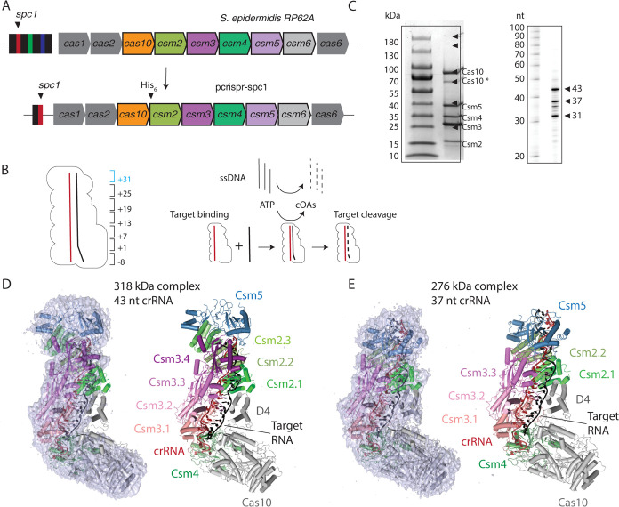
Fig 1 Overall architecture of two oligomeric states of S. epidermidis Cas10-Csm revealed by cryo-EM. (A) A schematic of the S. epidermidis Type III-A crispr locus. Cas10-Csm purification was facilitated by the construction of the plasmid pcrispr-spc1. (B) The numbering scheme describing positions within the crRNA-target duplex is shown. The six-nucleotide region beginning at +31 (blue) is present in the 43 nt crRNA complex but not the 37 nt crRNA complex. A schematic of steps in the interference reaction is shown. Target RNA binding activates single-stranded DNA (ssDNA) cleavage and cyclic oligoadenylates (cOAs) synthesis activities of Cas10-Csm. (C) SDS-PAGE and urea-PAGE analysis of purified Cas10-Csm. Cas10* denotes a band that has been identified by mass spectrometry as a truncated Cas10. CrRNA extracted from purified SeCas10-Csm possesses lengths of 31, 37 and 43 nt. (D) The larger complex consists of a 43 nt crRNA, three copies of Csm2 and four copies of Csm3. The cryo-EM density map is shown at left revealing that very little density is present for Cas10 domain 4 (D4) and Csm2.1. The map is contoured at level 3.0 σ. (E) The smaller complex consists of a 37 nt crRNA, two copies of Csm2 and three copies of Csm3. Density contoured at 3.0 σ is shown. In contrast to the larger complex, density is observed for Cas10 domain 4 and Csm2.1. A molecular model of the 276 kDa complex has been deposited with the PDB, code 8d06.
Fig 1 Overall architecture of two oligomeric states of S. epidermidis Cas10-Csm revealed by cryo-EM. (A) A schematic of the S. epidermidis Type III-A crispr locus. Cas10-Csm purification was facilitated by the construction of the plasmid pcrispr-spc1. (B) The numbering scheme describing positions within the crRNA-target duplex is shown. The six-nucleotide region beginning at +31 (blue) is present in the 43 nt crRNA complex but not the 37 nt crRNA complex. A schematic of steps in the interference reaction is shown. Target RNA binding activates single-stranded DNA (ssDNA) cleavage and cyclic oligoadenylates (cOAs) synthesis activities of Cas10-Csm. (C) SDS-PAGE and urea-PAGE analysis of purified Cas10-Csm. Cas10* denotes a band that has been identified by mass spectrometry as a truncated Cas10. CrRNA extracted from purified SeCas10-Csm possesses lengths of 31, 37 and 43 nt. (D) The larger complex consists of a 43 nt crRNA, three copies of Csm2 and four copies of Csm3. The cryo-EM density map is shown at left revealing that very little density is present for Cas10 domain 4 (D4) and Csm2.1. The map is contoured at level 3.0 σ. (E) The smaller complex consists of a 37 nt crRNA, two copies of Csm2 and three copies of Csm3. Density contoured at 3.0 σ is shown. In contrast to the larger complex, density is observed for Cas10 domain 4 and Csm2.1. A molecular model of the 276 kDa complex has been deposited with the PDB, code 8d06.CC BY · PubMed Central
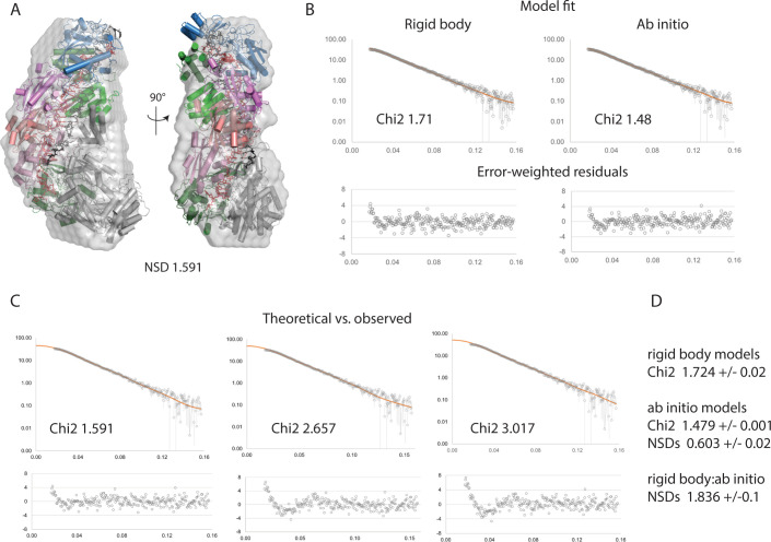
Fig 2 Support for Cas10-Csm complex stoichiometry from SEC-SAXS. (A) Multiple ab initio and rigid body models were calculated from the averaged peak frames of the SEC-SAXS run. Representative ab initio (gray envelope) and rigid body (cartoon) models are shown superposed (via SASREF). (B) The fits of the models from panel A (orange) to the experimental scattering curve (gray) are shown along with error-weighted residuals (lower panels). The rigid body model fit is shown at left, the ab initio model, at right. (C) EM structures reported here, overlaid with a representative ab initio model from our SAXS analysis. (D) CRYSOL was used to generate theoretical scattering curves from the EM models reported in this study, and these theoretical curves (orange) were scaled to the experimental SAXS data (gray). From left to right, the models shown are the 276 kDa complex, the 318 kDa complex, and the 318 kDa complex minus Csm2 subunits. As in B, error-weighted residuals are shown below each plot. Throughout this figure normalized spatial discrepancy is abbreviated NSD.
Fig 2 Support for Cas10-Csm complex stoichiometry from SEC-SAXS. (A) Multiple ab initio and rigid body models were calculated from the averaged peak frames of the SEC-SAXS run. Representative ab initio (gray envelope) and rigid body (cartoon) models are shown superposed (via SASREF). (B) The fits of the models from panel A (orange) to the experimental scattering curve (gray) are shown along with error-weighted residuals (lower panels). The rigid body model fit is shown at left, the ab initio model, at right. (C) EM structures reported here, overlaid with a representative ab initio model from our SAXS analysis. (D) CRYSOL was used to generate theoretical scattering curves from the EM models reported in this study, and these theoretical curves (orange) were scaled to the experimental SAXS data (gray). From left to right, the models shown are the 276 kDa complex, the 318 kDa complex, and the 318 kDa complex minus Csm2 subunits. As in B, error-weighted residuals are shown below each plot. Throughout this figure normalized spatial discrepancy is abbreviated NSD.CC BY · PubMed Central
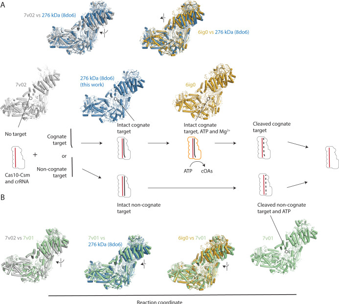
Fig 3 Placing the structure of the Cas10-Csm bound to intact, cognate target RNA on the reaction coordinate. (A) A diagram of the states of Cas10-Csm shows that it can exist unbound to target RNA, bound to cognate target RNA leading to the activation of cOA synthesis and bound to cleaved, cognate target RNA after the Csm3 mediated RNase reaction. Alternatively, Cas10-Csm can bind to non-cognate target RNA, which possesses base-pairing with crRNA in the 5’ tag-3’ flank region which leads to Csm3-mediated RNase activity but not cOA synthesis. Superpositions of PDB code 8do6, reported in this work, with an unbound S. epidermidis Cas10-Csm (7v02) via the Csm4 protein reveals target binding causes a rotation of Csm2 clockwise and toward Cas10. Superposition of 8do6 with S. thermophilus Cas10-Csm bound to cognate target RNA, two ATP and two Mg2+ reveals its Csm2 proteins are located slightly closer to Cas10 than 8do6. (B) A comparison of S. epidermidis Cas10-Csm unbound to target (7v02) and bound to a non-cognate target (7v01) displays clockwise and downward rotation of Csm2. Comparison of 7v01 with 8do6 or 6ig0 shows all three structures are similar, however, the Csm2 proteins of 8do6 are positioned slightly closer to 6ig0.
Fig 3 Placing the structure of the Cas10-Csm bound to intact, cognate target RNA on the reaction coordinate. (A) A diagram of the states of Cas10-Csm shows that it can exist unbound to target RNA, bound to cognate target RNA leading to the activation of cOA synthesis and bound to cleaved, cognate target RNA after the Csm3 mediated RNase reaction. Alternatively, Cas10-Csm can bind to non-cognate target RNA, which possesses base-pairing with crRNA in the 5’ tag-3’ flank region which leads to Csm3-mediated RNase activity but not cOA synthesis. Superpositions of PDB code 8do6, reported in this work, with an unbound S. epidermidis Cas10-Csm (7v02) via the Csm4 protein reveals target binding causes a rotation of Csm2 clockwise and toward Cas10. Superposition of 8do6 with S. thermophilus Cas10-Csm bound to cognate target RNA, two ATP and two Mg2+ reveals its Csm2 proteins are located slightly closer to Cas10 than 8do6. (B) A comparison of S. epidermidis Cas10-Csm unbound to target (7v02) and bound to a non-cognate target (7v01) displays clockwise and downward rotation of Csm2. Comparison of 7v01 with 8do6 or 6ig0 shows all three structures are similar, however, the Csm2 proteins of 8do6 are positioned slightly closer to 6ig0.CC BY · PubMed Central
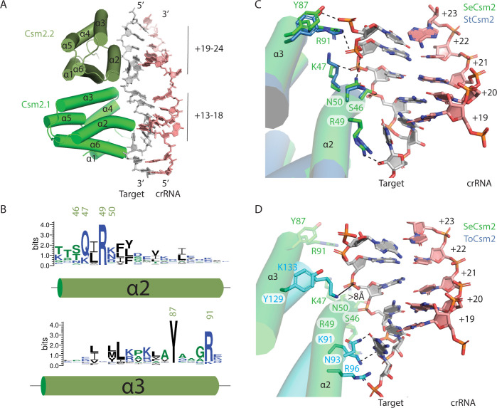
Fig 4 Conserved and idiosyncratic interactions of Csm2 with target RNA. (A) Helices α2 and α3 of S. epidermidis Csm2 (SeCsm2) form extensive interactions with target RNA in the major groove of the target-crRNA duplex. (B) A sequence logo depicting the conservation of α2 and α3 of Csm2. Residues of interest are numbered according to the SeCsm2 sequence. (C) A superposition of S. thermophilus Csm2 (StCsm2), given by PDB code 6ifu, with SeCsm2 was performed using the phosphate atoms of six nucleotides of target RNA (+19 to +24 positions). The superposition reveals that α2 and α3 in SeCsm2 and StCsm2 interact with target RNA in a similar manner. The target RNA-crRNA duplex depicted is from the SeCas10-Csm structure. (D) A superposition of T. onnurineus Csm2 (ToCsm2), given by PDB code 6MUS, with SeCsm2 was performed as in (C) which reveals that α2 and α3 in ToCsm2 are translated in space relative to SeCsm2, form fewer interactions with target RNA and these differ from the pattern seen in StCsm2 and SeCsm2. K91 of ToCsm2 forms a cation-pi interaction with the target-RNA nucleotide at position +18, analogous to the interaction of SeCsm2 R49 with the +18 position.
Fig 4 Conserved and idiosyncratic interactions of Csm2 with target RNA. (A) Helices α2 and α3 of S. epidermidis Csm2 (SeCsm2) form extensive interactions with target RNA in the major groove of the target-crRNA duplex. (B) A sequence logo depicting the conservation of α2 and α3 of Csm2. Residues of interest are numbered according to the SeCsm2 sequence. (C) A superposition of S. thermophilus Csm2 (StCsm2), given by PDB code 6ifu, with SeCsm2 was performed using the phosphate atoms of six nucleotides of target RNA (+19 to +24 positions). The superposition reveals that α2 and α3 in SeCsm2 and StCsm2 interact with target RNA in a similar manner. The target RNA-crRNA duplex depicted is from the SeCas10-Csm structure. (D) A superposition of T. onnurineus Csm2 (ToCsm2), given by PDB code 6MUS, with SeCsm2 was performed as in (C) which reveals that α2 and α3 in ToCsm2 are translated in space relative to SeCsm2, form fewer interactions with target RNA and these differ from the pattern seen in StCsm2 and SeCsm2. K91 of ToCsm2 forms a cation-pi interaction with the target-RNA nucleotide at position +18, analogous to the interaction of SeCsm2 R49 with the +18 position.CC BY · PubMed Central
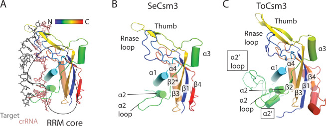
Fig 5 Variation in the secondary structure of Csm3. (A) The hydrophobic core of Csm3 is composed of an RNA recognition motif (RRM core). The secondary structure of Csm3 is colored by the position of each element on the chain from the N-terminus (blue) to the C-terminus (red). (B) The secondary structure of S. epidermidis Csm3 (SeCsm3) is shown with key functional components highlighted: The RNase loop contains Asp 32 which coordinates a metal to catalyze target RNA cleavage, α1 forms key interactions with crRNA, the α2 loop interacts with crRNA and lastly the thumb, composed of a β-hairpin protrudes through the crRNA-target RNA duplex interrupting it. β2* indicates that SeCsm3 adopts only β-strand like geometry at the position of β2 in the canonical RRM fold. (C) The secondary structure of T. onnurineus Csm3 (ToCsm3) differs from SeCsm3 because it contains additional elements of interest including the α2’ helix and the α2’ loop which make contacts to the target RNA that do not occur in SeCsm3. The structure depicted is taken from PDB code 6mus.
Fig 5 Variation in the secondary structure of Csm3. (A) The hydrophobic core of Csm3 is composed of an RNA recognition motif (RRM core). The secondary structure of Csm3 is colored by the position of each element on the chain from the N-terminus (blue) to the C-terminus (red). (B) The secondary structure of S. epidermidis Csm3 (SeCsm3) is shown with key functional components highlighted: The RNase loop contains Asp 32 which coordinates a metal to catalyze target RNA cleavage, α1 forms key interactions with crRNA, the α2 loop interacts with crRNA and lastly the thumb, composed of a β-hairpin protrudes through the crRNA-target RNA duplex interrupting it. β2* indicates that SeCsm3 adopts only β-strand like geometry at the position of β2 in the canonical RRM fold. (C) The secondary structure of T. onnurineus Csm3 (ToCsm3) differs from SeCsm3 because it contains additional elements of interest including the α2’ helix and the α2’ loop which make contacts to the target RNA that do not occur in SeCsm3. The structure depicted is taken from PDB code 6mus.CC BY · PubMed Central
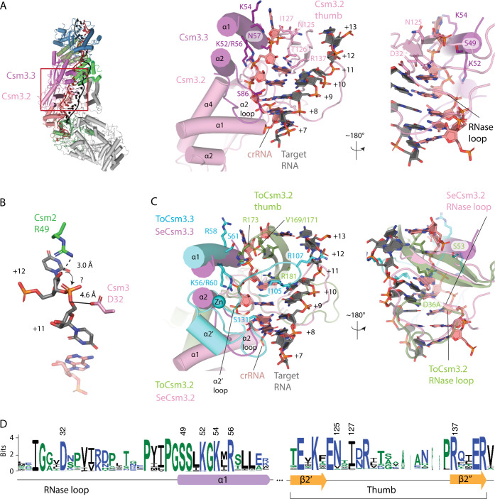
Fig 6 S. epidermidis Csm3 only contacts crRNA, however, T. onnurineus Csm3 contacts both crRNA and target RNA, utilizing the α2’ loop. (A) A red rectangle marks the area of Cas10-Csm which is magnified to the right. The detailed interactions of S. epidermidis Csm3 with crRNA are shown. A basic patch in α-helix 1 interacts with the terminal paired and the unpaired crRNA nucleotides of the repeating hexa-nucleotide structure. The S86 residue in the α2 loop hydrogen bonds to the minor groove of the duplex and residues N125-I127 of the Csm3.2 thumb form van der Waal’s interactions and hydrogen bonds with crRNA. An extensive loop structure that follows β-strand 1 of Csm3 and sits on the major groove of the crRNA-target duplex, is termed the RNase loop because it contains the catalytically essential residue D32. (B) Csm2 R49 and Csm3 D32 are positioned to catalyze target RNA cleavage but the mechanism of cleavage, in-line attack (arrow) or hydrolysis is unclear. (C) A superposition of T. onnurineus Csm3 (ToCsm3), given by PDB code 6mus, is shown revealing similar interactions of α-helix 1 and the α2 loop with crRNA in the two structures. However, ToCsm3 uses the α2’ loop, a structure not present in SeCsm3, to form hydrogen bonds (I105) and electrostatic interactions (R107) to target RNA. Additionally, the ToCsm3 thumb interacts differently with crRNA utilizing an electrostatic interaction via R173 that is absent in SeCsm3. The crRNA-target duplex shown is from PDB code 6mus. (D) A sequence logo depicting the conservation of Csm3 residues of interest is given. Numbering of the logo is according to the SeCsm3 sequence.
Fig 6 S. epidermidis Csm3 only contacts crRNA, however, T. onnurineus Csm3 contacts both crRNA and target RNA, utilizing the α2’ loop. (A) A red rectangle marks the area of Cas10-Csm which is magnified to the right. The detailed interactions of S. epidermidis Csm3 with crRNA are shown. A basic patch in α-helix 1 interacts with the terminal paired and the unpaired crRNA nucleotides of the repeating hexa-nucleotide structure. The S86 residue in the α2 loop hydrogen bonds to the minor groove of the duplex and residues N125-I127 of the Csm3.2 thumb form van der Waal’s interactions and hydrogen bonds with crRNA. An extensive loop structure that follows β-strand 1 of Csm3 and sits on the major groove of the crRNA-target duplex, is termed the RNase loop because it contains the catalytically essential residue D32. (B) Csm2 R49 and Csm3 D32 are positioned to catalyze target RNA cleavage but the mechanism of cleavage, in-line attack (arrow) or hydrolysis is unclear. (C) A superposition of T. onnurineus Csm3 (ToCsm3), given by PDB code 6mus, is shown revealing similar interactions of α-helix 1 and the α2 loop with crRNA in the two structures. However, ToCsm3 uses the α2’ loop, a structure not present in SeCsm3, to form hydrogen bonds (I105) and electrostatic interactions (R107) to target RNA. Additionally, the ToCsm3 thumb interacts differently with crRNA utilizing an electrostatic interaction via R173 that is absent in SeCsm3. The crRNA-target duplex shown is from PDB code 6mus. (D) A sequence logo depicting the conservation of Csm3 residues of interest is given. Numbering of the logo is according to the SeCsm3 sequence.CC BY · PubMed Central
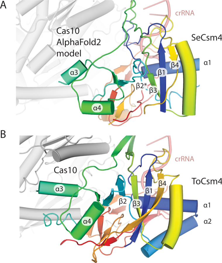
Fig 7 Csm4 possesses similar secondary structure elements in T. onnurineus and S. epidermidis. (A) The RNA recognition motif core of the Cas5 family member, SeCsm4, is elaborated to contact the 5’ tag of crRNA and Cas10. An AlphaFold2 model of SeCas10 is docked into the density present in S. epidermidis cryo-EM maps. (B) The secondary structure elements of ToCsm4 are similar to SeCsm4 but α3 has more extensive interactions with Cas10.
Fig 7 Csm4 possesses similar secondary structure elements in T. onnurineus and S. epidermidis. (A) The RNA recognition motif core of the Cas5 family member, SeCsm4, is elaborated to contact the 5’ tag of crRNA and Cas10. An AlphaFold2 model of SeCas10 is docked into the density present in S. epidermidis cryo-EM maps. (B) The secondary structure elements of ToCsm4 are similar to SeCsm4 but α3 has more extensive interactions with Cas10.CC BY · PubMed Central
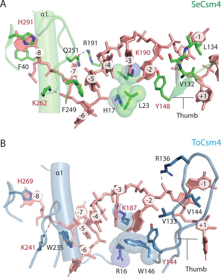
Fig 8 A detailed comparison of the interactions of SeCsm4 and ToCsm4 with crRNA. (A) SeCsm4 residues forming interactions with the crRNA 5’-tag that are likely critical for recognition shown as sticks. Three residues that form van der Waals interaction with crRNA and sterically position it are shown as spheres. Residues that are conserved between SeCsm4 and ToCsm4 are labeled in red. The thumb, β-hairpin-like element, that interrupts the co-axial nucleotide stack is labeled. The first α-helix of Csm4 is marked as α1. (B) ToCsm4 residues forming critical interactions with crRNA are shown as sticks or spheres. The β-hairpin-like element thumb is labeled as is α-helix 1, α1.
Fig 8 A detailed comparison of the interactions of SeCsm4 and ToCsm4 with crRNA. (A) SeCsm4 residues forming interactions with the crRNA 5’-tag that are likely critical for recognition shown as sticks. Three residues that form van der Waals interaction with crRNA and sterically position it are shown as spheres. Residues that are conserved between SeCsm4 and ToCsm4 are labeled in red. The thumb, β-hairpin-like element, that interrupts the co-axial nucleotide stack is labeled. The first α-helix of Csm4 is marked as α1. (B) ToCsm4 residues forming critical interactions with crRNA are shown as sticks or spheres. The β-hairpin-like element thumb is labeled as is α-helix 1, α1.CC BY · PubMed Central
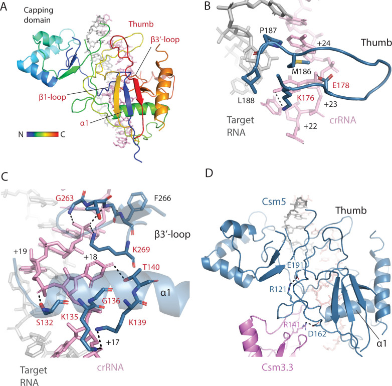
Fig 9 Most RNA-interacting regions of Csm5 are conserved and an electrostatic interaction may allow cross-talk between two regions. (A) SeCsm5 bound to the crRNA-target duplex is shown emphasizing regions that contact the duplex. Four of the five regions are conserved (red) across diverse Csm5 homologs. The secondary structure elements of Csm5 (β1, α1 and β3’) are named according to their position in the core, RRM domain—capping domain elements were bypassed. (B) The SeCsm5 Thumb region interacts with the major groove of the duplex by hydrogen-bonding and van der Waals interactions. Residues conserved across SeCsm5 homologs are colored red. (C) Helix-α1 and the β3’-loop interact with the duplex surrounding the flipped +18 nucleotide. (D) Electrostatic interactions between R121 and E191 may conformationally couple the helix-α1 and thumb components of SeCsm5. The interaction of SeCsm5 D162 with Csm3 R141 has been shown to be critical for crRNA maturation.
Fig 9 Most RNA-interacting regions of Csm5 are conserved and an electrostatic interaction may allow cross-talk between two regions. (A) SeCsm5 bound to the crRNA-target duplex is shown emphasizing regions that contact the duplex. Four of the five regions are conserved (red) across diverse Csm5 homologs. The secondary structure elements of Csm5 (β1, α1 and β3’) are named according to their position in the core, RRM domain—capping domain elements were bypassed. (B) The SeCsm5 Thumb region interacts with the major groove of the duplex by hydrogen-bonding and van der Waals interactions. Residues conserved across SeCsm5 homologs are colored red. (C) Helix-α1 and the β3’-loop interact with the duplex surrounding the flipped +18 nucleotide. (D) Electrostatic interactions between R121 and E191 may conformationally couple the helix-α1 and thumb components of SeCsm5. The interaction of SeCsm5 D162 with Csm3 R141 has been shown to be critical for crRNA maturation.CC BY · PubMed Central
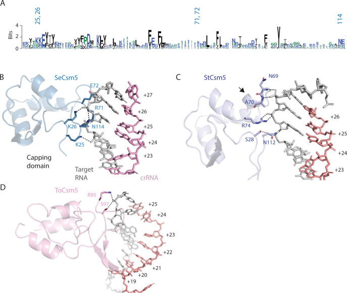
Fig 10 The capping domain of Csm5 diverges among Csm5 proteins. (A) A sequence logo of the capping domain with the positions of SeCsm5 residues that interact with target RNA numbered. The logo indicates there is limited sequence conservation within the capping domain. (B) Electrostatic and hydrogen bonding interactions between SeCsm5 and target RNA are indicated by dashed lines. (C) The interactions of the StCsm5 capping domain with target RNA are indicated. StCsm5 contains a helical segment, indicated by an arrow, absent in the other Csm5 structures. (D) Only two residues in the capping domain of the ToCsm5 structure reported in PDB code 6mus contact target.
Fig 10 The capping domain of Csm5 diverges among Csm5 proteins. (A) A sequence logo of the capping domain with the positions of SeCsm5 residues that interact with target RNA numbered. The logo indicates there is limited sequence conservation within the capping domain. (B) Electrostatic and hydrogen bonding interactions between SeCsm5 and target RNA are indicated by dashed lines. (C) The interactions of the StCsm5 capping domain with target RNA are indicated. StCsm5 contains a helical segment, indicated by an arrow, absent in the other Csm5 structures. (D) Only two residues in the capping domain of the ToCsm5 structure reported in PDB code 6mus contact target.CC BY · PubMed Central

Verification

Claims the paper makes

The claims and statistics of this paper haven’t been checked yet.

Run the check — free, no account needed. It reads the open-access text and checks the sources it cites.

A few free checks a day · papers already verified open instantly · results are public

Self-check

Percentages, sums and reported differences re-computed from the paper’s own numbers.

The self-check needs the paper’s full text.

Citations

Cites

Research works printed but not linked: 54

  1. 10.1016/j.cell.2015.12.035 · DOI · Paper page
  2. 10.1146/annurev-genet-022120-112523 · DOI
  3. 10.1038/s41579-019-0299-x · DOI · Paper page
  4. 10.1371/journal.pbio.3001481 · DOI
  5. 10.1038/nrmicro3569 · DOI · Paper page
  6. 10.1126/science.aao0100 · DOI · Paper page
  7. 10.1038/nature23467 · DOI · Paper page
  8. 10.1016/j.molcel.2014.09.027 · DOI
  9. 10.1016/j.molcel.2014.10.005 · DOI
  10. 10.1101/gad.273722.115 · DOI
  11. 10.1016/j.xcrm.2021.100319 · DOI
  12. 10.1038/s41467-021-25977-7 · DOI
  13. 10.1038/s41467-021-25337-5 · DOI
  14. 10.1093/nar/gkab1190 · DOI
  15. 10.1016/j.molcel.2013.09.013 · DOI
  16. 10.1016/j.molcel.2013.09.008 · DOI
  17. 10.1126/science.aaa4535 · DOI
  18. 10.1016/j.molcel.2015.03.018 · DOI · Paper page
  19. 10.1016/j.molcel.2020.07.008 · DOI
  20. 10.1016/j.cell.2018.10.052 · DOI
  21. 10.1038/s42003-022-03187-1 · DOI
  22. 10.1016/j.molcel.2018.11.007 · DOI
  23. 10.1038/s41422-019-0151-x · DOI
  24. 10.1016/j.str.2022.05.013 · DOI
  25. 10.1126/science.1165771 · DOI · Paper page
  26. 10.1038/nature08703 · DOI · Paper page
  27. 10.1074/jbc.M113.499244 · DOI
  28. 10.21769/BioProtoc.2353 · DOI
  29. 10.1261/rna.070417.119 · DOI
  30. 10.1038/nmeth.4169 · DOI · Paper page
  31. 10.1038/s41592-019-0575-8 · DOI · Paper page
  32. 10.1016/j.jsb.2021.107702 · DOI · Paper page
  33. 10.1038/s41592-020-00990-8 · DOI
  34. 10.1107/S2059798318006551 · DOI · Paper page
  35. 10.1107/S0907444910007493 · DOI · Paper page
  36. 10.1038/s41592-022-01488-1 · DOI · Paper page
  37. 10.1002/pro.3330 · DOI · Paper page
  38. 10.1107/S0021889803012779 · DOI
  39. 10.1006/pmed.1996.0081 · DOI
  40. 10.1016/S0006-3495(99)77121-3 · DOI
  41. 10.1529/biophysj.105.064154 · DOI
  42. 10.1107/S1600576717007786 · DOI
  43. 10.1107/S0021889800014126 · DOI · Paper page
  44. 10.1101/gr.849004 · DOI · Paper page
  45. 10.1093/nar/gkaa913 · DOI · Paper page
  46. 10.1016/j.molcel.2018.11.008 · DOI
  47. 10.1126/science.1256328 · DOI
  48. 10.1074/jbc.RA119.008728 · DOI
  49. 10.1093/nar/gkw891 · DOI
  50. Chou-Zheng L , Hatoum-Aslan A. Critical roles for ‘housekeeping’ nucleases in Type III CRISPR-Cas immunity. Biorxiv. 2022. Epub July 19, 2022. doi: https://doi.org/10.1101/2022.07.18.500432. · DOI
  51. 10.7554/eLife.45393 · DOI
  52. 10.1016/j.celrep.2019.02.029 · DOI
  53. 10.1128/JB.01130-13 · DOI
  54. 10.1016/j.cell.2015.04.027 · DOI · Paper page

Metadata

Authors
Paraan, M., Nasef, M., Chou-Zheng, L., Khweis, S. A., Schoeffler, A. J., Hatoum-Aslan, A., Stagg, S. M., & Dunkle, J. A.
Affiliations
  • New York Structural Biology Center (US)
  • University of Alabama (US)
  • University of Illinois Urbana-Champaign (US)
  • Loyola University New Orleans (US)
  • Florida State University (US)
Corresponding author
  • Stagg, Scott M.
  • Dunkle, Jack A.
Venue
PLoS ONE
Publisher
openRxiv
Published
2022-11-04
Volume
18
Issue
6
Pages
e0287461-e0287461
ISSN
1932-6203 (ISSN-L)
OpenAlex
W4381839396
Cited by
10
References
1
Citations by year
2026: 3 · 2025: 3 · 2024: 3 · 2023: 1
FWCI
0.89
Type
journal article
Language
en
Open access
Licence
cc-by
Locations
  • PLoS ONE (publishedVersion, cc-by)
  • bioRxiv (Cold Spring Harbor Laboratory) (acceptedVersion, cc-by)
  • PubMed Central (publishedVersion, cc-by)
  • PubMed (publishedVersion)
  • DOAJ (DOAJ: Directory of Open Access Journals) (submittedVersion, cc-by-sa)
Funders
  • U.S. Department of Energy (S10OD018483, P30 GM124169, GM124169)
  • Simons Foundation (SF 349247)
  • National Institutes of Health (SF349247, 1s10od018483-01, GM 103310, GM124169)
  • National Institute of General Medical Sciences (P41GM103310, GM139171, SF349247, GM124169, GM142966, GM103310, S10OD018483, P30GM124169, U24GM139171, R35GM142966)
  • Basic Energy Sciences (S10OD018483)
  • Biological and Environmental Research (P30 GM124169, S10OD018483)
  • Lawrence Berkeley National Laboratory (S10OD018483)
Topics
  • Life Sciences › Biochemistry, Genetics and Molecular Biology › Molecular Biology › CRISPR and Genetic Engineering
  • Life Sciences › Biochemistry, Genetics and Molecular Biology › Genetics › Bacterial Genetics and Biotechnology
  • Life Sciences › Biochemistry, Genetics and Molecular Biology › Molecular Biology › Biochemical and Structural Characterization
Keywords
crRNA, Staphylococcus epidermidis, cryo-EM structure, oligomeric state, small-angle X-ray scattering, structural comparison, RNA interference, genome editing
MeSH terms
RNA, Guide, CRISPR-Cas Systems, RNA, Bacterial, Multiprotein Complexes, CRISPR-Cas Systems, CRISPR-Associated Proteins

About these checks

Registry, retraction, Cites and statistics results are computed, not judged, with one exception: a printed reference that no index lists is read once by a model before it counts as a citation error. Claims are extracted by a model and kept only when their quote appears verbatim in the text; each cited source is then checked, with a model’s help, for whether it says what the claim relies on. Those verdicts can be wrong — read the claim’s quote and the cited source before relying on one.

Statistics and arithmetic are recomputed, not judged.

Confidence bands are shown only for fields calibrated on our annotated test set. None is calibrated yet, so no band is shown.

What counts as a claim ›How we measure confidence ›How we check this ›

Are you an author, or do you see a problem with this page? Request a review

Data sources

Bibliographic data: Semantic Scholar, OpenAlex, Crossref.

Contains information from the Semantic Scholar Academic Graph Datasets, made available under the ODC Attribution License (ODC-BY 1.0).

Semantic Scholar’s data is described in: Kinney, R., et al. (2023). The Semantic Scholar Open Data Platform. arXiv:2301.10140.