PreprintCited by 0CRISPR and Genetic EngineeringBacterial Genetics and BiotechnologyBiochemical and Structural Characterization

The DOI matches its Crossref record; 2 of the 2 references it cites were found in a registry or an index.

Branched DNA Processing by a Thermostable CAS-Cas4 from Thermococcus onnurineus: Expanding Biochemical Landscape of Nuclease Activity

Open access
Jain, Muskan, Pattnayak, Asish Kumar, Aggarwal, Sakshi, Rai, Praveen, J, Kavya, Chandrayan, Sanjeev, Goel, Manisha, Gaur, Vineet·2024·Journal of Biological Chemistry

Preprint — published as 10.1016/j.jbc.2025.110701

Abstract

The adaptive immune function of CRISPR-Cas systems in bacteria and archaea is mediated through CRISPR-Associated Proteins (Cas). The adaptation module, typically involving Cas1, Cas2, and Cas4, helps integrate viral spacer sequences into the host genome. Cas4 proteins are classified into two types based on neighboring genes: CAS-Cas4, flanked by other cas genes, and Solo-Cas4, which exist independently. While CAS-Cas4 proteins are implicated in adaptation, they remain biochemically uncharacterized in archaea, unlike archaeal Solo-Cas4 proteins. This study biochemically characterizes TON_0321, a CAS-Cas4 protein from the Type IV-C CRISPR cassette of Thermococcus onnurineus. TON_0321 exhibits 5' to 3' exonuclease activity and unique structure-dependent endonuclease activity, shedding light on CAS-Cas4 functional diversity. A distinct spatial organization of the catalytic site, angled with the positively charged patch on the protein surface, enables TON_0321 to recognize branching points in DNA substrates. Furthermore, this spatial arrangement facilitates cleavage 2 to 3 nucleotides away from the branch point in the 5' direction, demonstrating structure-specific endonuclease activity.

Overview

The claims and statistics of this paper haven’t been checked yet.

Run the check — free, no account needed. It reads the open-access text and checks the sources it cites.

A few free checks a day · papers already verified open instantly · results are public

Integrity signals

Registry

Crossref · 10.1101/2024.11.29.625776

Cites

2 of 2 references verified0 citation errors · 0 not checkable

Statistics

Figures

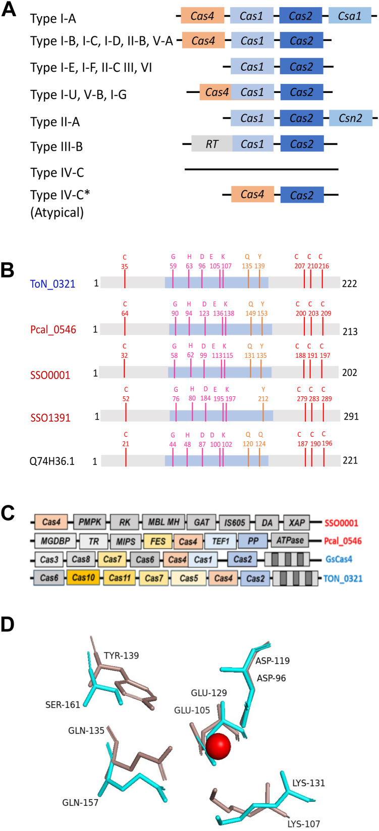
Figure 1 Diversity among Cas4 proteins.A, block diagram showing the typical organization of adaptation module in different CRISPR subtypes. Adaptation module usually consists of Cas1, Cas2, and Cas4 proteins. It may contain other proteins in specific subtypes. A few of the CRISPR subtypes does not include Cas4. The typical type IV-C CRISPR subtype does not contain adaptation module. The atypical type IV-C CRISPR cassette from Thermococcus onnurineus NA1 shows the presence of Cas2 and Cas4 adaptation proteins. Type I-A, I-B, III-B: different CRISPR subtypes; type IV-C: typical type IV-C cassette; type IV-C∗: atypical case of T. onnurineus NA1. B, a cartoon depicting conserved amino acid residues among various characterized Cas4 proteins: TON_0321 (T. onnurineus NA1), Pcal_0546 (Pyrobaculum calidifontis), SSO0001 (Sulfolobus solfataricus), SSO1391 (S. solfataricus), and GsCas4 (Geobacter sulfurreducens). Four cysteines involved in chelating iron–sulfur cluster are shown in red. RecB active site residues are shown in pink and orange; the blue-shaded region depicts the RecB-like domain of Cas4 proteins. C, a schematic showing the difference in gene cassettes of SSO0001 from S. solfataricus, Pcal_0546 from P. calidifontis, GsCas4 from G. sulfurreducens, and TON_0321 from T. onnurineus NA1. D, comparison of the catalytic sites of λ exonuclease (cyan; Protein Data Bank code: 1AVQ) and TON_0321 (red, model) depicting the conservation of active site residues. The metal ion at the active site is shown as a sphere. RecB, recombination protein B.
Figure 1 Diversity among Cas4 proteins.A, block diagram showing the typical organization of adaptation module in different CRISPR subtypes. Adaptation module usually consists of Cas1, Cas2, and Cas4 proteins. It may contain other proteins in specific subtypes. A few of the CRISPR subtypes does not include Cas4. The typical type IV-C CRISPR subtype does not contain adaptation module. The atypical type IV-C CRISPR cassette from Thermococcus onnurineus NA1 shows the presence of Cas2 and Cas4 adaptation proteins. Type I-A, I-B, III-B: different CRISPR subtypes; type IV-C: typical type IV-C cassette; type IV-C∗: atypical case of T. onnurineus NA1. B, a cartoon depicting conserved amino acid residues among various characterized Cas4 proteins: TON_0321 (T. onnurineus NA1), Pcal_0546 (Pyrobaculum calidifontis), SSO0001 (Sulfolobus solfataricus), SSO1391 (S. solfataricus), and GsCas4 (Geobacter sulfurreducens). Four cysteines involved in chelating iron–sulfur cluster are shown in red. RecB active site residues are shown in pink and orange; the blue-shaded region depicts the RecB-like domain of Cas4 proteins. C, a schematic showing the difference in gene cassettes of SSO0001 from S. solfataricus, Pcal_0546 from P. calidifontis, GsCas4 from G. sulfurreducens, and TON_0321 from T. onnurineus NA1. D, comparison of the catalytic sites of λ exonuclease (cyan; Protein Data Bank code: 1AVQ) and TON_0321 (red, model) depicting the conservation of active site residues. The metal ion at the active site is shown as a sphere. RecB, recombination protein B.CC BY · PubMed Central
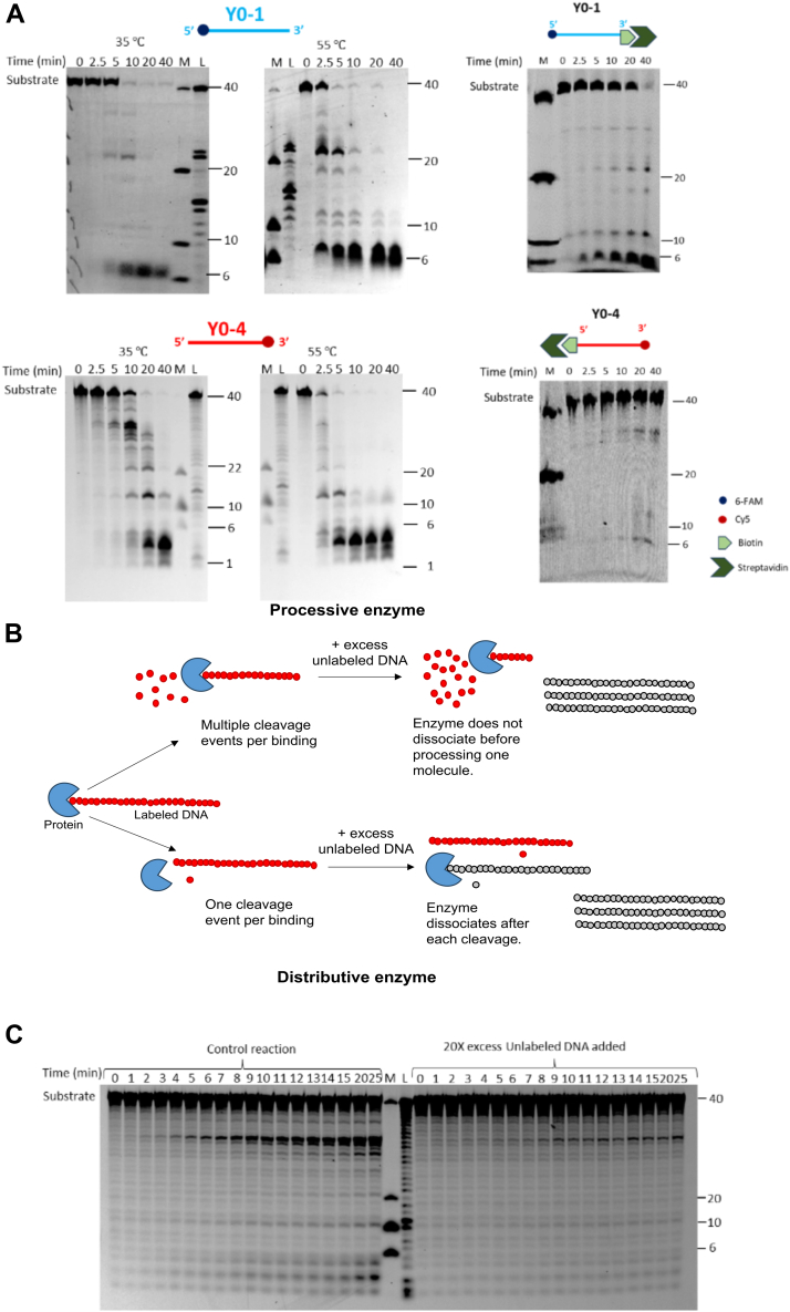
Figure 2 Exonuclease activity of TON_0321 on ssDNA.A, the catalytic activity of TON_0321 on ssDNA labeled at 5′-end with 6-FAM (upper panel) (Y0-1) and ssDNA labeled with Cy5 at 3′-end (lower panel) (Y0-2). The reaction products were resolved on an 18% TBE–urea PAGE. Upper panel gels were scanned for 6-FAM signal, and the lower panel gels were scanned for Cy5 signal. The activity of TON_0321 on ssDNA labeled at 5′ end with 6-FAM, with biotin at 3′ end (Y0-1_biotin), and ssDNA labeled with Cy5 at 3′ end, and with biotin at 5′ end (Y0-4_biotin). The biotinylated oligos were incubated with 2.5 times molar excess of streptavidin to completely block the biotinylated end of the oligo. The biotin–streptavidin-conjugated oligos were used as substrates for activity assay. The reaction products were resolved on an 18% TBE–urea PAGE. Gels were scanned for 6-FAM and Cy5 signals. M represents a marker made from mixing synthetic oligonucleotides of different sizes (40, 20, 10, and 6 nucleotides), and L represents a ladder made from DNase digestion of the 40-mer substrate. All experiments were done in triplicates (Figs. S8, S9). For experiments with ssDNA labeled with Cy5 at 3′end (lower panel), samples for 35 °C and 55 °C were run on the same gel with a common ladder and marker. B, a schematic illustrating the processing of DNA substrate by processive and distributive enzymes. Processive enzymes perform multiple cleavage cycles on a single-bound substrate before releasing it, preventing its binding to a new, unlabeled substrate introduced midreaction. Distributive enzymes catalyze a single cleavage per substrate binding, detaching afterward and becoming available to interact with subsequently added substrate molecules. C, TON_0321 acts in a distributive manner. An activity assay reaction was set with 3′ Cy5-labeled ssDNA, and formation of products was observed over different time points. In a parallel reaction, 20-fold excess unlabeled DNA of the same sequence was added after 2 min of time point. A decrease in the cleavage of the labeled product in comparison to the control reaction indicated that TON_0321 can dissociate from its substrate and catalyze the reaction in a distributive manner. The reaction products were resolved on an 18% TBE–urea PAGE. The gels were scanned for Cy5 signal. M represents a marker made from mixing synthetic oligonucleotides of different sizes (40, 20, 10, and 6 nucleotides), and L represents a ladder made from DNase digestion of the 40 -er substrate. All experiments were done in triplicates (Fig. S11). Cy5, cyanine-5 fluorophore; 6-FAM, 6-carboxyfluorescein; TBE, Tris–borate–EDTA.
Figure 2 Exonuclease activity of TON_0321 on ssDNA.A, the catalytic activity of TON_0321 on ssDNA labeled at 5′-end with 6-FAM (upper panel) (Y0-1) and ssDNA labeled with Cy5 at 3′-end (lower panel) (Y0-2). The reaction products were resolved on an 18% TBE–urea PAGE. Upper panel gels were scanned for 6-FAM signal, and the lower panel gels were scanned for Cy5 signal. The activity of TON_0321 on ssDNA labeled at 5′ end with 6-FAM, with biotin at 3′ end (Y0-1_biotin), and ssDNA labeled with Cy5 at 3′ end, and with biotin at 5′ end (Y0-4_biotin). The biotinylated oligos were incubated with 2.5 times molar excess of streptavidin to completely block the biotinylated end of the oligo. The biotin–streptavidin-conjugated oligos were used as substrates for activity assay. The reaction products were resolved on an 18% TBE–urea PAGE. Gels were scanned for 6-FAM and Cy5 signals. M represents a marker made from mixing synthetic oligonucleotides of different sizes (40, 20, 10, and 6 nucleotides), and L represents a ladder made from DNase digestion of the 40-mer substrate. All experiments were done in triplicates (Figs. S8, S9). For experiments with ssDNA labeled with Cy5 at 3′end (lower panel), samples for 35 °C and 55 °C were run on the same gel with a common ladder and marker. B, a schematic illustrating the processing of DNA substrate by processive and distributive enzymes. Processive enzymes perform multiple cleavage cycles on a single-bound substrate before releasing it, preventing its binding to a new, unlabeled substrate introduced midreaction. Distributive enzymes catalyze a single cleavage per substrate binding, detaching afterward and becoming available to interact with subsequently added substrate molecules. C, TON_0321 acts in a distributive manner. An activity assay reaction was set with 3′ Cy5-labeled ssDNA, and formation of products was observed over different time points. In a parallel reaction, 20-fold excess unlabeled DNA of the same sequence was added after 2 min of time point. A decrease in the cleavage of the labeled product in comparison to the control reaction indicated that TON_0321 can dissociate from its substrate and catalyze the reaction in a distributive manner. The reaction products were resolved on an 18% TBE–urea PAGE. The gels were scanned for Cy5 signal. M represents a marker made from mixing synthetic oligonucleotides of different sizes (40, 20, 10, and 6 nucleotides), and L represents a ladder made from DNase digestion of the 40 -er substrate. All experiments were done in triplicates (Fig. S11). Cy5, cyanine-5 fluorophore; 6-FAM, 6-carboxyfluorescein; TBE, Tris–borate–EDTA.CC BY · PubMed Central
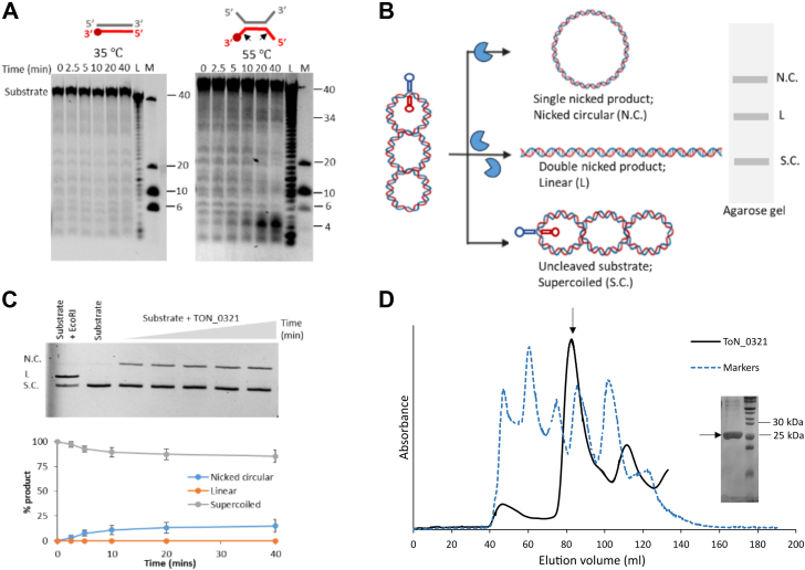
Figure 3 Endonuclease activity of TON_0321.A, catalytic activity of TON_0321 protein on dsDNA with one strand labeled with Cy5 at the 3′-end at two different temperatures, 35 °C and 55 °C. The reaction products were resolved on an 18% TBE–urea PAGE. The gels were scanned for Cy5 signal. M represents a marker made from mixing synthetic oligonucleotides of different sizes (40, 10, and 6 nucleotides), and L represents a ladder made from DNase digestion of the 40-mer substrate. All experiments were done in triplicates (Fig. S12). The major cleavage site in the schematic of the DNA substrate is marked by a solid arrow. B, a schematic diagram showing the cruciform assay using the double-stranded plasmid pIRbke8mut containing an inverted repeat sequence that forms a cruciform-type extrusion. The cruciform-like structure on the plasmid contains two sites for EcoRI restriction endonuclease (marked by asterisk). The purified plasmid is in a supercoiled (S.C.) state, which on single nick forms nicked circular (N.C.) DNA, and two simultaneous nicks produce a linear (L) form of DNA. C, ethidium bromide stained 0.8% agarose gel showing results of cruciform assay carried out with TON_0321 protein. The lane with EcoRI represents positive control and with substrate alone represents negative control. All experiments were done in triplicates (Fig. S14). The graph in the lower panel shows the quantitation of different forms of DNA formed in the cruciform assay along with standard errors calculated from three independent experiments. The reaction was carried out for 40 min. Aliquots were taken out at different time points (0, 2.5, 5, 10, 20, and 40 min). D, size-exclusion chromatogram of TON_0321 (black) purified on a HiLoad 16/600 Superdex 200 pg gel filtration column along with the gel filtration markers (broken blue) (vitamin B12, myoglobin, ovalbumin, gamma globulin, and thyroglobulin). The peak corresponding to the TON_0321 protein is marked by an arrow. The fractions from this peak were run on a 12% SDS gel. The molecular weight of TON_0321 protein was calculated using a standard curve of markers (Fig. S17C). Cy5, cyanine-5 fluorophore; TBE, Tris–borate–EDTA.
Figure 3 Endonuclease activity of TON_0321.A, catalytic activity of TON_0321 protein on dsDNA with one strand labeled with Cy5 at the 3′-end at two different temperatures, 35 °C and 55 °C. The reaction products were resolved on an 18% TBE–urea PAGE. The gels were scanned for Cy5 signal. M represents a marker made from mixing synthetic oligonucleotides of different sizes (40, 10, and 6 nucleotides), and L represents a ladder made from DNase digestion of the 40-mer substrate. All experiments were done in triplicates (Fig. S12). The major cleavage site in the schematic of the DNA substrate is marked by a solid arrow. B, a schematic diagram showing the cruciform assay using the double-stranded plasmid pIRbke8mut containing an inverted repeat sequence that forms a cruciform-type extrusion. The cruciform-like structure on the plasmid contains two sites for EcoRI restriction endonuclease (marked by asterisk). The purified plasmid is in a supercoiled (S.C.) state, which on single nick forms nicked circular (N.C.) DNA, and two simultaneous nicks produce a linear (L) form of DNA. C, ethidium bromide stained 0.8% agarose gel showing results of cruciform assay carried out with TON_0321 protein. The lane with EcoRI represents positive control and with substrate alone represents negative control. All experiments were done in triplicates (Fig. S14). The graph in the lower panel shows the quantitation of different forms of DNA formed in the cruciform assay along with standard errors calculated from three independent experiments. The reaction was carried out for 40 min. Aliquots were taken out at different time points (0, 2.5, 5, 10, 20, and 40 min). D, size-exclusion chromatogram of TON_0321 (black) purified on a HiLoad 16/600 Superdex 200 pg gel filtration column along with the gel filtration markers (broken blue) (vitamin B12, myoglobin, ovalbumin, gamma globulin, and thyroglobulin). The peak corresponding to the TON_0321 protein is marked by an arrow. The fractions from this peak were run on a 12% SDS gel. The molecular weight of TON_0321 protein was calculated using a standard curve of markers (Fig. S17C). Cy5, cyanine-5 fluorophore; TBE, Tris–borate–EDTA.CC BY · PubMed Central
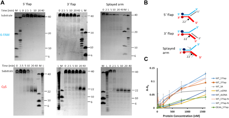
Figure 4 Endonuclease activity of TON_0321 on branched DNA substrates.A, the catalytic activity of TON_0321 protein on branched DNA molecules: 5′ flap, 3′ flap, and splayed arm. Each substrate has two labels: one strand labeled at 5′-end with 6-FAM and another strand labeled at 3′-end with Cy5. The reaction products were resolved on an 18% TBE–urea PAGE. The same gel was scanned for 6-FAM signal (upper panel) and for the Cy5 signal (lower panel). M represents a marker made from mixing synthetic oligonucleotides of different sizes (40, 10, and 6 nucleotides), and L represents a ladder made from DNase digestion of the 40-mer substrate. All experiments were done in triplicates (Fig. S19). B, the schematic of various branched DNA substrates shows the major cleavage sites as solid arrows and the minor sites as broken arrows. C, binding study of TON_0321 with different DNA substrates: ssDNA (light blue), dsDNA (orange), 5′ flap (gray), 3′ flap (yellow), and splayed arm (dark blue) using fluorescence anisotropy. DEAA_5′ flap represents binding analysis of catalytically dead TON_0321D96A/E105A in the presence of a 5′ flap substrate. 6-FAM-labeled substrates were used for anisotropy experiments. The Y-axis shows a change in anisotropy (A–A0), where A is observed anisotropy and A0 is anisotropy of DNA substrate alone. Cy5, cyanine-5 fluorophore; 6-FAM, 6-carboxyfluorescein; TBE, Tris–borate–EDTA.
Figure 4 Endonuclease activity of TON_0321 on branched DNA substrates.A, the catalytic activity of TON_0321 protein on branched DNA molecules: 5′ flap, 3′ flap, and splayed arm. Each substrate has two labels: one strand labeled at 5′-end with 6-FAM and another strand labeled at 3′-end with Cy5. The reaction products were resolved on an 18% TBE–urea PAGE. The same gel was scanned for 6-FAM signal (upper panel) and for the Cy5 signal (lower panel). M represents a marker made from mixing synthetic oligonucleotides of different sizes (40, 10, and 6 nucleotides), and L represents a ladder made from DNase digestion of the 40-mer substrate. All experiments were done in triplicates (Fig. S19). B, the schematic of various branched DNA substrates shows the major cleavage sites as solid arrows and the minor sites as broken arrows. C, binding study of TON_0321 with different DNA substrates: ssDNA (light blue), dsDNA (orange), 5′ flap (gray), 3′ flap (yellow), and splayed arm (dark blue) using fluorescence anisotropy. DEAA_5′ flap represents binding analysis of catalytically dead TON_0321D96A/E105A in the presence of a 5′ flap substrate. 6-FAM-labeled substrates were used for anisotropy experiments. The Y-axis shows a change in anisotropy (A–A0), where A is observed anisotropy and A0 is anisotropy of DNA substrate alone. Cy5, cyanine-5 fluorophore; 6-FAM, 6-carboxyfluorescein; TBE, Tris–borate–EDTA.CC BY · PubMed Central
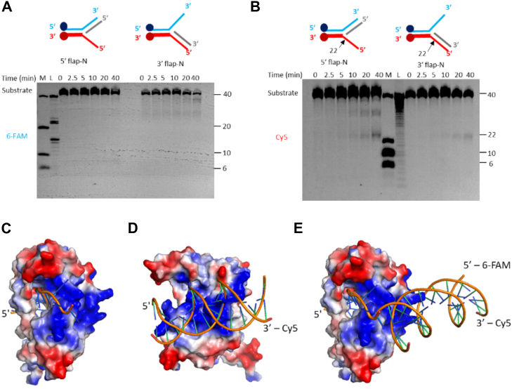
Figure 5 TON_0321 is a secondary structure–selective endonuclease. The catalytic activity of TON_0321 protein on branched DNA molecules, 5′ flap-N and 3′ flap-N, with each having one strand labeled at 5′-end with 6-FAM and another strand labeled at 3′-end with Cy5. The reaction products were resolved on 18% TBE–urea PAGE. The gels were scanned for (A) 6-FAM signal and (B) Cy5 signal. M represents a marker made from mixing synthetic oligonucleotides of different sizes (40, 20, 10, and 6 nucleotides), and L represents a ladder made from DNase digestion of the 40-mer substrate. All experiments were done in triplicates (Fig. S22). The major cleavage site in the schematic of branched DNA substrates is marked by a solid arrow. DNA binding model of TON_0321 with (C) ssDNA, (D) dsDNA, and (E) splayed arm substrate. The bound DNA was modeled by superimposing the catalytic domain of TON_0321 with the bound structure of GsCas4 (Protein Data Bank code: 7MI4). Cy5, cyanine-5 fluorophore; 6-FAM, 6-carboxyfluorescein; TBE, Tris–borate–EDTA.
Figure 5 TON_0321 is a secondary structure–selective endonuclease. The catalytic activity of TON_0321 protein on branched DNA molecules, 5′ flap-N and 3′ flap-N, with each having one strand labeled at 5′-end with 6-FAM and another strand labeled at 3′-end with Cy5. The reaction products were resolved on 18% TBE–urea PAGE. The gels were scanned for (A) 6-FAM signal and (B) Cy5 signal. M represents a marker made from mixing synthetic oligonucleotides of different sizes (40, 20, 10, and 6 nucleotides), and L represents a ladder made from DNase digestion of the 40-mer substrate. All experiments were done in triplicates (Fig. S22). The major cleavage site in the schematic of branched DNA substrates is marked by a solid arrow. DNA binding model of TON_0321 with (C) ssDNA, (D) dsDNA, and (E) splayed arm substrate. The bound DNA was modeled by superimposing the catalytic domain of TON_0321 with the bound structure of GsCas4 (Protein Data Bank code: 7MI4). Cy5, cyanine-5 fluorophore; 6-FAM, 6-carboxyfluorescein; TBE, Tris–borate–EDTA.CC BY · PubMed Central

Verification

Claims the paper makes

The claims and statistics of this paper haven’t been checked yet.

Run the check — free, no account needed. It reads the open-access text and checks the sources it cites.

A few free checks a day · papers already verified open instantly · results are public

Self-check

Percentages, sums and reported differences re-computed from the paper’s own numbers.

The self-check needs the paper’s full text.

Citations

Cites

Show all 2 references

Research works printed but not linked: 66

  1. 10.1016/j.visres.2023.108317 · DOI
  2. 10.1620/tjem.2023.J011 · DOI
  3. 10.1016/j.ggedit.2024.100031 · DOI
  4. 10.1186/s13059-023-02907-y · DOI
  5. 10.1007/s11033-023-08240-8 · DOI
  6. 10.1093/nar/gkac1192 · DOI · Paper page
  7. 2023, Recent advances in CRISPR-based genome editing technology and its applications in cardiovascular research, Mil. Med. Res, 10, 12
  8. 10.1186/s12943-022-01518-8 · DOI · Paper page
  9. 10.1016/j.sbi.2022.102466 · DOI
  10. 10.1016/j.cell.2009.07.040 · DOI · Paper page
  11. 10.1146/annurev-genet-110711-155447 · DOI
  12. 10.1126/science.1159689 · DOI · Paper page
  13. 10.1126/science.1225829 · DOI · Paper page
  14. 10.1038/nature09523 · DOI
  15. Jackson, S.A. , McKenzie, R.E. , Fagerlund, R.D. , Kieper, S.N. , Fineran, P.C. and Brouns, S.J.J. (2017) CRISPR-Cas: adapting to change. Science (80 -. )., 356, eaal5056. · DOI
  16. 10.1038/nrmicro.2015.14 · DOI
  17. 10.1038/s41586-021-03951-z · DOI
  18. 10.1016/j.molcel.2018.03.003 · DOI
  19. Zhang, Z. , Pan, S. , Liu, T. , Li, Y. and Peng, N. (2019) Cas4 Nucleases Can Effect Specific Integration of CRISPR Spacers. 201(12), 10–1128. · DOI
  20. 10.1016/j.celrep.2018.02.103 · DOI
  21. 10.1016/j.molcel.2018.05.002 · DOI
  22. 10.1093/nar/gks382 · DOI
  23. 10.1093/nar/gkn175 · DOI
  24. 10.1093/nar/28.18.3417 · DOI
  25. 10.1016/S1097-2765(00)80267-1 · DOI
  26. 10.1073/pnas.97.12.6306 · DOI
  27. 10.1093/emboj/17.5.1526 · DOI
  28. 10.1111/j.1742-4658.2009.07374.x · DOI
  29. 10.1186/s12862-017-0890-6 · DOI
  30. 10.1021/ja408729b · DOI
  31. 10.1093/nar/gku797 · DOI
  32. 10.1371/journal.pone.0047232 · DOI
  33. 10.1016/j.mib.2017.05.008 · DOI · Paper page
  34. 10.1093/nar/gkx612 · DOI
  35. 10.1016/j.molcel.2022.09.030 · DOI · Paper page
  36. Dhingra, Y. , Sashital, D.G. and Carver, R.J. Cas4/1 dual nuclease activities enable prespacer maturation and directional integration in 1 a type I-G CRISPR-Cas system. J. Biol. Chem., 299(9). · DOI
  37. 10.1093/nar/gkz217 · DOI
  38. 10.7554/elife.39324 · DOI · Paper page
  39. 10.1101/gad.585310 · DOI
  40. 10.1093/nar/13.5.1443 · DOI
  41. 10.1093/nar/gkh340 · DOI · Paper page
  42. 10.1093/MOLBEV/MSAB120 · DOI
  43. 10.1074/jbc.M808526200 · DOI
  44. 10.1093/NAR/GKAB301 · DOI
  45. 10.1038/nrmicro3569 · DOI · Paper page
  46. 10.1093/nar/gks216 · DOI · Paper page
  47. 10.1146/annurev-genet-022120-112523 · DOI
  48. 10.1038/nature15386 · DOI
  49. 10.1038/nsmb.3289 · DOI
  50. 10.1038/nature14237 · DOI · Paper page
  51. 10.1038/nature24020 · DOI
  52. 10.1017/S0033583510000181 · DOI · Paper page
  53. Zhang, J. , Mccabe, K.A. and Bell, C.E. Crystal structures of λ exonuclease in complex with DNA suggest an electrostatic ratchet mechanism for processivity. Proc. Natl. Acad. Sci., 108(29), 11872–11877. · DOI
  54. 1997, Toroidal structure of λ-exonuclease, Science, 80, 1824
  55. 10.4161/15384101.2014.950892 · DOI
  56. 10.1529/biophysj.107.110866 · DOI
  57. 10.1006/jmbi.1997.1128 · DOI
  58. 10.1016/j.pep.2009.09.003 · DOI
  59. 10.1038/s41586-020-2018-1 · DOI
  60. 10.1093/nar/gkac239 · DOI
  61. 10.1016/j.celrep.2015.02.019 · DOI
  62. 10.1093/nar/gkz842 · DOI
  63. 10.3389/fmicb.2021.666584 · DOI
  64. 10.1093/nar/gkz1197 · DOI
  65. 10.1146/annurev-biochem-072511-122603 · DOI
  66. Vallur, A.C. and Maizels, N. (2010) Distinct activities of exonuclease 1 and flap endonuclease 1 at telomeric G4 DNA. PLoS One, 5. · DOI

Metadata

Authors
Jain, M., Pattnayak, A. K., Aggarwal, S., Rai, P., J, K., Chandrayan, S., Goel, M., & Gaur, V.
Affiliations
  • University of Delhi (IN)
  • National Institute of Plant Genome Research (IN)
  • Institute of Chemical Technology (IN)
Corresponding author
  • Goel, Manisha
  • Gaur, Vineet
Venue
Journal of Biological Chemistry
Publisher
openRxiv
Published
2024-11-29
Volume
301
Issue
10
Pages
110701-110701
ISSN
0021-9258 (ISSN-L)
OpenAlex
W4414142587
Cited by
0
References
2
FWCI
0.00
Type
journal article
Language
en
Open access
Locations
  • Journal of Biological Chemistry (publishedVersion, cc-by)
  • bioRxiv (Cold Spring Harbor Laboratory) (acceptedVersion)
  • Europe PMC (PubMed Central) (publishedVersion, cc-by)
  • PubMed (publishedVersion)
Funders
  • Department of Biotechnology, Ministry of Science and Technology, India (BT/RLF/Re-entry/27/2017)
  • Department of Science and Technology, Ministry of Science and Technology, India
  • University of Delhi (IOE /2021/12/ FRP, IOE/2023-2024/12/FRP)
  • Montana Institute on Ecosystems
  • Science and Engineering Research Board (CRG/2019/001310, CRG/2020/000335)
Topics
  • Life Sciences › Biochemistry, Genetics and Molecular Biology › Molecular Biology › CRISPR and Genetic Engineering
  • Life Sciences › Biochemistry, Genetics and Molecular Biology › Genetics › Bacterial Genetics and Biotechnology
  • Life Sciences › Biochemistry, Genetics and Molecular Biology › Molecular Biology › Biochemical and Structural Characterization
Keywords
Thermococcus onnurineus, CRISPR adaptation, 5' to 3' exonuclease, structure-specific endonuclease, branched DNA
MeSH terms
Endonucleases, Enzyme Stability, DNA, Archaeal, Thermococcus, Archaeal Proteins, Catalytic Domain, CRISPR-Cas Systems, CRISPR-Associated Proteins

About these checks

Registry, retraction, Cites and statistics results are computed, not judged, with one exception: a printed reference that no index lists is read once by a model before it counts as a citation error. Claims are extracted by a model and kept only when their quote appears verbatim in the text; each cited source is then checked, with a model’s help, for whether it says what the claim relies on. Those verdicts can be wrong — read the claim’s quote and the cited source before relying on one.

Statistics and arithmetic are recomputed, not judged.

Confidence bands are shown only for fields calibrated on our annotated test set. None is calibrated yet, so no band is shown.

What counts as a claim ›How we measure confidence ›How we check this ›

Are you an author, or do you see a problem with this page? Request a review

How to cite

Cite CoChat for the checks on this page, and the paper for its own findings.

This page
CoChat. (2026). Citation check: Branched DNA Processing by a Thermostable CAS-Cas4 from Thermococcus onnurineus: Expanding Biochemical Landscape of Nuclease Activity. https://app.cochat.ai/papers/10.1101/2024.11.29.625776
The paper
Jain, M., Pattnayak, A. K., Aggarwal, S., Rai, P., J, K., Chandrayan, S., Goel, M., & Gaur, V. (2024). Branched DNA Processing by a Thermostable CAS-Cas4 from Thermococcus onnurineus: Expanding Biochemical Landscape of Nuclease Activity. Journal of Biological Chemistry. https://doi.org/10.1101/2024.11.29.625776