Student or educator? Sign up with your school email and get 6 months of Pro free.
PreprintCited by 1RNA Interference and Gene DeliveryPlant Molecular Biology ResearchRNA Research and Splicing

The DOI matches its Crossref record; 1 of the 1 references it cites were found in a registry or an index.

Clade-wide fungal proteome analysis reveals structure–function conservation in divergent Dicer proteins

Open access
Melet, Lorena, Canan, Jonathan, Villalobos, Pablo, Vidal-Veuthey, Boris, González-Toro, Fabian, Orellana, Ivana, Rivas-Pardo, J. Andrés, Cárdenas, Juan P., Moraga, Carol, Castro-Fernandez, Víctor, Johnson, Nathan R., Vidal, Elena A.·2025·Computational and Structural Biotechnology Journal

Preprint — published as 10.1016/j.csbj.2025.11.009 · Paper page

Abstract

Dicers (Dcrs) are central proteins involved in the biogenesis of small RNAs (sRNAs) in eukaryotes. Most of the knowledge on Dcr structure, function and evolution comes from studies conducted in animal and plant species. Comparatively, much less is known in fungi, which are a genetically and ecologically diverse group with important roles in ecosystems, agriculture, medicine, and biotechnology. While canonical Dcrs in plants and animals contain a well-defined domain architecture, most fungal Dcrs with experimentally validated functions lack one or more identifiable canonical domains, raising questions about how RNA-binding and precise sRNA processing is retained. Here, we conducted the most extensive survey of fungal Dcr proteins, analyzing 1,593 proteomes across eight phyla. We found a diversity of Dcr domain architectures, with some of them lacking an identifiable PAZ, Helicase, and/or double-stranded RNA binding domains. Phylogenetic analyses showed that different Dcr classes are distributed across distinct clades that often align with fungal taxonomic groups. Despite the lack of canonical domain architectures, we found that fungal Dcrs fold into a characteristic L-shaped structure and show PAZ-like folds, even in proteins without detectable PAZ sequences. Molecular docking and electrostatic analyses further indicate that these divergent Dcrs maintain key RNA-binding surfaces for proper sRNA processing. Our results indicate a remarkable evolutionary plasticity of Dcr in fungi, showing that essential sRNA processing functions can be retained through structural conservation, and highlighting fungi as models to study the modular evolution of the RNAi machinery in eukaryotes. Significance statement: Dicer (Dcr) proteins are central to RNA interference (RNAi), a gene regulatory mechanism conserved across eukaryotes. However, current models of Dcr structure, function, and evolution are largely based on studies in animals and plants. Here, we present the most comprehensive analysis to date of Dcr proteins in fungi, a diverse eukaryotic group including many societally important pathogens and symbiotes which are reliant on RNAi. Our findings reveal that despite widespread divergence from canonical Dcr architecture, fungal Dcrs conserve critical folds and RNA-binding features, further suggesting that core RNAi functions are maintained. This work establishes fungi as key models for studying the evolution and functional robustness of the RNAi machinery, offering broader insight into the diversity and plasticity of sRNA biogenesis pathways across eukaryotes.

Overview

The claims and statistics of this paper haven’t been checked yet.

Run the check — free, no account needed. It reads the open-access text and checks the sources it cites.

A few free checks a day · papers already verified open instantly · results are public

Integrity signals

Registry

Crossref · 10.1101/2025.04.28.651110

Cites

1 of 1 references verified0 citation errors · 0 not checkable

Statistics

Figures

Graphical abstract
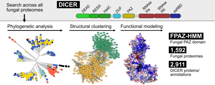
Graphical abstract CC BY · PubMed Central
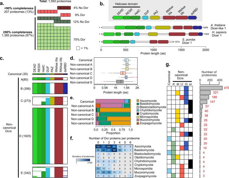
Fig. 1 Domain-based classification and distribution of Dicer proteins across fungal phyla. (a) Summary of the 1592 annotated fungal proteomes analyzed for the identification of Dcr proteins, showing the percentage of < 90 % and ≥ 90 % BUSCO completeness proteomes. No Dcr: no Dcr detected by HMMER analysis; Dcr: Dcr detected by HMMER analysis (b) Representative domain architectures of known Dcr proteins from Arabidopsis thaliana, Homo sapiens, and Schizosaccharomyces pombe, illustrating the conserved domain composition: DEAD, ResIII, Helicase C (HeliC), DUF283, PAZ, RNase IIIa and IIIb, and dsRNA-binding domain (dsRBD). Protein lengths are shown in amino acids (aa). (c) Classification of all predicted fungal Dcr proteins into six categories based on the presence or absence of key domains. Canonical Dcrs retain a full complement of conserved domains, while non-canonical groups exhibit partial or divergent architectures. Each file represents a Dcr protein, and the colors represent whether a domain was detected by HMMER (green shades: Helicase subdomains; bright cyan: DUF283 domain; olive yellow: PAZ domain; brick red: RNaseIII domain; indigo blue: dsRBD domain). White color indicates that the specific domain was not detected by HMMER. (d) Average protein length (in aa) per Dcr category. (e) Distribution of Dcr categories across fungal phyla, highlighting differences in domain conservation and structural diversity among lineages. (f) Variability in Dcr family expansion across phyla. Numbers in the boxes represent the number of proteomes per phyla containing 0–6 Dcr proteins. (g) Combinations of Dcr categories present in fungal proteomes. Each row shows the presence (colored square) or absence (white square) of canonical and non-canonical Dcrs (A–E) in the proteome. Bars on the right represent the number of proteomes presenting a given combination of Dcr categories (total number in red).
Fig. 1 Domain-based classification and distribution of Dicer proteins across fungal phyla. (a) Summary of the 1592 annotated fungal proteomes analyzed for the identification of Dcr proteins, showing the percentage of < 90 % and ≥ 90 % BUSCO completeness proteomes. No Dcr: no Dcr detected by HMMER analysis; Dcr: Dcr detected by HMMER analysis (b) Representative domain architectures of known Dcr proteins from Arabidopsis thaliana, Homo sapiens, and Schizosaccharomyces pombe, illustrating the conserved domain composition: DEAD, ResIII, Helicase C (HeliC), DUF283, PAZ, RNase IIIa and IIIb, and dsRNA-binding domain (dsRBD). Protein lengths are shown in amino acids (aa). (c) Classification of all predicted fungal Dcr proteins into six categories based on the presence or absence of key domains. Canonical Dcrs retain a full complement of conserved domains, while non-canonical groups exhibit partial or divergent architectures. Each file represents a Dcr protein, and the colors represent whether a domain was detected by HMMER (green shades: Helicase subdomains; bright cyan: DUF283 domain; olive yellow: PAZ domain; brick red: RNaseIII domain; indigo blue: dsRBD domain). White color indicates that the specific domain was not detected by HMMER. (d) Average protein length (in aa) per Dcr category. (e) Distribution of Dcr categories across fungal phyla, highlighting differences in domain conservation and structural diversity among lineages. (f) Variability in Dcr family expansion across phyla. Numbers in the boxes represent the number of proteomes per phyla containing 0–6 Dcr proteins. (g) Combinations of Dcr categories present in fungal proteomes. Each row shows the presence (colored square) or absence (white square) of canonical and non-canonical Dcrs (A–E) in the proteome. Bars on the right represent the number of proteomes presenting a given combination of Dcr categories (total number in red).CC BY · PubMed Central
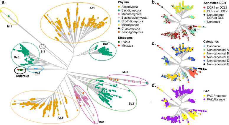
Fig. 2 Phylogenetic and functional classification of fungal Dcr proteins. (a) Maximum-likelihood phylogenetic tree showing the distribution of Dcr proteins across fungal phyla, as well as representative outgroups from plants and metazoans. Colored ovals highlight major clades identified within fungal lineages, labeled as As1–As2 (Ascomycota), Ba1–Ba3 (Basidiomycota), Mu1–Mu2 (Mucoromycota and Zoopagomycota), Bl1 (Blastocladiomycota) or Ch1 (Chytridiomycota), based on their phylogenetic position. (b) Naming of Dcr proteins according to UniProtKB, distinguishing Dcr1/Dcl1 (Dark red), Dcr2/Dcl2 (yellow), unnumbered Dcr/Dcl entries (black), and proteins with no assigned name (unnamed, light green). (c) Classification of fungal Dcrs into canonical and non-canonical categories (A–E), based on domain composition. (d) Mapping of canonical PAZ domain presence (yellow) or absence (purple) across the phylogeny, according to HMMER detection.
Fig. 2 Phylogenetic and functional classification of fungal Dcr proteins. (a) Maximum-likelihood phylogenetic tree showing the distribution of Dcr proteins across fungal phyla, as well as representative outgroups from plants and metazoans. Colored ovals highlight major clades identified within fungal lineages, labeled as As1–As2 (Ascomycota), Ba1–Ba3 (Basidiomycota), Mu1–Mu2 (Mucoromycota and Zoopagomycota), Bl1 (Blastocladiomycota) or Ch1 (Chytridiomycota), based on their phylogenetic position. (b) Naming of Dcr proteins according to UniProtKB, distinguishing Dcr1/Dcl1 (Dark red), Dcr2/Dcl2 (yellow), unnumbered Dcr/Dcl entries (black), and proteins with no assigned name (unnamed, light green). (c) Classification of fungal Dcrs into canonical and non-canonical categories (A–E), based on domain composition. (d) Mapping of canonical PAZ domain presence (yellow) or absence (purple) across the phylogeny, according to HMMER detection.CC BY · PubMed Central
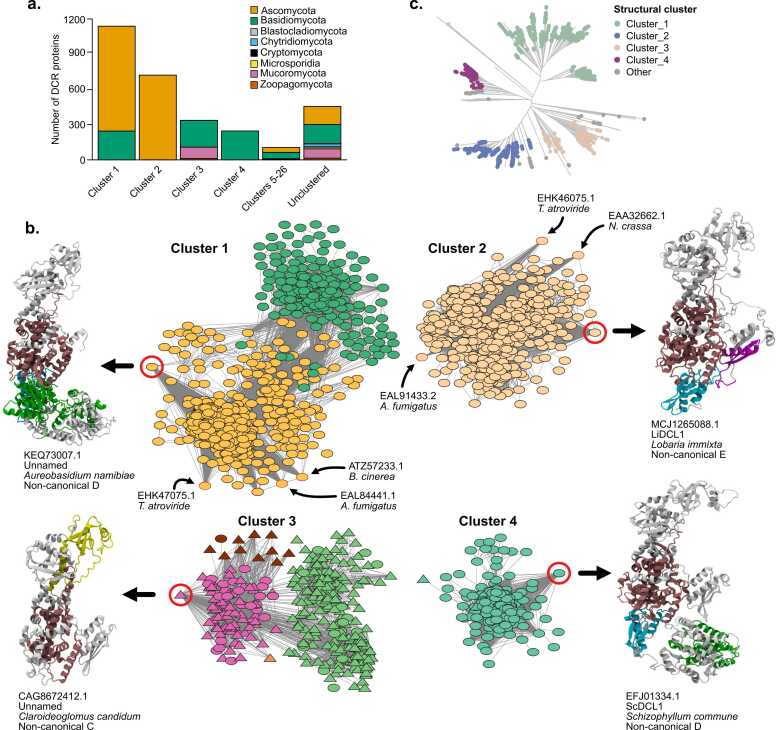
Fig. 3 Structural similarity network analysis reveals strong structural conservation of Dcr proteins. (a) Taxonomic composition of each structural cluster, shown as the number of Dcr proteins per fungal phylum. (b) Structural similarity network of predicted Dcr proteins, clustered based on TM-score ≥ 0.9. Each node represents a protein structure; edges indicate pairwise structural similarity (only edges of TM-score 0.9 or more are shown). Node colors indicate the clade classification defined in Fig. 2A (e.g., As1, As2, Ba1–Ba3, Mu1–Mu2), allowing direct comparison between structural and phylogenetic groupings. The most connected node (hub) from selected modules is shown with a red circle and its corresponding AlphaFold-predicted 3D structure is represented on the side, including protein code, Dicer naming according to UniProtKB (unnamed: no name available), the species name and Dcr group (according to Fig. 1 classification). Proteins displayed correspond to representative members of non-canonical Dcr groups B, C, and E. Structural domains are colored as follows: PAZ (yellow), Helicase (green), RNase III (dark red/burgundy), DUF283 (cyan), and dsRBD (purple). Regions without domain predictions are shown in grey. Triangular nodes correspond to Dcr proteins with a canonical PAZ domain (cPAZ); oval nodes indicate Dcrs with non-canonical PAZ domains. (c) Phylogenetic tree of all Dcr proteins, with tips colored according to structural cluster membership, illustrating the correspondence between structural similarity and evolutionary lineage. “Other” corresponds to clusters 5–26.
Fig. 3 Structural similarity network analysis reveals strong structural conservation of Dcr proteins. (a) Taxonomic composition of each structural cluster, shown as the number of Dcr proteins per fungal phylum. (b) Structural similarity network of predicted Dcr proteins, clustered based on TM-score ≥ 0.9. Each node represents a protein structure; edges indicate pairwise structural similarity (only edges of TM-score 0.9 or more are shown). Node colors indicate the clade classification defined in Fig. 2A (e.g., As1, As2, Ba1–Ba3, Mu1–Mu2), allowing direct comparison between structural and phylogenetic groupings. The most connected node (hub) from selected modules is shown with a red circle and its corresponding AlphaFold-predicted 3D structure is represented on the side, including protein code, Dicer naming according to UniProtKB (unnamed: no name available), the species name and Dcr group (according to Fig. 1 classification). Proteins displayed correspond to representative members of non-canonical Dcr groups B, C, and E. Structural domains are colored as follows: PAZ (yellow), Helicase (green), RNase III (dark red/burgundy), DUF283 (cyan), and dsRBD (purple). Regions without domain predictions are shown in grey. Triangular nodes correspond to Dcr proteins with a canonical PAZ domain (cPAZ); oval nodes indicate Dcrs with non-canonical PAZ domains. (c) Phylogenetic tree of all Dcr proteins, with tips colored according to structural cluster membership, illustrating the correspondence between structural similarity and evolutionary lineage. “Other” corresponds to clusters 5–26.CC BY · PubMed Central
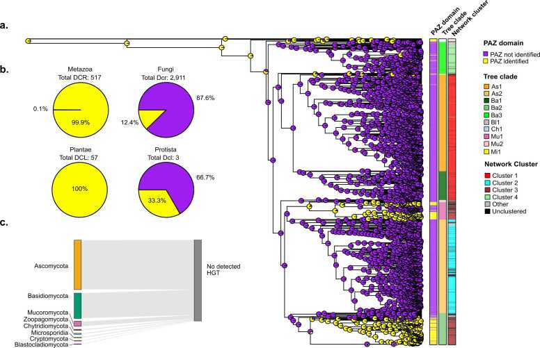
Fig. 4 Evolutionary reconstruction of the PAZ domain in fungal Dcr proteins. (a) Phylogenetic tree of fungal Dcr proteins with ancestral state reconstruction for the canonical PAZ domain. Pie charts at internal nodes represent the inferred probability of PAZ domain presence (yellow) or absence (purple) using maximum likelihood estimation. Additional metadata layers include the presence/absence of a canonical PAZ domain, phylogenetic group according to Fig. 2(e.g., As1, Ba2, Mu1), and structural cluster as defined in Fig. 3. (b) Proportion of Dcr proteins with a canonical PAZ domain across eukaryotic groups Metazoa, Fungi, Plantae and Protista, highlighting variation in PAZ domain conservation. (c) Horizontal gene transfer (HGT) analysis. No recent inter-kingdom HGT events involving Dcr proteins were detected between fungi and plants, animals, or protists.
Fig. 4 Evolutionary reconstruction of the PAZ domain in fungal Dcr proteins. (a) Phylogenetic tree of fungal Dcr proteins with ancestral state reconstruction for the canonical PAZ domain. Pie charts at internal nodes represent the inferred probability of PAZ domain presence (yellow) or absence (purple) using maximum likelihood estimation. Additional metadata layers include the presence/absence of a canonical PAZ domain, phylogenetic group according to Fig. 2(e.g., As1, Ba2, Mu1), and structural cluster as defined in Fig. 3. (b) Proportion of Dcr proteins with a canonical PAZ domain across eukaryotic groups Metazoa, Fungi, Plantae and Protista, highlighting variation in PAZ domain conservation. (c) Horizontal gene transfer (HGT) analysis. No recent inter-kingdom HGT events involving Dcr proteins were detected between fungi and plants, animals, or protists.CC BY · PubMed Central
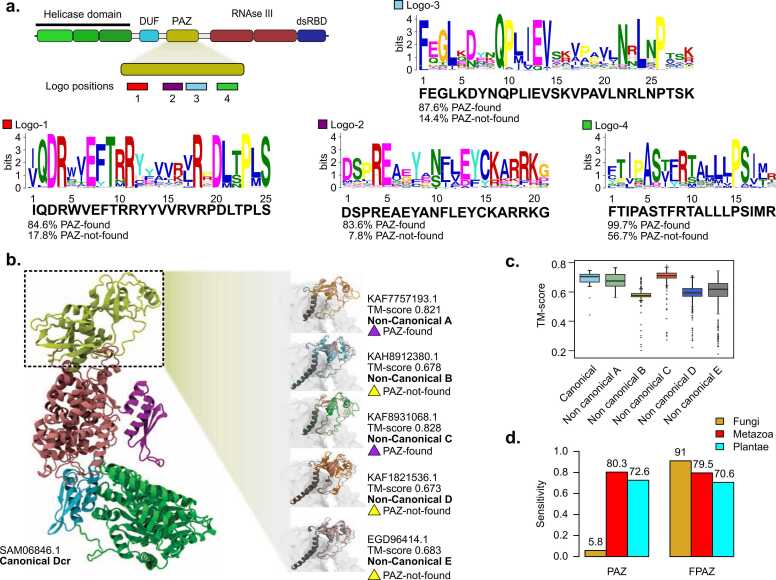
Fig. 5 Motif conservation and structural variability of PAZ domains in fungal Dicer-like proteins. (a) Conserved sequence motifs identified within the canonical PAZ domain using MEME Suite. Schematic representation (top) shows the approximate location of four conserved motifs (Logos 1–4) along the PAZ region. Sequence logos illustrate consensus motifs (below logos) and their relative frequency in proteins where the PAZ domain was detected (PAZ-found) versus those where it was not (PAZ-not-found). (b) Structural comparison of PAZ domains across Dcr categories. The full-length AlphaFold model of a canonical Dcr (SAM06846.1, Dicer-like 1 from Absidia glauca) is shown on the left, with the PAZ domain highlighted in yellow. Right panel: Representative AlphaFold structural models of PAZ domains from non-canonical categories A (KAF7757193.1, Dicer-like 1 from Entomophthora muscae), B (KAH8912380.1, unnamed protein from Coniochaeta sp. PMI_546), C (KAF8931068.1, Dicer-like 1 from Dissophora ornata), D (KAF1821536.1, unnamed protein from Dissoconium aciculare) and E (EGD96414.1, Dicer-like 1 from Trichophyton tonsurans), annotated with TM-score values and presence/absence of a recognizable cPAZ domain. PAZ regions in non-canonical Dcrs are color-coded according to their category (A–E). Structural alignment was performed using the PAZ domain of SAM06846.1 as reference to compare the spatial conservation of PAZ-like regions across non-canonical Dcrs. (c) Boxplot of TM-scores comparing structural similarity of PAZ domains across canonical and non-canonical Dcr categories. Canonical domains show consistently higher structural conservation. (d) Sensitivity comparison of the canonical PAZ HMM (PAZ) versus the fungal-specific FPAZ-HMM (FPAZ) across Eukaryotic groups.
Fig. 5 Motif conservation and structural variability of PAZ domains in fungal Dicer-like proteins. (a) Conserved sequence motifs identified within the canonical PAZ domain using MEME Suite. Schematic representation (top) shows the approximate location of four conserved motifs (Logos 1–4) along the PAZ region. Sequence logos illustrate consensus motifs (below logos) and their relative frequency in proteins where the PAZ domain was detected (PAZ-found) versus those where it was not (PAZ-not-found). (b) Structural comparison of PAZ domains across Dcr categories. The full-length AlphaFold model of a canonical Dcr (SAM06846.1, Dicer-like 1 from Absidia glauca) is shown on the left, with the PAZ domain highlighted in yellow. Right panel: Representative AlphaFold structural models of PAZ domains from non-canonical categories A (KAF7757193.1, Dicer-like 1 from Entomophthora muscae), B (KAH8912380.1, unnamed protein from Coniochaeta sp. PMI_546), C (KAF8931068.1, Dicer-like 1 from Dissophora ornata), D (KAF1821536.1, unnamed protein from Dissoconium aciculare) and E (EGD96414.1, Dicer-like 1 from Trichophyton tonsurans), annotated with TM-score values and presence/absence of a recognizable cPAZ domain. PAZ regions in non-canonical Dcrs are color-coded according to their category (A–E). Structural alignment was performed using the PAZ domain of SAM06846.1 as reference to compare the spatial conservation of PAZ-like regions across non-canonical Dcrs. (c) Boxplot of TM-scores comparing structural similarity of PAZ domains across canonical and non-canonical Dcr categories. Canonical domains show consistently higher structural conservation. (d) Sensitivity comparison of the canonical PAZ HMM (PAZ) versus the fungal-specific FPAZ-HMM (FPAZ) across Eukaryotic groups.CC BY · PubMed Central
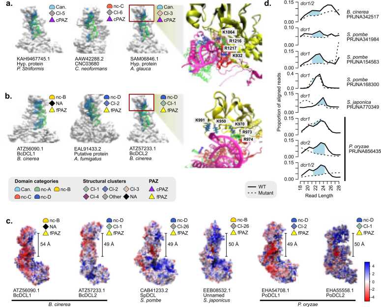
Fig. 6 Structural and functional characterization of RNA-binding interfaces in canonical and non-canonical fungal Dcr proteins. (a–b) Surface representations of models of full-length Dcr proteins (gray) in complex with a pre-cleaved dsRNA substrate that was obtained from the PDB model of D. melanogaster Dicer (pdb_00007w0f) (green), showing the RNA-binding interfaces (blue). (a) Canonical Dcr proteins containing a canonical PAZ domain (cPAZ), grouped by structural cluster and domain category (Can.: canonical, nc-A-E: non-canonical A-E). (b) Non-canonical Dcrs possessing fungal-specific PAZ regions (fPAZ), grouped by structural cluster and domain category. Representative residues involved in interaction with RNA (lysines and arginines) are shown in atomic detail, with the side chains located within 3 Å of the RNA. Red boxes highlight the cPAZ or fPAZ region that anchors the 3′ end of the dsRNA. "Other" includes proteins belonging to minor structural clusters outside the four major groups. "NA" includes proteins that did not meet the clustering thresholds (≥90 % TM-score similarity and pLDDT ≥70). (c) Electrostatic surface potential is shown for each model, with red indicating negative charge and blue indicating positive charge (scale in kT/e). The measured distance between the fPAZ region and the RNase III catalytic site (in Å) is shown, suggesting its possible role in determining the length of small RNA (sRNA). (d) Read length distribution of the sRNA libraries. Solid lines represent WT, dashed lines correspond to dcr mutants. Light blue peaks indicate sRNA species lost in the mutant background.
Fig. 6 Structural and functional characterization of RNA-binding interfaces in canonical and non-canonical fungal Dcr proteins. (a–b) Surface representations of models of full-length Dcr proteins (gray) in complex with a pre-cleaved dsRNA substrate that was obtained from the PDB model of D. melanogaster Dicer (pdb_00007w0f) (green), showing the RNA-binding interfaces (blue). (a) Canonical Dcr proteins containing a canonical PAZ domain (cPAZ), grouped by structural cluster and domain category (Can.: canonical, nc-A-E: non-canonical A-E). (b) Non-canonical Dcrs possessing fungal-specific PAZ regions (fPAZ), grouped by structural cluster and domain category. Representative residues involved in interaction with RNA (lysines and arginines) are shown in atomic detail, with the side chains located within 3 Å of the RNA. Red boxes highlight the cPAZ or fPAZ region that anchors the 3′ end of the dsRNA. "Other" includes proteins belonging to minor structural clusters outside the four major groups. "NA" includes proteins that did not meet the clustering thresholds (≥90 % TM-score similarity and pLDDT ≥70). (c) Electrostatic surface potential is shown for each model, with red indicating negative charge and blue indicating positive charge (scale in kT/e). The measured distance between the fPAZ region and the RNase III catalytic site (in Å) is shown, suggesting its possible role in determining the length of small RNA (sRNA). (d) Read length distribution of the sRNA libraries. Solid lines represent WT, dashed lines correspond to dcr mutants. Light blue peaks indicate sRNA species lost in the mutant background.CC BY · PubMed Central

Verification

Claims the paper makes

The claims and statistics of this paper haven’t been checked yet.

Run the check — free, no account needed. It reads the open-access text and checks the sources it cites.

A few free checks a day · papers already verified open instantly · results are public

Self-check

Percentages, sums and reported differences re-computed from the paper’s own numbers.

The self-check needs the paper’s full text.

Citations

Cites

Research works printed but not linked: 80

  1. 10.1038/nature03315 · DOI · Paper page
  2. 10.1038/nature03957 · DOI
  3. 10.1126/science.1074973 · DOI · Paper page
  4. 10.1038/ncomms6488 · DOI · Paper page
  5. 10.1126/science.aar4142 · DOI
  6. M. Wang , et al., Bidirectional cross-kingdom RNAi and fungal uptake of external RNAs confer plant protection. Nature Plants 2, 16151 (2016). · DOI · Paper page
  7. 10.1016/S0092-8674(02)00908-X · DOI
  8. 10.1016/S0092-8674(03)00759-1 · DOI · Paper page
  9. 10.1016/j.str.2009.08.013 · DOI
  10. 10.1038/nsmb.2268 · DOI
  11. 10.1038/nsmb.1673 · DOI · Paper page
  12. 10.1016/j.molcel.2015.03.012 · DOI
  13. 10.1126/science.aaq0921 · DOI
  14. 10.1016/j.molcel.2011.02.005 · DOI
  15. 10.1007/s11103-004-6853-5 · DOI · Paper page
  16. 10.1261/rna.1965310 · DOI
  17. 10.1016/j.cell.2013.03.024 · DOI · Paper page
  18. 10.1093/emboj/cdf582 · DOI · Paper page
  19. 10.1038/nature02519 · DOI · Paper page
  20. 10.1126/science.1121638 · DOI · Paper page
  21. J. Choi , et al., funRNA: a fungi-centered genomics platform for genes encoding key components of RNAi. BMC Genomics 15, S14 (2014). · DOI
  22. 10.1126/science.1176945 · DOI · Paper page
  23. 10.3852/13-171 · DOI
  24. 10.1016/j.cub.2021.01.074 · DOI · Paper page
  25. 10.1146/annurev-micro-022020-051835 · DOI
  26. 10.1007/s13225-018-0401-0 · DOI · Paper page
  27. 10.1007/s13225-024-00540-z · DOI
  28. 10.1016/j.meegid.2018.08.011 · DOI
  29. 10.1128/MCB.24.6.2536-2545.2004 · DOI
  30. 10.1074/jbc.M408259200 · DOI
  31. 10.1146/annurev-micro-092611-150138 · DOI · Paper page
  32. 10.1128/EC.00191-09 · DOI
  33. 10.3389/fbinf.2023.1178926 · DOI · Paper page
  34. 10.15252/embr.202357215 · DOI
  35. 10.1093/molbev/mss263 · DOI
  36. 10.1007/s13225-023-00525-4 · DOI
  37. N. Jespersen , L. Monrroy , J. Barandun , “Impact of Genome Reduction in Microsporidia” in Microsporidia, Experientia Supplementum., L. M. Weiss , A. W. Reinke , Eds. (Springer International Publishing, 2022), pp. 1–42. · DOI
  38. 10.1111/mpp.13269 · DOI
  39. 10.1126/science.1239705 · DOI
  40. 10.1261/rna.079350.122 · DOI
  41. 10.1111/mmi.12261 · DOI
  42. 2024, Argonaute and Dicer are essential for communication between Trichoderma atroviride and fungal hosts during mycoparasitism, Microbiol Spectr, 12, e03165
  43. J. M. Villalobos-Escobedo , et al., Trichoderma atroviride hyphal regeneration and conidiation depend on cell-signaling processes regulated by a microRNA-like RNA. Microbial Genomics 8 (2022). · DOI
  44. 10.1016/j.molcel.2010.04.005 · DOI · Paper page
  45. 10.1093/nar/gkv416 · DOI
  46. 10.1093/nar/gkx407 · DOI
  47. 10.1016/j.molcel.2014.01.003 · DOI
  48. 10.1128/EC.05109-11 · DOI
  49. 10.1007/s10577-013-9388-2 · DOI
  50. 10.1038/ng1253 · DOI · Paper page
  51. 10.1016/j.cub.2005.07.024 · DOI
  52. 10.1093/nar/gkab238 · DOI
  53. 10.1038/s41477-021-01000-1 · DOI
  54. A. J. Malik , C. Puente-Lelievre , N. Matzke , D. B. Ascher , On use of tertiary structure characters in hidden Markov models for protein fold prediction. [Preprint] (2024). Available at: http://biorxiv.org/lookup/doi/10.1101/2024.04.08.588419 [Accessed 19 May 2025]. · DOI
  55. T. Benavides , C. Bystroff , HMMSTRTM: A hidden Markov model for local structure prediction in globular and membrane associated proteins. [Preprint] (2023). Available at: http://biorxiv.org/lookup/doi/10.1101/2023.02.08.527695 [Accessed 19 May 2025]. · DOI
  56. 10.1006/jmbi.2000.3837 · DOI
  57. M. Seppey , M. Manni , E. M. Zdobnov , “BUSCO: Assessing Genome Assembly and Annotation Completeness” in Gene Prediction, Methods in Molecular Biology., M. Kollmar , Ed. (Springer New York, 2019), pp. 227–245. · DOI
  58. 10.1094/MPMI-11-16-0236-R · DOI
  59. 10.1093/nar/gky448 · DOI · Paper page
  60. 10.1093/bioinformatics/btu031 · DOI · Paper page
  61. 10.1038/S41592-022-01488-1 · DOI · Paper page
  62. 10.1093/nar/gkh381 · DOI
  63. 10.1021/ct200133y · DOI
  64. 10.1021/ct100578z · DOI · Paper page
  65. 10.1073/pnas.181342398 · DOI
  66. 10.1093/nar/gky430 · DOI
  67. 10.1101/gr.1239303 · DOI · Paper page
  68. 10.1006/jmbi.2000.4042 · DOI
  69. 10.1093/nar/22.22.4673 · DOI · Paper page
  70. 10.1371/journal.pbio.3001007 · DOI · Paper page
  71. 10.1093/molbev/msaa015 · DOI · Paper page
  72. 10.1111/j.2041-210X.2011.00169.x · DOI · Paper page
  73. Q. Zhu , M. Kosoy , K. Dittmar , HGTector: an automated method facilitating genome-wide discovery of putative horizontal gene transfers. (2014). · DOI
  74. 10.1038/nmeth.3176 · DOI · Paper page
  75. 10.1002/jcc.23354 · DOI · Paper page
  76. J. C. Phillips , et al., Scalable molecular dynamics on CPU and GPU architectures with NAMD. The Journal of Chemical Physics 153 (2020). · DOI
  77. 10.1093/molbev/mst010 · DOI · Paper page
  78. N. R. Johnson , F. Gonzalez-Toro , B. Bernal Gomez , Class-agnostic annotation of small RNAs balances sensitivity and specificity in diverse organisms. [Preprint] (2025). Available at: http://biorxiv.org/lookup/doi/10.1101/2025.01.25.634868 [Accessed 23 April 2025]. · DOI
  79. 10.1534/g3.116.030452 · DOI
  80. 10.1002/pro.3923 · DOI · Paper page

Metadata

Authors
Melet, L., Canan, J., Villalobos, P., Vidal-Veuthey, B., González-Toro, F., Orellana, I., Rivas-Pardo, J. A., Cárdenas, J. P., Moraga, C., Castro-Fernandez, V., Johnson, N. R., & Vidal, E. A.
Affiliations
  • Universidad Mayor (CL)
  • University of Chile (CL)
  • Centro de Genómica y Bioinformática, Universidad Mayor, 8580745 Santiago, Chile.
  • Millennium Science Initiative (CL)
  • Departamento de Biología, Facultad de Ciencias, Universidad de Chile, 7800003 Santiago, Chile
Corresponding author
  • Johnson, Nathan R.
  • Vidal, Elena A.
Venue
Computational and Structural Biotechnology Journal
Publisher
openRxiv
Published
2025-05-01
Volume
27
Pages
5020-5035
ISSN
2001-0370 (ISSN-L)
OpenAlex
W4415972686
Cited by
1
References
1
Citations by year
2026: 1
FWCI
0.30
Type
journal article
Language
en
Open access
Locations
  • Computational and Structural Biotechnology Journal (publishedVersion, cc-by)
  • bioRxiv (Cold Spring Harbor Laboratory) (acceptedVersion, cc-by)
  • Europe PMC (PubMed Central) (publishedVersion, cc-by)
  • PubMed (publishedVersion)
  • DOAJ (DOAJ: Directory of Open Access Journals) (submittedVersion, cc-by-sa)
  • Universidad de Chile (submittedVersion, cc-by-nc-nd)
Funders
  • Universidad Mayor
  • Agenția Națională pentru Cercetare și Dezvoltare (FOVI230159, 2021\u201321211564, 2022\u201321221055, NCN2024_047)
  • Agencia Nacional de Investigación y Desarrollo (11251927, 1221667, 1221064, NCN2024_047, 2021\u201321211564, 3230600, 11200209, ICN17_022, FOVI230159, 11220727, 2022\u201321221055)
  • Fondo Nacional de Desarrollo Científico y Tecnológico (11220727, 3230600, 11200209, 11251927, IUP23\u201326, 1221667, ICN17_022, 1221064)
  • Universidad de O'Higgins
Topics
  • Life Sciences › Biochemistry, Genetics and Molecular Biology › Molecular Biology › RNA Interference and Gene Delivery
  • Life Sciences › Agricultural and Biological Sciences › Plant Science › Plant Molecular Biology Research
  • Life Sciences › Biochemistry, Genetics and Molecular Biology › Molecular Biology › RNA Research and Splicing
Keywords
fungi, small RNA biogenesis, RNA interference, PAZ domain, OB-fold, domain architecture, protein evolution, fungal proteome, phylogenetic analysis, molecular simulation

About these checks

Registry, retraction, Cites and statistics results are computed, not judged, with one exception: a printed reference that no index lists is read once by a model before it counts as a citation error. Claims are extracted by a model and kept only when their quote appears verbatim in the text; each cited source is then checked, with a model’s help, for whether it says what the claim relies on. Those verdicts can be wrong — read the claim’s quote and the cited source before relying on one.

Statistics and arithmetic are recomputed, not judged.

Confidence bands are shown only for fields calibrated on our annotated test set. None is calibrated yet, so no band is shown.

What counts as a claim ›How we measure confidence ›How we check this ›

Are you an author, or do you see a problem with this page? Request a review

Data sources

Bibliographic data: Semantic Scholar, OpenAlex, Crossref.

Contains information from the Semantic Scholar Academic Graph Datasets, made available under the ODC Attribution License (ODC-BY 1.0).

Semantic Scholar’s data is described in: Kinney, R., et al. (2023). The Semantic Scholar Open Data Platform. arXiv:2301.10140.