

The Allen Cell and Structure Segmenter: a new open source toolkit for segmenting 3D intracellular structures in fluorescence microscopy images
Open accessThe DOI matches its Crossref record; 41 of the 42 references it cites were found in a registry or an index, and 1 are not checked yet.
Abstract
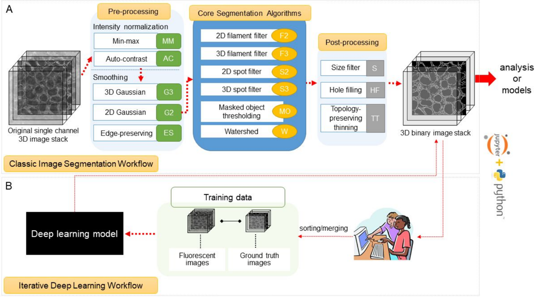
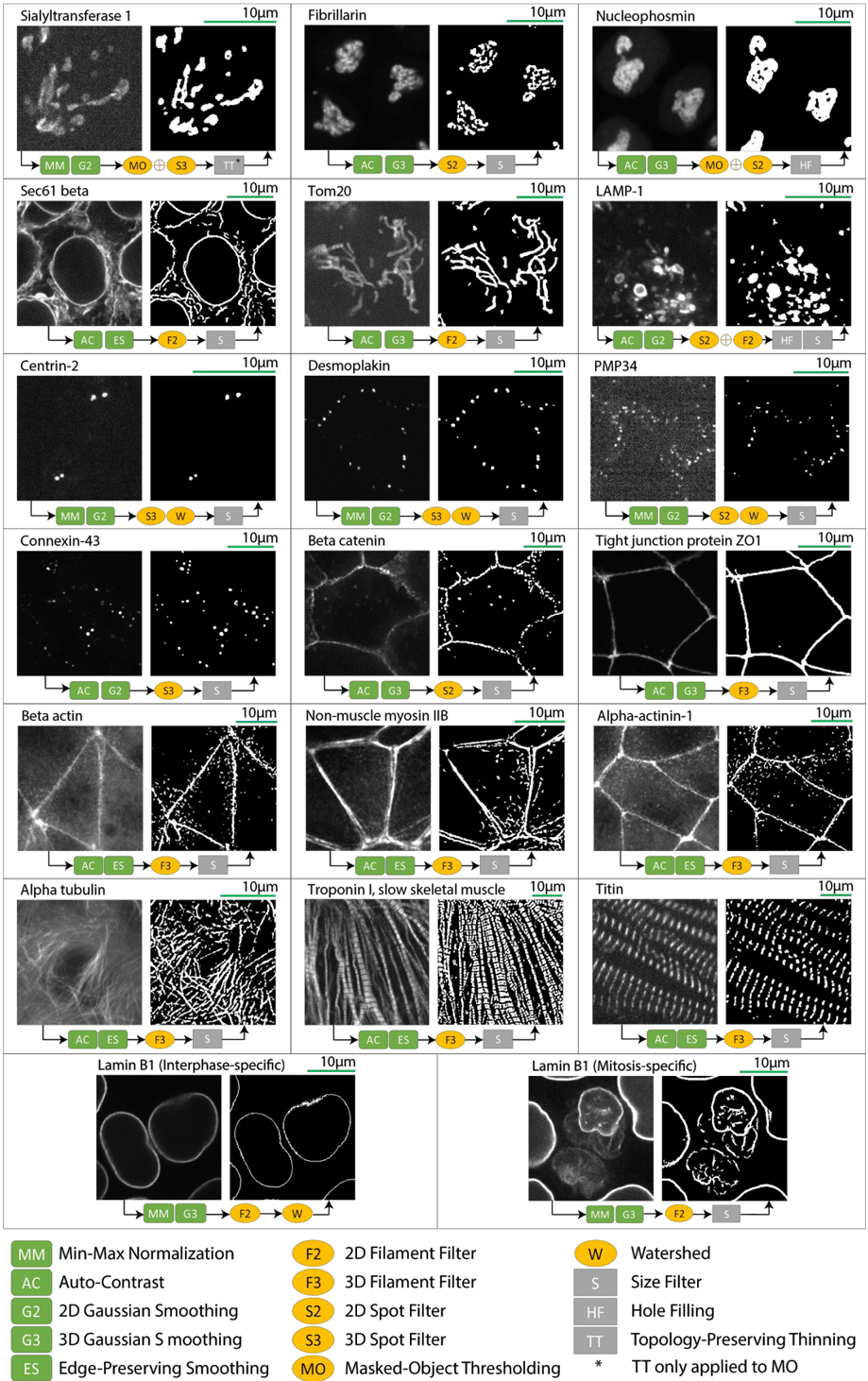
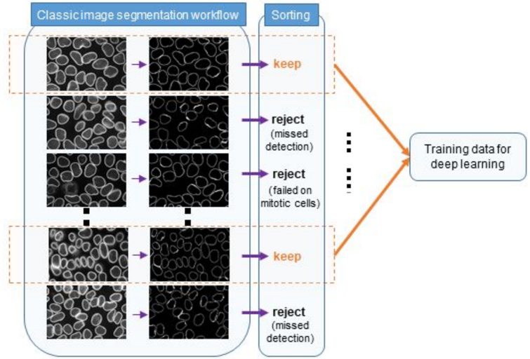
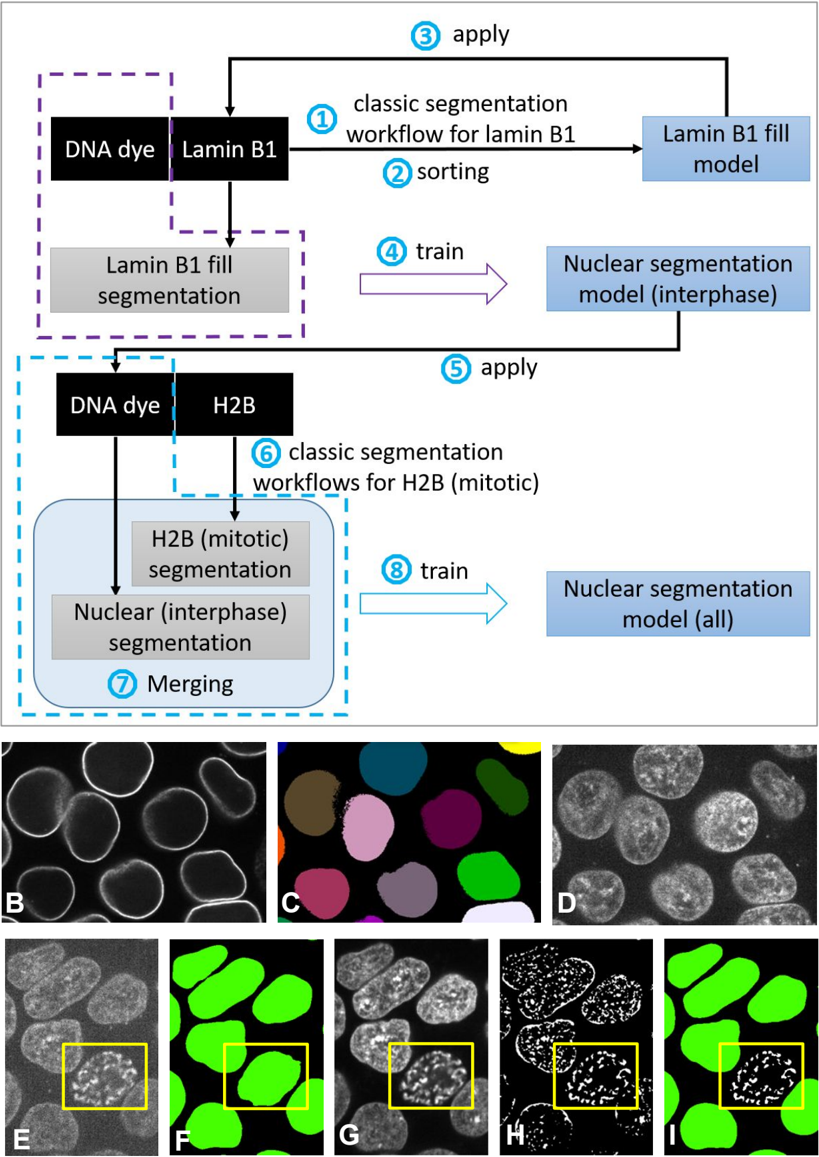
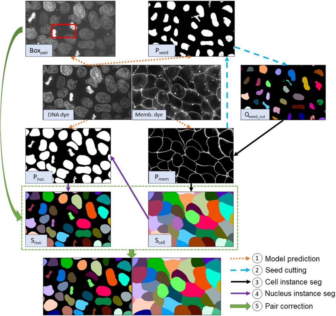
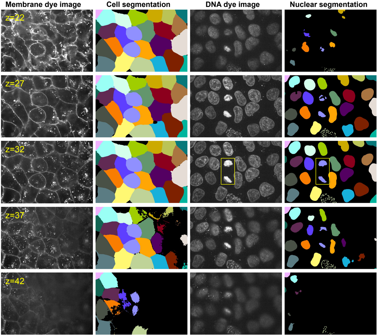
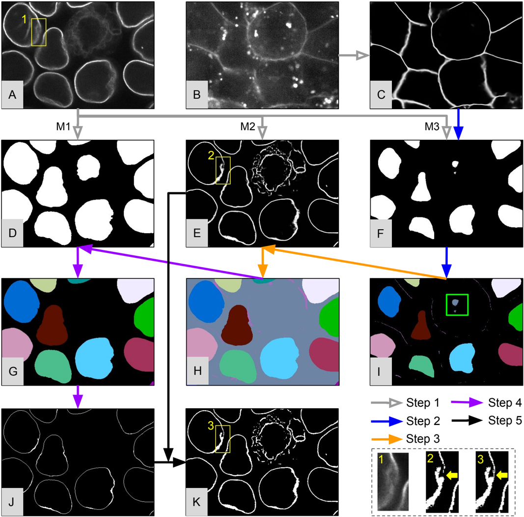
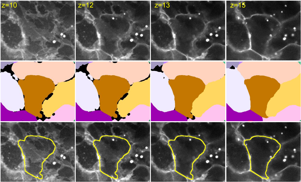
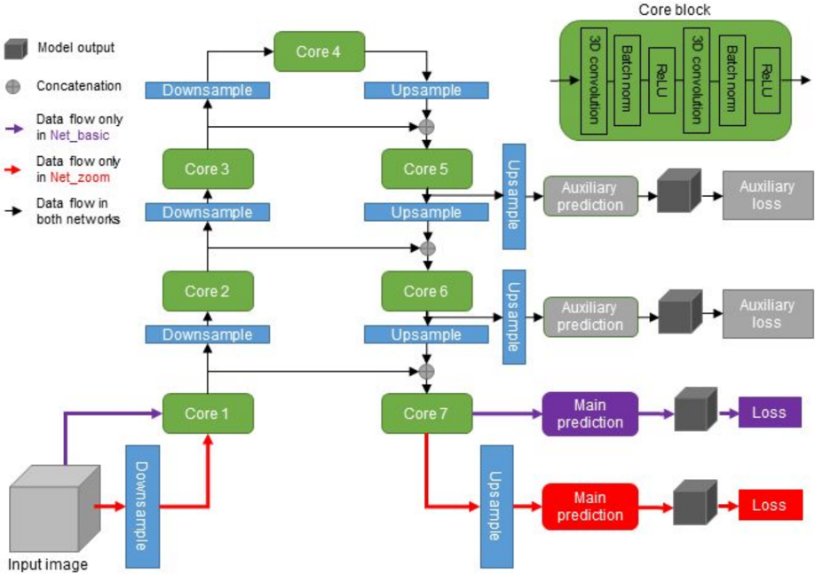
A continuing challenge in quantitative cell biology is the accurate and robust 3D segmentation of structures of interest from fluorescence microscopy images in an automated, reproducible, and widely accessible manner for subsequent interpretable data analysis. We describe the Allen Cell and Structure Segmenter (Segmenter), a Python-based open source toolkit developed for 3D segmentation of cells and intracellular structures in fluorescence microscope images. This toolkit brings together classic image segmentation and iterative deep learning workflows first to generate initial high-quality 3D intracellular structure segmentations and then to easily curate these results to generate the ground truths for building robust and accurate deep learning models. The toolkit takes advantage of the high-replicate 3D live cell image data collected at the Allen Institute for Cell Science of over 30 endogenous fluorescently tagged human induced pluripotent stem cell (hiPSC) lines. Each cell line represents a different intracellular structure with one or more distinct localization patterns within undifferentiated hiPS cells and hiPSC-derived cardiomyocytes. The Segmenter consists of two complementary elements, a classic image segmentation workflow with a restricted set of algorithms and parameters and an iterative deep learning segmentation workflow. We created a collection of 20 classic image segmentation workflows based on 20 distinct and representative intracellular structure localization patterns as a “lookup table” reference and starting point for users. The iterative deep learning workflow can take over when the classic segmentation workflow is insufficient. Two straightforward “human-in-the-loop” curation strategies convert a set of classic image segmentation workflow results into a set of 3D ground truth images for iterative model training without the need for manual painting in 3D. The deep learning model architectures used in this toolkit were designed and tested specifically for 3D fluorescence microscope images and implemented as readable scripts. The Segmenter thus leverages state of the art computer vision algorithms in an accessible way to facilitate their application by the experimental biology researcher. We include two useful applications to demonstrate how we used the classic image segmentation and iterative deep learning workflows to solve more challenging 3D segmentation tasks. First, we introduce the ‘Training Assay’ approach, a new experimental-computational co-design concept to generate more biologically accurate segmentation ground truths. We combined the iterative deep learning workflow with three Training Assays to develop a robust, scalable cell and nuclear instance segmentation algorithm, which could achieve accurate target segmentation for over 98% of individual cells and over 80% of entire fields of view. Second, we demonstrate how to extend the lamin B1 segmentation model built from the iterative deep learning workflow to obtain more biologically accurate lamin B1 segmentation by utilizing multi-channel inputs and combining multiple ML models. The steps and workflows used to develop these algorithms are generalizable to other similar segmentation challenges. More information, including tutorials and code repositories, are available at allencell.org/segmenter.
Overview
The claims and statistics of this paper haven’t been checked yet.
Free: it reads the open-access text and checks the first 5 claims that cite a source we can check. Create a free account to check every claim.
Integrity signals
Registry
Cites
Statistics
Figures




Verification
Claims the paper makes
The claims and statistics of this paper haven’t been checked yet.
Free: it reads the open-access text and checks the first 5 claims that cite a source we can check. Create a free account to check every claim.
Claims other papers make about this one
Each is checked against this paper’s own text: does it say what the citing paper says it does?
The cited source says what the claim attributes to it.
In the citing paper
“required deep-learning-based segmentation approaches to create a robust, scalable and highly accurate 3D cell and nuclear segmentation algorithm15 (Methods)”
From Integrated intracellular organization and its variations in human iPS cells (2023) · cites this paper as [15]
Self-check
Percentages, sums and reported differences re-computed from the paper’s own numbers.
The self-check needs the paper’s full text.
Citations
Cites
Adam: A Method for Stochastic Optimization
Kingma, D. P., & Ba, J. (2014) · arXiv · doi:10.48550/arxiv.1412.6980 · Cited by 98,946
Faster R-CNN: Towards Real-Time Object Detection with Region Proposal Networks
Ren, S., He, K., Girshick, R., & Sun, J. (2017) · IEEE Transactions on Pattern Analysis and Machine Intelligence · doi:10.1109/tpami.2016.2577031 · Cited by 73,472
Fiji: an open-source platform for biological-image analysis
Schindelin, J., Arganda-Carreras, I., Frise, E., Kaynig, V., Longair, M., Pietzsch, T., Preibisch, S., Rueden, C., Saalfeld, S., Schmid, B., et al. (2012) · Nature Methods · doi:10.1038/nmeth.2019 · Cited by 66,890
Fully convolutional networks for semantic segmentation
Long, J., Shelhamer, E., & Darrell, T. (2015) · 2015 IEEE Conference on Computer Vision and Pattern Recognition (CVPR) · doi:10.1109/cvpr.2015.7298965 · Cited by 38,834
SciPy 1.0: fundamental algorithms for scientific computing in Python
Virtanen, P., Gommers, R., Oliphant, T. E., Haberland, M., Reddy, T., Cournapeau, D., Burovski, E., Peterson, P., Weckesser, W., Bright, J., et al. (2020) · Nature Methods · doi:10.1038/s41592-019-0686-2 · Cited by 38,430
Harris, C. R., Millman, K. J., van der Walt, S. J., Gommers, R., Virtanen, P., Cournapeau, D., Wieser, E., Taylor, J., Berg, S., Smith, N. J., et al. (2020) · Nature · doi:10.1038/s41586-020-2649-2 · Cited by 23,826
Harris, C., Millman, K., van der Walt, S., Gommers, R., Virtanen, P., Cournapeau, D., Wieser, E., Taylor, J., Berg, S., Smith, N., et al. (2020) · RePEc: Research Papers in Economics · Cited by 17,844
Scale-space and edge detection using anisotropic diffusion
Perona, P., & Malik, J. (1990) · IEEE Transactions on Pattern Analysis and Machine Intelligence · doi:10.1109/34.56205 · Cited by 12,119
SciPy 1.0: fundamental algorithms for scientific computing in Python
Virtanen, P., Gommers, R., Oliphant, T. E., Haberland, M., Reddy, T., Cournapeau, D., Burovski, E., Peterson, P., Weckesser, W., Bright, J., et al. (2019) · Apollo - University of Cambridge Repository · doi:10.17863/cam.64134 · Cited by 10,460
Yushkevich, P. A., Piven, J., Hazlett, H. C., Smith, R. G., Ho, S., Gee, J. C., & Gerig, G. (2006) · NeuroImage · doi:10.1016/j.neuroimage.2006.01.015 · Cited by 8,868 · cited 1×
“Some algorithms are available as collections of basic functions in several open platforms, including the widely used ImageJ [Schindelin et al., 2012], CellProfiler [Carpenter et al., 2006, McQuin et al., 2018], Icy [De Chaumont et al., 2012], and ITK-SNAP [Yushkevich et al., 2006].”
Method
Cited by
Cellpose: a generalist algorithm for cellular segmentation
Stringer, C., Wang, T., Michaelos, M., & Pachitariu, M. (2021) · Nature Methods · doi:10.1038/s41592-020-01018-x · Cited by 4,369
Deep learning for cellular image analysis
Moen, E., Bannon, D., Kudo, T., Graf, W., Covert, M., & Van Valen, D. (2019) · Nature Methods · doi:10.1038/s41592-019-0403-1 · Cited by 1,225
Understanding metric-related pitfalls in image analysis validation
Reinke, A., Tizabi, M. D., Baumgartner, M., Eisenmann, M., Heckmann-Nötzel, D., Kavur, A. E., Rädsch, T., Sudre, C. H., Acion, L., Antonelli, M., et al. (2024) · Nature Methods · doi:10.1038/s41592-023-02150-0 · Cited by 241
Self-citationIntegrated intracellular organization and its variations in human iPS cells
Viana, M. P., Chen, J., Knijnenburg, T. A., Vasan, R., Yan, C., Arakaki, J. E., Bailey, M., Berry, B., Borensztejn, A., Brown, E. M., et al. (2023) · Nature · doi:10.1038/s41586-022-05563-7 · Cited by 132
Self-citationNucleus segmentation: towards automated solutions
Hollandi, R., Moshkov, N., Paavolainen, L., Tasnádi, E., Piccinini, F., & Horváth, P. (2022) · Trends in Cell Biology · doi:10.1016/j.tcb.2021.12.004 · Cited by 92
Microtubule-based transport is essential to distribute RNA and nascent protein in skeletal muscle
Denes, L. T., Kelley, C. P., & Wang, E. T. (2021) · Nature Communications · doi:10.1038/s41467-021-26383-9 · Cited by 79
Maynard, K. R., Tippani, M., Takahashi, Y., Phan, B. N., Hyde, T. M., Jaffe, A. E., & Martinowich, K. (2020) · Nucleic Acids Research · doi:10.1093/nar/gkaa312 · Cited by 83
Cell-DINO: Self-supervised image-based embeddings for cell fluorescent microscopy
Moutakanni, T., Couprie, C., Yi, S., Doron, M., Chen, Z. S., Moshkov, N., Gardes, E., Caron, M., Touvron, H., Joulin, A., et al. (2025) · PLoS Computational Biology · doi:10.1371/journal.pcbi.1013828 · Cited by 74
Spangenberg, P., Hagemann, N., Squire, A., Förster, N., Krauß, S. D., Qi, Y., Yusuf, A. M., Wang, J., Grüneboom, A., Kowitz, L., et al. (2023) · Cell Reports Methods · doi:10.1016/j.crmeth.2023.100436 · Cited by 67
Self-citationExploring single cells in space and time during tissue development, homeostasis and regeneration
Mayr, U., Serra, D., & Liberali, P. (2019) · Development · doi:10.1242/dev.176727 · Cited by 63
Sinclair, J. W., Hoying, D. R., Bresciani, E., Nogare, D. D., Needle, C. D., Berger, A., Wu, W., Bishop, K. S., ELKAHLOUN, A. G., Chitnis, A. B., et al. (2021) · npj Regenerative Medicine · doi:10.1038/s41536-021-00163-x · Cited by 63
Grancharova, T., Gerbin, K. A., Rosenberg, A. B., Roco, C. M., Arakaki, J. E., DeLizo, C. M., Dinh, S. Q., Donovan-Maiye, R. M., Hirano, M., Nelson, A. M., et al. (2021) · Scientific Reports · doi:10.1038/s41598-021-94732-1 · Cited by 61
Lefebvre, A. E. Y. T., Sturm, G., Lin, T., Stoops, E. H., López, M. P., Kaufmann-Malaga, B., & Hake, K. (2025) · Nature Methods · doi:10.1038/s41592-025-02612-7 · Cited by 61
Epithelial tissue confinement inhibits cell growth and leads to volume-reducing divisions
Devany, J. M., Falk, M. J., Holt, L. J., Murugan, A., & Gardel, M. L. (2023) · Developmental Cell · doi:10.1016/j.devcel.2023.05.018 · Cited by 60
Gerbin, K. A., Grancharova, T., Donovan-Maiye, R. M., Hendershott, M. C., Anderson, H. G., Brown, J. M., Chen, J., Dinh, S. Q., Gehring, J. L., Johnson, G. R., et al. (2021) · Cell Systems · doi:10.1016/j.cels.2021.05.001 · Cited by 59
Self-citationAutomatic segmentation of mitochondria and endolysosomes in volumetric electron microscopy data
Mekuč, M. Ž., Bohak, C., Hudoklin, S., Kim, B. H., Romih, R., Kim, M. Y., & Marolt, M. (2020) · Computers in Biology and Medicine · doi:10.1016/j.compbiomed.2020.103693 · Cited by 56
The Importance of Computational Modeling in Stem Cell Research
del Sol, A., & Jung, S. (2020) · Trends in biotechnology · doi:10.1016/j.tibtech.2020.07.006 · Cited by 49
Jelli, E., Ohmura, T., Netter, N., Abt, M., Jiménez‐Siebert, E., Neuhaus, K., Rode, D. K. H., Nadell, C. D., & Drescher, K. (2023) · Molecular Microbiology · doi:10.1111/mmi.15064 · Cited by 48
Nogare, D. E. D., Natesh, N. R., Vishwasrao, H. D., Shroff, H., & Chitnis, A. B. (2020) · eLife · doi:10.7554/elife.58251 · Cited by 47
Evaluation of cell segmentation methods without reference segmentations
Chen, H., & Murphy, R. F. (2023) · Molecular Biology of the Cell · doi:10.1091/mbc.e22-08-0364 · Cited by 40
Single-cell image analysis to explore cell-to-cell heterogeneity in isogenic populations
Ušaj, M. M., Yeung, C. H. L., Friesen, H., Boone, C. M., & Andrews, B. (2021) · Cell Systems · doi:10.1016/j.cels.2021.05.010 · Cited by 46
A deep generative model of 3D single-cell organization
Donovan-Maiye, R. M., Brown, J. M., Chan, C. K., Ding, L., Yan, C., Gaudreault, N., Theriot, J. A., Maleckar, M. M., Knijnenburg, T. A., & Johnson, G. R. (2022) · PLOS Computational Biology · doi:10.1371/journal.pcbi.1009155 · Cited by 44
Self-citationBurgess, J., Nirschl, J., Zanellati, M. C., Lozano, A., Cohen, S., & Yeung, S. Y. (2024) · Nature Communications · doi:10.1038/s41467-024-45362-4 · Cited by 44
Mesoscale microscopy and image analysis tools for understanding the brain
Tyson, A. L., & Margrie, T. W. (2021) · Progress in Biophysics and Molecular Biology · doi:10.1016/j.pbiomolbio.2021.06.013 · Cited by 43
Narrative online guides for the interpretation of digital-pathology images and tissue-atlas data
Rashid, R., Chen, Y., Hoffer, J., Muhlich, J. L., Lin, J., Krueger, R., Pfister, H., Mitchell, R. N., Santagata, S., & Sorger, P. K. (2021) · Nature Biomedical Engineering · doi:10.1038/s41551-021-00789-8 · Cited by 43
Universal consensus 3D segmentation of cells from 2D segmented stacks
Zhou, F., Marin, Z., Yapp, C., Zou, Q., Nanes, B. A., Daetwyler, S., Jamieson, A. R., Islam, M. T., Jenkins, E. M., Gihana, G. M., et al. (2025) · Nature Methods · doi:10.1038/s41592-025-02887-w · Cited by 38
Ryder, P. V., Fang, J., & Lerit, D. A. (2020) · The Journal of Cell Biology · doi:10.1083/jcb.202004101 · Cited by 38
Major nuclear locales define nuclear genome organization and function beyond A and B compartments
Gholamalamdari, O., van Schaik, T., Wang, Y., Kumar, P., Zhang, L., Zhang, Y., Gonzalez, G. A. H., Vouzas, A. E., Zhao, P. A., Gilbert, D. M., et al. (2024) · eLife · doi:10.7554/elife.99116 · Cited by 35
Understanding metric-related pitfalls in image analysis validationPreprint
Reinke, A., Tizabi, M. D., Baumgartner, M., Eisenmann, M., Heckmann-Nötzel, D., Kavur, A. E., Rädsch, T., Sudre, C. H., Ación, L., Antonelli, M., et al. (2023) · IT University Of Copenhagen (IT University of Copenhagen) · doi:10.48550/arxiv.2302.01790 · Cited by 30
Self-citationDeep learning in fluorescence imaging and analysis
Mao, J., & He, H. (2024) · Journal of intelligent medicine. · doi:10.1002/jim4.17 · Cited by 28
Shen, Z., Belcheva, K. T., Jelcic, M., Hui, K. L., Katikaneni, A., & Niethammer, P. (2021) · Proceedings of the National Academy of Sciences · doi:10.1073/pnas.2112390119 · Cited by 27
Progress and new challenges in image-based profiling
Serrano, E., Peters, J., Wagner, J., Graham, R. E., Chen, Z., Feng, B. Y., Miranda, G., Kalinin, A. A., Vulliard, L., Tomkinson, J., et al. (2026) · Molecular Systems Biology · doi:10.1038/s44320-026-00197-7 · Cited by 26
MitoTNT: Mitochondrial Temporal Network Tracking for 4D live-cell fluorescence microscopy data
Wang, Z., Natekar, P., Tea, C., Tamir, S., Hakozaki, H., & Schöneberg, J. (2023) · PLoS Computational Biology · doi:10.1371/journal.pcbi.1011060 · Cited by 26
Chen, J., Viana, M. P., & Rafelski, S. M. (2023) · Nature Methods · doi:10.1038/s41592-023-01881-4 · Cited by 27
Self-citationGregor, B. W., Coston, M. E., Adams, E. M., Arakaki, J., Borensztejn, A., Do, T. P., Fuqua, M. A., Haupt, A., Hendershott, M. C., Leung, W. W., et al. (2023) · Nature Protocols · doi:10.1038/s41596-023-00912-w · Cited by 25
Self-citationPhase-separated condensates of metabolic complexes in living cells: Purinosome and glucosome
An, S., Jeon, M., Kennedy, E. L., & Kyoung, M. (2019) · Methods in enzymology on CD-ROM/Methods in enzymology · doi:10.1016/bs.mie.2019.06.013 · Cited by 23
Neurons and astrocytes have distinct organelle signatures and responses to stress
Rhoads, S. N., Dong, W., Hsu, C.-H., Mfulama, N. R., Yarlagadda, R., Ragusa, J. V., Ye, M. Y., Henrie, A., Zanellati, M. C., Diering, G. H., et al. (2025) · Cell Reports · doi:10.1016/j.celrep.2025.116280 · Cited by 23
Endoplasmic reticulum disruption stimulates nuclear membrane mechanotransduction
Shen, Z., Gelashvili, Z., & Niethammer, P. (2025) · Nature Cell Biology · doi:10.1038/s41556-025-01820-9 · Cited by 21
Bridging scales: From cell biology to physiology using in situ single-cell technologies
Nagle, M. P., Tam, G. S., Maltz, E., Hemminger, Z., & Wollman, R. (2021) · Cell Systems · doi:10.1016/j.cels.2021.03.002 · Cited by 17
Implicit neural image field for biological microscopy image compression
Dai, G., Zhang, R., Wuwu, Q., Tseng, C.-C., Zhou, Y., Wang, S., Qian, S., Lu, M., Tuz, A. A., Gunzer, M., et al. (2025) · Nature Computational Science · doi:10.1038/s43588-025-00889-4 · Cited by 16
Self-citationImproving and evaluating deep learning models of cellular organization
Sun, H., Fu, X., Abraham, S., Shen, J., & Murphy, R. F. (2022) · Bioinformatics · doi:10.1093/bioinformatics/btac688 · Cited by 17
Polarimetric and Electrical Structure of the 19 May 2013 Edmond–Carney, Oklahoma, Tornadic Supercell
Sharma, M., Tanamachi, R. L., Bruning, E. C., & Calhoun, K. M. (2021) · Monthly Weather Review · doi:10.1175/mwr-d-20-0280.1 · Cited by 16
Zheng, W., Mu, H., Chen, Z., Liu, J., Xia, D., Cheng, Y., Jing, Q., Lau, P.-M., Tang, J., Bi, G., et al. (2024) · National Science Review · doi:10.1093/nsr/nwae109 · Cited by 14
In silico labeling in cell biology: Potential and limitations
Elmalam, N., Nedava, L. B., & Zaritsky, A. (2024) · Current Opinion in Cell Biology · doi:10.1016/j.ceb.2024.102378 · Cited by 14
Hewitt, M. N., Cruz, I. A., Linbo, T. H., & Raible, D. W. (2024) · Development · doi:10.1242/dev.202251 · Cited by 14
Rethinking deep learning in bioimaging through a data centric lens
Cao, J., Wenzel, J., Zhang, S., Lampe, J., Wang, H., Yao, J., Zhang, Z., Zhao, S., Zhou, Y., Chen, C., et al. (2025) · npj Imaging · doi:10.1038/s44303-025-00092-0 · Cited by 13
Self-citationChoi, Y. K., Feng, L., Jeong, W., & Kim, J. (2024) · Brain Informatics · doi:10.1186/s40708-024-00228-9 · Cited by 13
Quantitative analysis of subcellular distributions with an open-source, object-based tool
Ryder, P. V., & Lerit, D. A. (2020) · Biology Open · doi:10.1242/bio.055228 · Cited by 12
Imaging developmental cell cycles
Kohrman, A. Q., Kim, R., & Pósfai, E. (2021) · Biophysical Journal · doi:10.1016/j.bpj.2021.04.035 · Cited by 11
Orb-dependent polyadenylation contributes to PLP expression and centrosome scaffold assembly
Fang, J., & Lerit, D. A. (2022) · Development · doi:10.1242/dev.200426 · Cited by 11
Applications and Challenges of Machine Learning to Enable Realistic Cellular Simulations
Vasan, R., Rowan, M. P., Lee, C. T., Johnson, G. R., Rangamani, P., & Holst, M. (2020) · Frontiers in Physics · doi:10.3389/fphy.2019.00247 · Cited by 11
Chao, J. T., Roskelley, C. D., & Loewen, C. J. R. (2021) · BMC Bioinformatics · doi:10.1186/s12859-021-04117-4 · Cited by 10
A community-based approach to image analysis of cells, tissues and tumors
CSBC/PS-ON Image Analysis Working Group, Vizcarra, J. C., Burlingame, E. A., Hug, C. B., Goltsev, Y., White, B. S., Tyson, D. R., & Sokolov, A. (2021) · Computerized Medical Imaging and Graphics · doi:10.1016/j.compmedimag.2021.102013 · Cited by 10
Zebrafish macrophages convert physical wound signals into rapid vascular permeabilization
Gelashvili, Z., Shen, Z., Ma, Y., Jelcic, M., & Niethammer, P. (2026) · Nature Communications · doi:10.1038/s41467-026-68520-2 · Cited by 9
Wu, P., Barros-Becker, F., Ogelman, R., Camci, E. D., Linbo, T. H., Simon, J. A., Rubel, E. W., & Raible, D. W. (2024) · Frontiers in Neurology · doi:10.3389/fneur.2024.1480435 · Cited by 9
Automated tracking of S. pombe spindle elongation dynamics
Uzsoy, A. S. M., Zareiesfandabadi, P., Jennings, J. A., Kemper, A. F., & Elting, M. W. (2021) · Journal of Microscopy · doi:10.1111/jmi.13044 · Cited by 9
Leonardo: a toolset to correct sample-induced artifacts in light sheet microscopy imagesPreprint
Liu, Y., Müller, G. F., Kowitz, L., Chobola, T., Weiss, K. R., Maier, P., Luo, J., Roeßing, M., Stenzel, M., Grüneboom, A., et al. (2025) · bioRxiv (Cold Spring Harbor Laboratory) · doi:10.1101/2025.10.26.684661 · Cited by 9
Self-citationAn Overview of Open Source Deep Learning-Based Libraries for Neuroscience
Tshimanga, L. F., Pup, F. D., Corbetta, M., & Atzori, M. (2023) · Applied Sciences · doi:10.3390/app13095472 · Cited by 8
Interpretable representation learning for 3D multi-piece intracellular structures using point clouds
Vasan, R., Ferrante, A. J., Borensztejn, A., Frick, C. L., Garrison, P., Gaudreault, N., Mogre, S. S., Mohammed, F. S., Morris, B., Pires, G. G., et al. (2025) · Nature Methods · doi:10.1038/s41592-025-02729-9 · Cited by 7
Self-citationOn the risk of manual annotations in 3D confocal microscopy image segmentation
Sonneck, J., Zhao, S., & Chen, J. (2023) · IEEE International Conference on Computer Vision Workshops · doi:10.1109/iccvw60793.2023.00421 · Cited by 7
Self-citationThe PCM scaffold enables RNA localization to centrosomes
Fang, J., Tian, W., Quintanilla, M. A., Beach, J. R., & Lerit, D. A. (2025) · Molecular Biology of the Cell · doi:10.1091/mbc.e25-03-0117 · Cited by 7
MMV_Im2Im: an open-source microscopy machine vision toolbox for image-to-image transformation
Sonneck, J., Zhou, Y., & Chen, J. (2024) · GigaScience · doi:10.1093/gigascience/giad120 · Cited by 7
Self-citationDrosophila pericentrin‐like protein promotes the formation of primordial germ cells
Fang, J., & Lerit, D. A. (2019) · genesis · doi:10.1002/dvg.23347 · Cited by 7
OrNet - a Python Toolkit to Model the Diffuse Structure of Organelles as Social Networks
Fazli, M. S., Hill, M., Durden, A., Mattson, R., Loy, A., Reaves, B. J., Courtney, A. J., Quinn, F. D., Chennubhotla, C., & Quinn, S. P. (2020) · The Journal of Open Source Software · doi:10.21105/joss.01983 · Cited by 7
Automated hiPSC culture and sample preparation for 3D live cell microscopy
Coston, M. E., Gregor, B. W., Arakaki, J., Borensztejn, A., Do, T. P., Fuqua, M. A., Haupt, A., Hendershott, M. C., Leung, W., Mueller, I. A., et al. (2020) · doi:10.1101/2020.12.18.423371 · Cited by 6
Self-citation(2025) · doi:10.1016/j.ymeth.2025.07.007 · Cited by 6
Goyal, V., Schaub, N. J., Voss, T. C., & Hotaling, N. A. (2023) · BMC Bioinformatics · doi:10.1186/s12859-023-05486-8 · Cited by 6
Wang, N., Hu, L., & Walsh, A. J. (2024) · Computers in Biology and Medicine · doi:10.1016/j.compbiomed.2024.108846 · Cited by 6
Integrating AI with Cellular and Mechanobiology: Trends and Perspectives
Mohammad, S., Hossain, M. S., & Sarver, S. L. (2025) · Biophysica · doi:10.3390/biophysica5040062 · Cited by 5
From observation to understanding: A multi‐agent framework for smart microscopy
Kesavan, P. S., & Nordenfelt, P. (2026) · Journal of Microscopy · doi:10.1111/jmi.70063 · Cited by 5
Ortolan, D., Sathe, P. S., Volkov, A. I., Reichert, D., Sebastian, S., Maminishkis, A., Schaub, N. J., Ljungquist, B., Bose, D., Escoto, J. F., et al. (2026) · npj Artificial Intelligence · doi:10.1038/s44387-026-00074-6 · Cited by 5
Functional regulation of 4D metabolic network of multienzyme glucosome condensates with mitochondria
Kyoung, M., Kennedy, E. L., Jeon, M., Augustine, F., Chauhan, K. M., & An, S. (2024) · Biophysical Journal · doi:10.1016/j.bpj.2023.11.159 · Cited by 5
Gholamalamdari, O., van Schaik, T., Wang, Y., Kumar, P., Zhang, L., Zhang, Y., Gonzalez, G. A. H., Vouzas, A. E., Zhao, P. A., Gilbert, D. M., et al. (2024) · eLife · doi:10.7554/elife.99116.1 · Cited by 5
DMC-BrainMap is an open-source, end-to-end tool for multi-feature brain mapping in different species
Jung, F., Cao, X., Heymans, L., & Carlén, M. (2026) · Cell Reports Methods · doi:10.1016/j.crmeth.2026.101302 · Cited by 4
Whole-cell particle-based digital twin simulations from 4D lattice light-sheet microscopy data
Arkfeld, E., Wang, Z., Hakozaki, H., & Schöneberg, J. (2026) · Cell · doi:10.1016/j.cell.2026.06.010 · Cited by 4
Applications of Computer Vision and Machine Learning in Bioimaging
Argunşah, A. Ö., Erdil, E., & Ünay, D. (2025) · Springer eBooks · doi:10.1007/978-3-031-99410-4_28 · Cited by 3
Münker, L. J., Hohgardt, M., Albrecht, A., Pfennig, D., Tegtmeier, J., Holz, A., Zagrebelsky, M., Körte, M., & Walla, P. J. (2025) · npj Imaging · doi:10.1038/s44303-025-00094-y · Cited by 3
Gholamalamdari, O., van Schaik, T., Wang, Y., Kumar, P., Zhang, L., Zhang, Y., Gonzalez, G. A. H., Vouzas, A. E., Zhao, P. A., Gilbert, D. M., et al. (2025) · eLife · doi:10.7554/elife.99116.2 · Cited by 3
Enabling high-plex spectral imaging via DNA-barcoded signal tuning and panel optimizationPreprint
Reinhardt, R., Straka, T., Vierdag, W.-M., Jevdokimenko, K., Hecht, F., Pianfetti, E., Hudelmaier, T., Lai, H., Fouquet, W., Fahrbach, F. O., et al. (2026) · bioRxiv (Cold Spring Harbor Laboratory) · doi:10.64898/2026.03.18.709053 · Cited by 3
Christ, R., Siemes, D., Zhao, S., Widera, L., Spangenberg, P., Lill, J. K., Thiebes, S., Bottek, J., Borgards, L., Pinho, A. G., et al. (2025) · Journal of Neuroinflammation · doi:10.1186/s12974-025-03356-z · Cited by 2
Self-citationLi, H., Santis, M. C. D., Tucci, F. A., Tosoni, D., Zhang, P., Jenkins, M. L., Villari, G., Filippone, M. G., Guerrera, E., Tealdi, S., et al. (2026) · Journal of Clinical Investigation · doi:10.1172/jci192705 · Cited by 2
Deep learning for three-dimensional optical microscopy: a review
Zhang, G., Tong, S., Tang, X., & Chen, S. (2026) · Micro & Nano Manufacturing · doi:10.1007/s44374-026-00013-0 · Cited by 2
Memory-efficient semantic segmentation of large microscopy images using graph-based neural networks
Jain, A., Laidlaw, D. H., Bajcsy, P., & Singh, R. (2023) · Microscopy · doi:10.1093/jmicro/dfad049 · Cited by 2
Weakly supervised learning analysis of Aβ plaque distribution in the whole rat brain
Chen, Z., Zheng, W., Pang, K., Xia, D., Guo, L., Chen, X., Wu, F., & Wang, H. (2023) · Frontiers in Neuroscience · doi:10.3389/fnins.2022.1097019 · Cited by 2
Integrative modeling of the cell
Zhong, X., Zhao, J., & Sun, L. (2022) · Acta Biochimica et Biophysica Sinica · doi:10.3724/abbs.2022115 · Cited by 3
A data-driven active learning approach to reusing ML solutions in scientific applications
Hajiabadi, H., Gerking, C., Hilbert, L., & Koziolek, A. (2024) · Journal of Systems and Software · doi:10.1016/j.jss.2024.111986 · Cited by 2
Gholamalamdari, O., van Schaik, T., Wang, Y., Kumar, P., Zhang, L., Zhang, Y., Gonzalez, G. A. H., Vouzas, A. E., Zhao, P. A., Gilbert, D. M., et al. (2025) · eLife · doi:10.7554/elife.99116.3 · Cited by 2
Kong, D., Wang, Y., Guo, W., Bu, Y., Zhang, B., Duan, Y., Yue, X., Du, W., Zhong, Y., Chen, Y., et al. (2026) · arXiv (Cornell University) · doi:10.48550/arxiv.2603.20611 · Cited by 2
Vo-Phamhi, J. M., Yamauchi, K. A., & Gómez-Sjöberg, R. (2021) · PLOS Computational Biology · doi:10.1371/journal.pcbi.1009274 · Cited by 1
Labeling of nascent RNA in the C. elegans intestine
Gholamalamdari, O., & Weber, S. C. (2026) · PLoS ONE · doi:10.1371/journal.pone.0341598 · Cited by 1
Hailstock, T., Buehler, J., Robinson, B. V., Low, T. C. H., Hua, Y., Adebambo, T. H., Ahn, J., Govaerts, J. A., Schoborg, T. A., Lipshitz, H. D., et al. (2026) · Cell Reports · doi:10.1016/j.celrep.2026.117680 · Cited by 1
Bright 4B: Scaling Hyperspherical Learning for Segmentation in 3D Brightfield MicroscopyPreprint
Khan, A., Viana, M. P., Mishra, S., & Manjunath, B. S. (2025) · arXiv (Cornell University) · doi:10.48550/arxiv.2512.22423 · Cited by 1
Self-citationVisualization of Spatiotemporal Biosystems Dynamics Information
Kyoda, K., Ho, K. H. L., Itoga, H., Tohsato, Y., & Onami, S. (2020) · Journal of the Visualization Society of Japan · doi:10.3154/jvs.40.156_25
User-driven Data Exploration Integrating Bioimage Informatics and Visualization Technology
Natsukawa, H., Onoue, Y., & Koyamada, K. (2020) · Journal of the Visualization Society of Japan · doi:10.3154/jvs.40.156_29
Application of deep learning algorithms to single-cell segmentation and phenotypic profiling
Moshkov, N. (2022) · SZTE Repository of Dissertations (University of Szeged) · doi:10.14232/phd.11423
Banerjee, B., Raghav, P. K., Chadha, R., Raghav, A., Sengar, A., & Sengar, M. (2024) · Elsevier eBooks · doi:10.1016/b978-0-443-13222-3.00032-0
An image analysis toolbox for 3D vascular tracing and network topology
Cudmore, R. H., & Zawadzki, R. J. (2024) · Optical Coherence Tomography and Coherence Domain Optical Methods in Biomedicine · doi:10.1117/12.3001224
Gholamalamdari, O., van Schaik, T., Wang, Y., Kumar, P., Zhang, L., Zhang, Y., Gonzalez, G. A. H., Vouzas, A. E., Zhao, P. A., Gilbert, D. M., et al. (2024) · eLife · doi:10.7554/elife.99116.1.sa3
Metadata
- Authors
- Chen, J., Ding, L., Viana, M. P., Lee, H., Sluezwski, M. F., Morris, B., Hendershott, M. C., Yang, R., Mueller, I. A., & Rafelski, S. M.
- Affiliations
- Allen Institute for Cell Science (US)
- Corresponding author
- Rafelski, Susanne M.
- ORCID
- Venue
- bioRxiv (Cold Spring Harbor Laboratory)
- Publisher
- openRxiv
- Published
- 2018-12-08
- OpenAlex
- W2903709268
- Cited by
- 99
- References
- 41
- Citations by year
- 2026: 11 · 2025: 15 · 2024: 18 · 2023: 13 · 2022: 10 · 2021: 16 · 2020: 13 · 2019: 3
- Type
- posted content
- Language
- en
- Open access
- green
- doi.org
- Licence
- cc-by-nc
- Locations
- bioRxiv (Cold Spring Harbor Laboratory) (acceptedVersion, cc-by-nc)
- Topics
- Life Sciences › Biochemistry, Genetics and Molecular Biology › Biophysics › Cell Image Analysis Techniques
- Physical Sciences › Computer Science › Computer Vision and Pattern Recognition › Medical Image Segmentation Techniques
- Life Sciences › Biochemistry, Genetics and Molecular Biology › Biophysics › Advanced Fluorescence Microscopy Techniques
- Keywords
- 3D image segmentation, fluorescence microscopy, open source software, ground truth generation, cell instance segmentation, nuclei segmentation, lamin B1, hiPSC-derived cardiomyocytes, live-cell imaging
About these checks
Registry, retraction, Cites and statistics results are computed, not judged, with one exception: a printed reference that no index lists is read once by a model before it counts as a citation error. Claims are extracted by a model and kept only when their quote appears verbatim in the text; each cited source is then checked, with a model’s help, for whether it says what the claim relies on. Those verdicts can be wrong — read the claim’s quote and the cited source before relying on one.
Statistics and arithmetic are recomputed, not judged.
Confidence bands are shown only for fields calibrated on our annotated test set. None is calibrated yet, so no band is shown.
What counts as a claim ›How we measure confidence ›How we check this ›
Are you an author, or do you see a problem with this page? Request a review
Data sources
Bibliographic data: Semantic Scholar, OpenAlex, Crossref.
Contains information from the Semantic Scholar Academic Graph Datasets, made available under the ODC Attribution License (ODC-BY 1.0).
Semantic Scholar’s data is described in: Kinney, R., et al. (2023). The Semantic Scholar Open Data Platform. arXiv:2301.10140.
