

The DOI matches its Crossref record; 161 of the 162 references it cites were found in a registry or an index.
Role of TET enzymes in DNA methylation, development, and cancer
Open accessAbstract
The pattern of DNA methylation at cytosine bases in the genome is tightly linked to gene expression, and DNA methylation abnormalities are often observed in diseases. The ten eleven translocation (TET) enzymes oxidize 5-methylcytosines (5mCs) and promote locus-specific reversal of DNA methylation. TET genes, and especially TET2 , are frequently mutated in various cancers, but how the TET proteins contribute to prevent the onset and maintenance of these malignancies is largely unknown. Here, we highlight recent advances in understanding the physiological function of the TET proteins and their role in regulating DNA methylation and transcription. In addition, we discuss some of the key outstanding questions in the field.
Overview
The claims and statistics of this paper haven’t been checked yet.
Run the check — free, no account needed. It reads the open-access text and checks the sources it cites.
Integrity signals
Registry
Cites
Statistics
Figures
Verification
Claims the paper makes
The claims and statistics of this paper haven’t been checked yet.
Run the check — free, no account needed. It reads the open-access text and checks the sources it cites.
Claims other papers make about this one
Each is checked against this paper’s own text: does it say what the citing paper says it does?
The source backs part of the claim, not all of it — or backs it for a different population, condition or comparison.
In the citing paper
“Generally, 2OGO catalyze specific hydroxylation of substrates, which, in turn, leads to demethylations via other catalytic cycles or downstream pathway involving thymine-DNA-glycosylase, which catalyzed base excision with DNA base excision repair [101,120].”
From Vitamin C-Sources, Physiological Role, Kinetics, Deficiency, Use, Toxicity, and Determination (2021) · cites this paper as [120]
Experimental — accuracy not yet measuredIn the citing paper
“TET2 is frequently mutated or lost somatically in both myeloid and lymphoid malignancies63,64.”
From Targeting cancer vulnerabilities with high-dose vitamin C (2019) · cites this paper as [63]
Self-check
Percentages, sums and reported differences re-computed from the paper’s own numbers.
The self-check needs the paper’s full text.
Citations
Cites
Conversion of 5-Methylcytosine to 5-Hydroxymethylcytosine in Mammalian DNA by MLL Partner TET1
Tahiliani, M., Koh, K. P., Shen, Y., Pastor, W. A., Bandukwala, H., Brudno, Y., Agarwal, S., Iyer, L. M., Liu, D. R., Aravind, L., et al. (2009) · Science · doi:10.1126/science.1170116 · Cited by 5,836 · cited 1×
“Thus, it was remarkable when ten eleven translocation protein 1 (TET1) was discovered and shown to be able to modify methylcytosine and potentially erase DNAmethylation (Tahiliani et al. 2009).”
BackgroundGenomic and Epigenomic Landscapes of Adult De Novo Acute Myeloid Leukemia
The Cancer Genome Atlas Research Network (2013) · New England Journal of Medicine · doi:10.1056/nejmoa1301689 · Cited by 5,078
Age-related clonal hematopoiesis associated with adverse outcomes
Jaiswal, S., Fontanillas, P., Flannick, J., Manning, A., Grauman, P. V., Mar, B. G., Lindsley, R. C., Mermel, C. H., Burtt, N., Chavez, A., et al. (2014) · New England Journal of Medicine · doi:10.1056/nejmoa1408617 · Cited by 4,841 · cited 1×
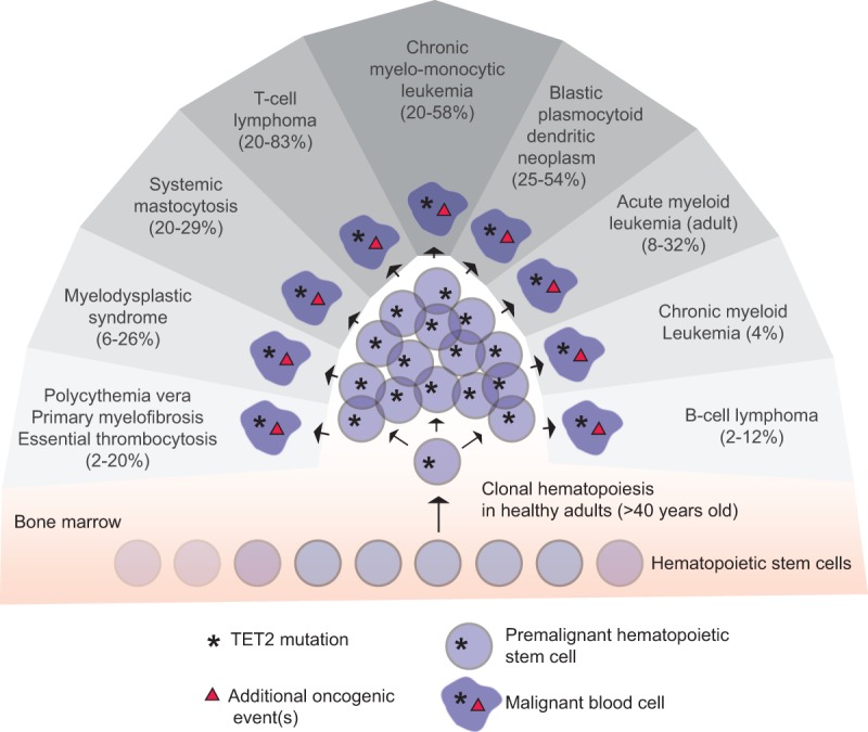
“Second, TET2 mutations are frequently observed in aged healthy individuals with clonal hematopoiesis and increase the propensity to develop hematopoieticmalignancies (Busque et al. 2012; Genovese et al. 2014; Jaiswal et al. 2014; Xie et al. 2014).”
BackgroundClonal hematopoiesis and blood-cancer risk inferred from blood DNA sequence
Genovese, G., Kähler, A. K., Handsaker, R. E., Lindberg, J., Rose, S. A., Bakhoum, S. F., Chambert, K., Mick, E., Neale, B. M., Fromer, M., et al. (2014) · New England Journal of Medicine · doi:10.1056/nejmoa1409405 · Cited by 3,531 · cited 1×
“Second, TET2 mutations are frequently observed in aged healthy individuals with clonal hematopoiesis and increase the propensity to develop hematopoieticmalignancies (Busque et al. 2012; Genovese et al. 2014; Jaiswal et al. 2014; Xie et al. 2014).”
BackgroundTet proteins can convert 5-methylcytosine to 5-formylcytosine and 5-carboxylcytosine
Ito, S., Shen, L., Dai, Q., Wu, S. C., Collins, L. B., Swenberg, J. A., He, C., & Zhang, Y. (2011) · Science · doi:10.1126/science.1210597 · Cited by 3,451 · cited 3×
“6-fold) substrates compared with the initial oxidation reaction of 5mC (Ito et al. 2011; Hu et al. 2015).”
BackgroundXu, W., Yang, H., Liu, Y., Yang, Y., Wang, P., Kim, S.-H., Ito, S., Yang, C., Wang, P., Xiao, M.-T., et al. (2011) · Cancer Cell · doi:10.1016/j.ccr.2010.12.014 · Cited by 2,875
A decade of exploring the cancer epigenome—biological and translational implications
Baylin, S. B., & Jones, P. A. (2011) · Nature Reviews Cancer · doi:10.1038/nrc3130 · Cited by 2,871
Tet-Mediated Formation of 5-Carboxylcytosine and Its Excision by TDG in Mammalian DNA
He, Y., Li, B.-Z., Li, Z., Liu, P., Wang, Y., Tang, Q., Ding, J., Jia, Y., Chen, Z., Li, L., et al. (2011) · Science · doi:10.1126/science.1210944 · Cited by 2,776 · cited 4×
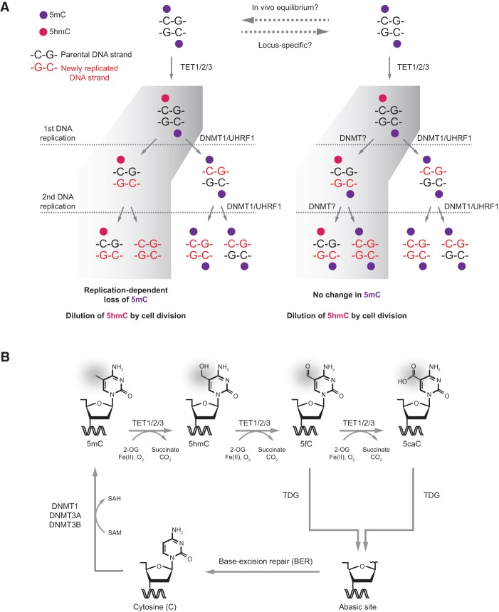
“Subsequent to the discovery of oxidized 5mC bases, it was demonstrated that TDG has no effect on 5hmC but shows robust recruitment and in vitro catalytic activity toward 5fC and 5caC (He et al. 2011; Maiti and Drohat 2011; Spruijt et al. 2013).”
BackgroundInfluentialFigueroa, M. E., Abdel-Wahab, O., Lu, C., Ward, P. S., Patel, J., Shih, A., Li, Y., Bhagwat, N., Vasanthakumar, A., Fernandez, H. F., et al. (2010) · Cancer Cell · doi:10.1016/j.ccr.2010.11.015 · Cited by 2,766
The Nuclear DNA Base 5-Hydroxymethylcytosine Is Present in Purkinje Neurons and the Brain
Kriaucionis, S., & Heintz, N. (2009) · Science · doi:10.1126/science.1169786 · Cited by 2,700 · cited 1×
“By comparison, the products of further TET-mediated oxidation, 5fC and 5caC, are observed at ∼10-fold to 100-fold lower levels than 5hmC and often close to or under the detection limit (Kriaucionis and Heintz 2009; Globisch et al. 2010; Szwagierczak et al. 2010; He et al. 2011; Ito et al. 2011; Pfaffeneder et al. 2011…”
Background
Cited by
Sorted by citations
Items 1,001–1,001 of 1,001
Metadata
- Authors
- Rasmussen, K. D., & Helin, K.
- Affiliations
- University of Copenhagen (DK)
- Corresponding author
- Helin, Kristian
- Venue
- Genes & Development
- Publisher
- Cold Spring Harbor Laboratory
- Published
- 2016-04-01
- Volume
- 30
- Issue
- 7
- Pages
- 733-750
- ISSN
- 0890-9369 (ISSN-L)
- 1549-5477
- PMC
- PMC4826392
- OpenAlex
- W2325267338
- Cited by
- 1,040
- References
- 162
- Citations by year
- 2026: 56 · 2025: 87 · 2024: 116 · 2023: 100 · 2022: 135 · 2021: 141 · 2020: 131 · 2019: 97 · 2018: 83 · 2017: 60 · 2016: 30
- Type
- posted content
- Language
- en
- Open access
- hybrid
- doi.org
- Licence
- cc-by-nc
- Locations
- Genes & Development (publishedVersion, cc-by-nc)
- PubMed Central (publishedVersion, cc-by-nc)
- PubMed (publishedVersion)
- Research at the University of Copenhagen (University of Copenhagen) (publishedVersion, cc-by-nc)
- HighWire Press Open Archive (submittedVersion)
- Funders
- Danish Medical Research Council (1333-00120B)
- European Research Council (294666_DNAMET)
- Danish Cancer Society
- Danish National Research Foundation (DNRF 82)
- Danish Council for Strategic Research
- Novo Nordisk Foundation
- Topics
- Life Sciences › Biochemistry, Genetics and Molecular Biology › Molecular Biology › Epigenetics and DNA Methylation
- Life Sciences › Biochemistry, Genetics and Molecular Biology › Molecular Biology › Genomics and Chromatin Dynamics
- Life Sciences › Biochemistry, Genetics and Molecular Biology › Molecular Biology › Cancer-related gene regulation
- Keywords
- TET enzymes, DNA methylation, 5-methylcytosine oxidation, TET2, demethylation, cancer, developmental regulation, transcriptional regulation, gene expression
- MeSH terms
- Animals, Cytosine, DNA-Binding Proteins, Humans, Neoplasms, Oxidation-Reduction, Proto-Oncogene Proteins, Gene Expression Regulation, Neoplastic, DNA Methylation, Epigenesis, Genetic, Dioxygenases
About these checks
Registry, retraction, Cites and statistics results are computed, not judged, with one exception: a printed reference that no index lists is read once by a model before it counts as a citation error. Claims are extracted by a model and kept only when their quote appears verbatim in the text; each cited source is then checked, with a model’s help, for whether it says what the claim relies on. Those verdicts can be wrong — read the claim’s quote and the cited source before relying on one.
Statistics and arithmetic are recomputed, not judged.
Confidence bands are shown only for fields calibrated on our annotated test set. None is calibrated yet, so no band is shown.
What counts as a claim ›How we measure confidence ›How we check this ›
Are you an author, or do you see a problem with this page? Request a review
