

The DOI matches its Crossref record; 35 of the 35 references it cites were found in a registry or an index.
SonoNet: Real-Time Detection and Localisation of Fetal Standard Scan Planes in Freehand Ultrasound
Open accessAbstract
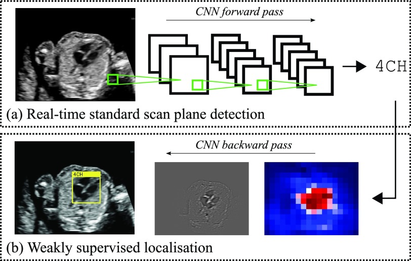
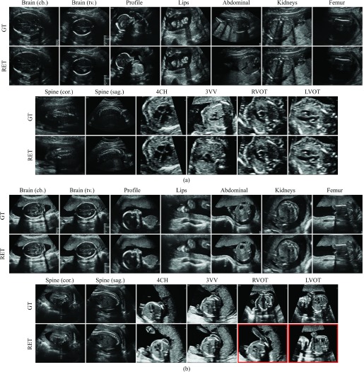
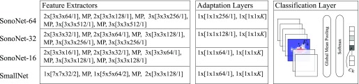
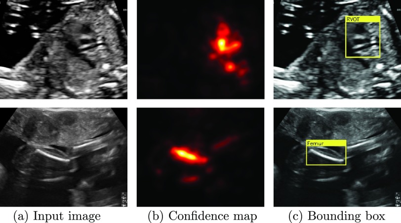
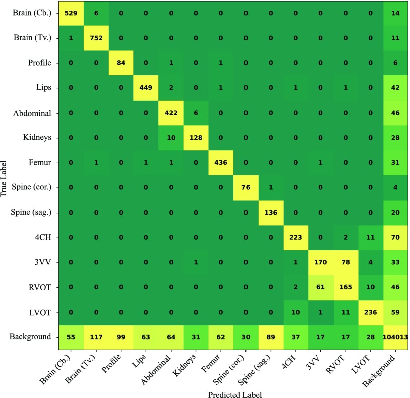
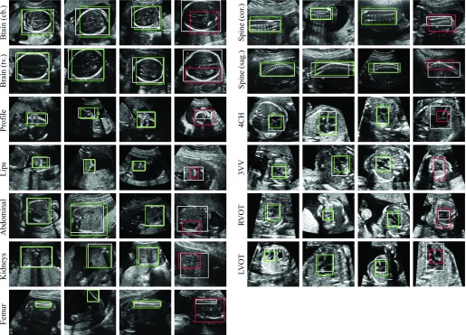
Identifying and interpreting fetal standard scan planes during 2-D ultrasound mid-pregnancy examinations are highly complex tasks, which require years of training. Apart from guiding the probe to the correct location, it can be equally difficult for a non-expert to identify relevant structures within the image. Automatic image processing can provide tools to help experienced as well as inexperienced operators with these tasks. In this paper, we propose a novel method based on convolutional neural networks, which can automatically detect 13 fetal standard views in freehand 2-D ultrasound data as well as provide a localization of the fetal structures via a bounding box. An important contribution is that the network learns to localize the target anatomy using weak supervision based on image-level labels only. The network architecture is designed to operate in real-time while providing optimal output for the localization task. We present results for real-time annotation, retrospective frame retrieval from saved videos, and localization on a very large and challenging dataset consisting of images and video recordings of full clinical anomaly screenings. We found that the proposed method achieved an average F1-score of 0.798 in a realistic classification experiment modeling real-time detection, and obtained a 90.09% accuracy for retrospective frame retrieval. Moreover, an accuracy of 77.8% was achieved on the localization task.
Overview
The claims and statistics of this paper haven’t been checked yet.
Run the check — free, no account needed. It reads the open-access text and checks the sources it cites.
Integrity signals
Registry
Cites
Statistics
Figures
Verification
Claims the paper makes
The claims and statistics of this paper haven’t been checked yet.
Run the check — free, no account needed. It reads the open-access text and checks the sources it cites.
Claims other papers make about this one
Each is checked against this paper’s own text: does it say what the citing paper says it does?
The cited source says what the claim attributes to it.
In the citing paper
“the possibility of real-time applicability [15,25,32,33]”
The check wasn’t confident either way, so it gives no verdict — never disputed.
In the citing paper
“Conventional assessments of fetal lung maturity rely on invasive amniocentesis and surface-level ultrasound scoring, achieving moderate predictive performance (AUC ≈ 0.82) but lacking dynamic integration of placental function (23, 24).”
From Placenta-pulmonary coupling-guided multimodal AI for fetal lung maturity staging and individualized glucocorticoid therapy (2026) · cites this paper as [23]
Experimental — accuracy not yet measuredIn the citing paper
“Baumgartner et al. [117,118] and Chen et al. [78,119] accomplished the detection of 13 fetal standard views (e.g., kidneys, brain, abdominal, spine, femur, and cardiac plane) and the fetal abdominal (or face and four-chamber view) standard plane in 2D US images through the transferred deep models, respectively.”
From Deep Learning in Medical Ultrasound Analysis: A Review (2019) · cites this paper as [118]
Self-check
Percentages, sums and reported differences re-computed from the paper’s own numbers.
The self-check needs the paper’s full text.
Citations
Cites
Deep Residual Learning for Image Recognition
He, K., Zhang, X., Ren, S., & Sun, J. (2016) · 2016 IEEE Conference on Computer Vision and Pattern Recognition (CVPR) · doi:10.1109/cvpr.2016.90 · Cited by 235,474
Very Deep Convolutional Networks for Large-Scale Image Recognition
Simonyan, K., & Zisserman, A. (2014) · arXiv · doi:10.48550/arxiv.1409.1556 · Cited by 114,148
ImageNet classification with deep convolutional neural networks
Krizhevsky, A., Sutskever, I., & Hinton, G. E. (2017) · Communications of the ACM · doi:10.1145/3065386 · Cited by 112,155
Batch Normalization: Accelerating Deep Network Training by Reducing Internal Covariate Shift
Ioffe, S., & Szegedy, C. (2015) · arXiv · doi:10.48550/arxiv.1502.03167 · Cited by 27,818
The pascal visual object classes (VOC) challenge
Everingham, M., Van Gool, L., Williams, C. K. I., Winn, J., & Zisserman, A. (2010) · International Journal of Computer Vision · doi:10.1007/s11263-009-0275-4 · Cited by 19,599
Batch Normalization: Accelerating Deep Network Training by Reducing Internal Covariate ShiftPreprint
Ioffe, S. (2024) · arXiv (Cornell University) · doi:10.57702/o9raffed · Cited by 15,273
He, H., & Garcia, E. A. (2009) · IEEE Transactions on Knowledge and Data Engineering · doi:10.1109/tkde.2008.239 · Cited by 10,359
Fast approximate energy minimization via graph cuts
Boykov, Y., Veksler, O., & Zabih, R. (2001) · IEEE Transactions on Pattern Analysis and Machine Intelligence · doi:10.1109/34.969114 · Cited by 6,988
Deep Inside Convolutional Networks: Visualising Image Classification Models and Saliency Maps
Simonyan, K., Vedaldi, A., & Zisserman, A. (2013) · arXiv · doi:10.48550/arxiv.1312.6034 · Cited by 5,370
Deep Residual Learning for Image Recognition
He, K., Zhang, X., Ren, S., & Sun, J. (2015) · arXiv · doi:10.48550/arxiv.1512.03385 · Cited by 4,196
Research works printed but not linked: 2
- 2015, Fetal Anomalie Screen Programme Handbook NHS Screening Programmes
- springett, 2014, British Isles Network of Congenital Anomaly Registers Congenital Anomaly Statistics 2010 England and Wales
Cited by
Sorted by citations
Attention gated networks: learning to leverage salient regions in medical images
Schlemper, J., Oktay, O., Schaap, M., Heinrich, M., Kainz, B., Glocker, B., & Rueckert, D. (2019) · Medical Image Analysis · doi:10.1016/j.media.2019.01.012 · Cited by 2,014
Self-citationRecent advances and clinical applications of deep learning in medical image analysis
Chen, X., Wang, X., Zhang, K., Fung, K.-M. A., Thai, T., Moore, K. N., Mannel, R. S., Liu, H., Zheng, B., & Qiu, Y. (2022) · Medical Image Analysis · doi:10.1016/j.media.2022.102444 · Cited by 1,180
Deep Learning in Medical Ultrasound Analysis: A Review
Liu, S., Wang, Y., Yang, X., Lei, B., Liu, L., Li, S. X., Ni, D., & Wang, T. (2019) · Engineering · doi:10.1016/j.eng.2018.11.020 · Cited by 798
Self-supervised learning for medical image analysis using image context restoration
Chen, L., Bentley, P., Mori, K., Misawa, K., Fujiwara, M., & Rueckert, D. (2019) · Medical Image Analysis · doi:10.1016/j.media.2019.101539 · Cited by 575
Self-citationA Novel Ultrasound Robot With Force/Torque Measurement and Control for Safe and Efficient Scanning
Bao, X., Wang, S., Zheng, L., Housden, R. J., Hajnal, J. V., & Rhode, K. (2023) · IEEE Transactions on Instrumentation and Measurement · doi:10.1109/tim.2023.3239925 · Cited by 432
Metadata
- Authors
- Baumgartner, C. F., Kamnitsas, K., Matthew, J., Fletcher, T. P., Smith, S., Koch, L. M., Kainz, B., & Rueckert, D.
- Affiliations
- Imperial College London (GB)
- Guy's Hospital (GB)
- King's College London (GB)
- Corresponding author
- Baumgartner, Christian F.
- ORCID
- Venue
- IEEE Transactions on Medical Imaging
- Published
- 2017-11
- Volume
- 36
- Issue
- 11
- Pages
- 2204-2215
- ISSN
- 0278-0062 (ISSN-L)
- 1558-254X
- PMC
- PMC6051487
- OpenAlex
- W2735582614
- Cited by
- 416
- References
- 34
- Citations by year
- 2026: 52 · 2025: 48 · 2024: 54 · 2023: 56 · 2022: 55 · 2021: 57 · 2020: 44 · 2019: 28 · 2018: 21 · 2017: 1
- FWCI
- 10.67
- Type
- journal article
- Language
- en
- Open access
- hybrid
- doi.org
- Locations
- IEEE Transactions on Medical Imaging (publishedVersion, cc-by)
- Europe PMC (PubMed Central) (publishedVersion, cc-by)
- PubMed Central (publishedVersion, cc-by)
- PubMed (publishedVersion)
- arXiv (Cornell University) (submittedVersion)
- Research Portal (King's College London) (submittedVersion, cc-by)
- Spiral (Imperial College London) (submittedVersion, cc-by)
- arXiv (Cornell University)
- Funders
- Wellcome Trust IEH Award (102431)
- Topics
- Physical Sciences › Computer Science › Computer Vision and Pattern Recognition › Medical Image Segmentation Techniques
- Health Sciences › Medicine › Health Informatics › Artificial Intelligence in Healthcare and Education
- Physical Sciences › Computer Science › Computer Vision and Pattern Recognition › Advanced Neural Network Applications
- Keywords
- real-time detection, weakly supervised learning, image-level labels, convolutional neural networks, bounding box localization
- MeSH terms
- Algorithms, Female, Humans, Image Processing, Computer-Assisted, Pregnancy, Video Recording, Ultrasonography, Prenatal, Neural Networks, Computer
About these checks
Registry, retraction, Cites and statistics results are computed, not judged, with one exception: a printed reference that no index lists is read once by a model before it counts as a citation error. Claims are extracted by a model and kept only when their quote appears verbatim in the text; each cited source is then checked, with a model’s help, for whether it says what the claim relies on. Those verdicts can be wrong — read the claim’s quote and the cited source before relying on one.
Statistics and arithmetic are recomputed, not judged.
Confidence bands are shown only for fields calibrated on our annotated test set. None is calibrated yet, so no band is shown.
What counts as a claim ›How we measure confidence ›How we check this ›
Are you an author, or do you see a problem with this page? Request a review
