

The DOI matches its Crossref record; 35 of the 35 references it cites were found in a registry or an index.
SonoNet: Real-Time Detection and Localisation of Fetal Standard Scan Planes in Freehand Ultrasound
Open accessAbstract
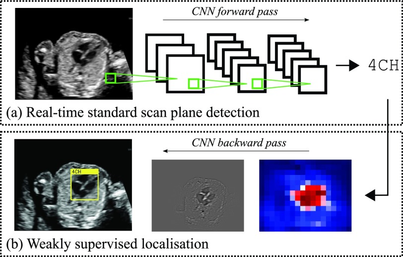
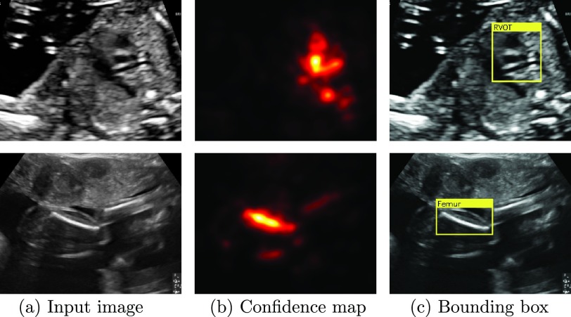
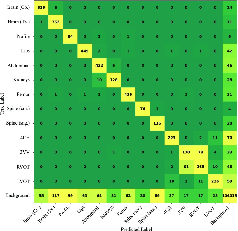
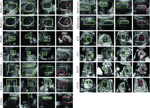
Identifying and interpreting fetal standard scan planes during 2-D ultrasound mid-pregnancy examinations are highly complex tasks, which require years of training. Apart from guiding the probe to the correct location, it can be equally difficult for a non-expert to identify relevant structures within the image. Automatic image processing can provide tools to help experienced as well as inexperienced operators with these tasks. In this paper, we propose a novel method based on convolutional neural networks, which can automatically detect 13 fetal standard views in freehand 2-D ultrasound data as well as provide a localization of the fetal structures via a bounding box. An important contribution is that the network learns to localize the target anatomy using weak supervision based on image-level labels only. The network architecture is designed to operate in real-time while providing optimal output for the localization task. We present results for real-time annotation, retrospective frame retrieval from saved videos, and localization on a very large and challenging dataset consisting of images and video recordings of full clinical anomaly screenings. We found that the proposed method achieved an average F1-score of 0.798 in a realistic classification experiment modeling real-time detection, and obtained a 90.09% accuracy for retrospective frame retrieval. Moreover, an accuracy of 77.8% was achieved on the localization task.
Overview
The claims and statistics of this paper haven’t been checked yet.
Run the check — free, no account needed. It reads the open-access text and checks the sources it cites.
Integrity signals
Registry
Cites
Statistics
Figures
Verification
Claims the paper makes
The claims and statistics of this paper haven’t been checked yet.
Run the check — free, no account needed. It reads the open-access text and checks the sources it cites.
Claims other papers make about this one
Each is checked against this paper’s own text: does it say what the citing paper says it does?
The cited source says what the claim attributes to it.
In the citing paper
“the possibility of real-time applicability [15,25,32,33]”
The check wasn’t confident either way, so it gives no verdict — never disputed.
In the citing paper
“Conventional assessments of fetal lung maturity rely on invasive amniocentesis and surface-level ultrasound scoring, achieving moderate predictive performance (AUC ≈ 0.82) but lacking dynamic integration of placental function (23, 24).”
From Placenta-pulmonary coupling-guided multimodal AI for fetal lung maturity staging and individualized glucocorticoid therapy (2026) · cites this paper as [23]
Experimental — accuracy not yet measuredIn the citing paper
“Baumgartner et al. [117,118] and Chen et al. [78,119] accomplished the detection of 13 fetal standard views (e.g., kidneys, brain, abdominal, spine, femur, and cardiac plane) and the fetal abdominal (or face and four-chamber view) standard plane in 2D US images through the transferred deep models, respectively.”
From Deep Learning in Medical Ultrasound Analysis: A Review (2019) · cites this paper as [118]
Self-check
Percentages, sums and reported differences re-computed from the paper’s own numbers.
The self-check needs the paper’s full text.
Citations
Cites
Deep Residual Learning for Image Recognition
He, K., Zhang, X., Ren, S., & Sun, J. (2016) · 2016 IEEE Conference on Computer Vision and Pattern Recognition (CVPR) · doi:10.1109/cvpr.2016.90 · Cited by 235,474
Very Deep Convolutional Networks for Large-Scale Image Recognition
Simonyan, K., & Zisserman, A. (2014) · arXiv · doi:10.48550/arxiv.1409.1556 · Cited by 114,148
ImageNet classification with deep convolutional neural networks
Krizhevsky, A., Sutskever, I., & Hinton, G. E. (2017) · Communications of the ACM · doi:10.1145/3065386 · Cited by 112,155
Batch Normalization: Accelerating Deep Network Training by Reducing Internal Covariate Shift
Ioffe, S., & Szegedy, C. (2015) · arXiv · doi:10.48550/arxiv.1502.03167 · Cited by 27,818
The pascal visual object classes (VOC) challenge
Everingham, M., Van Gool, L., Williams, C. K. I., Winn, J., & Zisserman, A. (2010) · International Journal of Computer Vision · doi:10.1007/s11263-009-0275-4 · Cited by 19,599
Batch Normalization: Accelerating Deep Network Training by Reducing Internal Covariate ShiftPreprint
Ioffe, S. (2024) · arXiv (Cornell University) · doi:10.57702/o9raffed · Cited by 15,273
He, H., & Garcia, E. A. (2009) · IEEE Transactions on Knowledge and Data Engineering · doi:10.1109/tkde.2008.239 · Cited by 10,359
Fast approximate energy minimization via graph cuts
Boykov, Y., Veksler, O., & Zabih, R. (2001) · IEEE Transactions on Pattern Analysis and Machine Intelligence · doi:10.1109/34.969114 · Cited by 6,988
Deep Inside Convolutional Networks: Visualising Image Classification Models and Saliency Maps
Simonyan, K., Vedaldi, A., & Zisserman, A. (2013) · arXiv · doi:10.48550/arxiv.1312.6034 · Cited by 5,370
Deep Residual Learning for Image Recognition
He, K., Zhang, X., Ren, S., & Sun, J. (2015) · arXiv · doi:10.48550/arxiv.1512.03385 · Cited by 4,196
Research works printed but not linked: 2
- 2015, Fetal Anomalie Screen Programme Handbook NHS Screening Programmes
- springett, 2014, British Isles Network of Congenital Anomaly Registers Congenital Anomaly Statistics 2010 England and Wales
Cited by
Sorted by citations
Attention gated networks: learning to leverage salient regions in medical images
Schlemper, J., Oktay, O., Schaap, M., Heinrich, M., Kainz, B., Glocker, B., & Rueckert, D. (2019) · Medical Image Analysis · doi:10.1016/j.media.2019.01.012 · Cited by 2,014
Self-citationRecent advances and clinical applications of deep learning in medical image analysis
Chen, X., Wang, X., Zhang, K., Fung, K.-M. A., Thai, T., Moore, K. N., Mannel, R. S., Liu, H., Zheng, B., & Qiu, Y. (2022) · Medical Image Analysis · doi:10.1016/j.media.2022.102444 · Cited by 1,180
Deep Learning in Medical Ultrasound Analysis: A Review
Liu, S., Wang, Y., Yang, X., Lei, B., Liu, L., Li, S. X., Ni, D., & Wang, T. (2019) · Engineering · doi:10.1016/j.eng.2018.11.020 · Cited by 798
Self-supervised learning for medical image analysis using image context restoration
Chen, L., Bentley, P., Mori, K., Misawa, K., Fujiwara, M., & Rueckert, D. (2019) · Medical Image Analysis · doi:10.1016/j.media.2019.101539 · Cited by 575
Self-citationA Novel Ultrasound Robot With Force/Torque Measurement and Control for Safe and Efficient Scanning
Bao, X., Wang, S., Zheng, L., Housden, R. J., Hajnal, J. V., & Rhode, K. (2023) · IEEE Transactions on Instrumentation and Measurement · doi:10.1109/tim.2023.3239925 · Cited by 432
Deep learning in ultrasound imaging
van Sloun, R. J. G., Cohen, R., & Eldar, Y. C. (2020) · Proceedings of the IEEE · doi:10.1109/jproc.2019.2932116 · Cited by 316
Arnaout, R., Curran, L., Zhao, Y., Levine, J. C., Chinn, E., & Moon-Grady, A. J. (2020) · doi:10.1101/2020.06.22.20137786 · Cited by 315
Burgos-Artizzu, X. P., Coronado-Gutiérrez, D., Valenzuela-Alcaraz, B., Bonet-Carne, E., Eixarch, E., Crispi, F., & Gratacós, E. (2020) · Scientific Reports · doi:10.1038/s41598-020-67076-5 · Cited by 240
Automatic Fetal Ultrasound Standard Plane Recognition Based on Deep Learning and IIoT
Pu, B., Li, K., Li, S., & Zhu, N. (2021) · IEEE Transactions on Industrial Informatics · doi:10.1109/tii.2021.3069470 · Cited by 221
A review on deep-learning algorithms for fetal ultrasound-image analysis
Fiorentino, M. C., Villani, F. P., Cosmo, M. D., Frontoni, E., & Moccia, S. (2022) · Medical Image Analysis · doi:10.1016/j.media.2022.102629 · Cited by 224
Robotic ultrasound imaging: State-of-the-art and future perspectives
Jiang, Z., Salcudean, S. E., & Navab, N. (2023) · Medical Image Analysis · doi:10.1016/j.media.2023.102878 · Cited by 213
Using deep-learning algorithms to classify fetal brain ultrasound images as normal or abnormal
Xie, H. N., Wang, N., He, M., Zhang, L. H., Cai, H. M., Xian, J. B., Lin, M. F., Zheng, J., & Yang, Y. Z. (2020) · Ultrasound in Obstetrics & Gynecology · doi:10.1002/uog.21967 · Cited by 176
A Survey on Multimodal Data-Driven Smart Healthcare Systems: Approaches and Applications
Cai, Q., Wang, H. H., Li, Z., & Liu, X. (2019) · IEEE Access · doi:10.1109/access.2019.2941419 · Cited by 172
Becker, A. S., Mueller, M., Stoffel, E., Marcon, M., Ghafoor, S., & Boss, A. (2018) · The British Journal of Radiology · doi:10.1259/bjr.20170576 · Cited by 157
Visual Feature Attribution Using Wasserstein GANs
Baumgartner, C. F., Koch, L. M., Tezcan, K. C., Ang, J. X., & Konukoğlu, E. (2018) · IEEE Computer Society Conference on Computer Vision and Pattern Recognition/Proceedings - IEEE Computer Society Conference on Computer Vision and Pattern Recognition/Proceedings · doi:10.1109/cvpr.2018.00867 · Cited by 155
Self-citationAn Overview of Systems and Techniques for Autonomous Robotic Ultrasound Acquisitions
Li, K., Xu, Y., & Meng, M. Q. ‐. (2021) · IEEE Transactions on Medical Robotics and Bionics · doi:10.1109/tmrb.2021.3072190 · Cited by 162
Detection of Cardiac Structural Abnormalities in Fetal Ultrasound Videos Using Deep Learning
Komatsu, M., Sakai, A., Komatsu, R., Matsuoka, R., Yasutomi, S., Shozu, K., Dozen, A., Machino, H., Hidaka, H., Arakaki, T., et al. (2021) · Applied Sciences · doi:10.3390/app11010371 · Cited by 151
Multi-task learning for quality assessment of fetal head ultrasound images
Lin, Z., Li, S., Ni, D., Liao, Y., Wen, H., Du, J., Chen, S., Wang, T., & Lei, B. (2019) · Medical Image Analysis · doi:10.1016/j.media.2019.101548 · Cited by 129
Østvik, A., Smistad, E., Aase, S. A., Haugen, B. O., & Løvstakken, L. (2018) · Ultrasound in Medicine & Biology · doi:10.1016/j.ultrasmedbio.2018.07.024 · Cited by 129
Zhang, J., Liu, M., Wang, L., Chen, S., Yuan, P., Li, J., Shen, S. G.-F., Tang, Z., Chen, K.-C., Xia, J. J., et al. (2019) · Medical Image Analysis · doi:10.1016/j.media.2019.101621 · Cited by 119
A Generic Quality Control Framework for Fetal Ultrasound Cardiac Four-Chamber Planes
Dong, J., Liu, S., Liao, Y., Wen, H., Lei, B., Li, S., & Wang, T. (2020) · IEEE Journal of Biomedical and Health Informatics · doi:10.1109/jbhi.2019.2948316 · Cited by 104
Use of artificial intelligence and deep learning in fetal ultrasound imaging
Ramirez Zegarra, R., & Ghi, T. (2023) · Ultrasound in Obstetrics & Gynecology · doi:10.1002/uog.26130 · Cited by 102
Robot-Assisted Medical Imaging: A Review
Salcudean, S. E., Moradi, H., Black, D., & Navab, N. (2022) · Proceedings of the IEEE · doi:10.1109/jproc.2022.3162840 · Cited by 101
Namburete, A. I. L., Xie, W., Yaqub, M., Zisserman, A., & Noble, J. A. (2018) · Medical Image Analysis · doi:10.1016/j.media.2018.02.006 · Cited by 95
Segmentation and classification in MRI and US fetal imaging: Recent trends and future prospects
Torrents-Barrena, J., Piella, G., Masoller, N., Gratacós, E., Eixarch, E., Ceresa, M., & Ballester, M. Á. G. (2019) · Medical Image Analysis · doi:10.1016/j.media.2018.10.003 · Cited by 97
Dhombres, F., Bonnard, J., Bailly, K., Maurice, P., Papageorghiou, A. T., & Jouannic, J.-M. (2022) · Journal of Medical Internet Research · doi:10.2196/35465 · Cited by 93
van den Heuvel, T. L. A., Petros, H., Santini, S., de Korte, C. L., & van Ginneken, B. (2019) · Ultrasound in Medicine & Biology · doi:10.1016/j.ultrasmedbio.2018.09.015 · Cited by 91
Kim, K. C., Cho, H. C., Jang, T. J., Choi, J. M., & Seo, J. K. (2020) · Computer Methods and Programs in Biomedicine · doi:10.1016/j.cmpb.2020.105833 · Cited by 88
Ultrasound Video Transformers for Cardiac Ejection Fraction Estimation
Reynaud, H., Vlontzos, A., Hou, B., Beqiri, A., Leeson, P., & Kainz, B. (2021) · Lecture notes in computer science · doi:10.1007/978-3-030-87231-1_48 · Cited by 86
Self-citationRecent Advances in Artificial Intelligence-Assisted Ultrasound Scanning
Tenajas, R., Miraut, D., Illana, C., Alonso-González, R., Arias-Valcayo, F., & Herraiz, J. L. (2023) · Applied Sciences · doi:10.3390/app13063693 · Cited by 87
Sinclair, M. D., Baumgartner, C. F., Matthew, J., Bai, W., Martinez, J. C., Li, Y., Smith, S., Knight, C. L., Kainz, B., Hajnal, J. V., et al. (2018) · Annual International Conference of the IEEE Engineering in Medicine and Biology Society (EMBC) · doi:10.1109/embc.2018.8512278 · Cited by 78
Self-citationDeep learning for ultrasound image caption generation based on object detection
Zeng, X., Wen, L., Liu, B., & Qi, X. (2019) · Neurocomputing · doi:10.1016/j.neucom.2018.11.114 · Cited by 79
Li, K., Wang, J., Xu, Y., Qin, H., Liu, D., Liu, L., & Meng, M. Q. ‐. (2021) · Proceedings - IEEE International Conference on Robotics and Automation/Proceedings · doi:10.1109/icra48506.2021.9561295 · Cited by 76
Xiao, Q., Brown, L. G., Foran, D. J., Nosher, J. L., & Hacihaliloglu, I. (2021) · International Journal of Computer Assisted Radiology and Surgery · doi:10.1007/s11548-020-02305-w · Cited by 72
Artificial intelligence in obstetric ultrasound: A scoping review.
Horgan, R., Nehme, L., & Abuhamad, A. (2023) · Prenatal Diagnosis · doi:10.1002/pd.6411 · Cited by 72
“Two studies evaluated AI systems to automatically detect 13 standard anatomic planes.(105,106) In the study by Matthews et al.”
MethodEfficient 3-D Near-Field MIMO-SAR Imaging for Irregular Scanning Geometries
Smith, J. W., & Torlak, M. (2022) · IEEE Access · doi:10.1109/access.2022.3145370 · Cited by 72
Sridar, P., Kumar, A., Quinton, A., Nanan, R., Kim, J., & Krishnakumar, R. (2019) · Ultrasound in Medicine & Biology · doi:10.1016/j.ultrasmedbio.2018.11.016 · Cited by 70
Model-Based and Data-Driven Strategies in Medical Image Computing
Rueckert, D., & Schnabel, J. A. (2019) · Proceedings of the IEEE · doi:10.1109/jproc.2019.2943836 · Cited by 76
Self-citationSakai, A., Komatsu, M., Komatsu, R., Matsuoka, R., Yasutomi, S., Dozen, A., Shozu, K., Arakaki, T., Machino, H., Asada, K., et al. (2022) · Biomedicines · doi:10.3390/biomedicines10030551 · Cited by 68
Standard fetal ultrasound plane classification based on stacked ensemble of deep learning models
Krishna, T. B., & Kokil, P. (2023) · Expert Systems with Applications · doi:10.1016/j.eswa.2023.122153 · Cited by 67
Lin, M., He, X., Guo, H., He, M., Zhang, L., Xian, J., Lei, T., Xu, Q., Zheng, J., Feng, J., et al. (2022) · Ultrasound in Obstetrics & Gynecology · doi:10.1002/uog.24843 · Cited by 65
Computer-aided diagnosis for fetal brain ultrasound images using deep convolutional neural networks
Xie, B., Lei, T., Wang, N., Cai, H., Xian, J., He, M., Zhang, L., & Xie, H. (2020) · International Journal of Computer Assisted Radiology and Surgery · doi:10.1007/s11548-020-02182-3 · Cited by 65
A review of image processing methods for fetal head and brain analysis in ultrasound images
Torres, H. R., Morais, P., Oliveira, B., Birdir, C., Rüdiger, M., Fonseca, J. C., & Vilaça, J. L. (2022) · Computer Methods and Programs in Biomedicine · doi:10.1016/j.cmpb.2022.106629 · Cited by 67
Artificial Intelligence in Prenatal Ultrasound Diagnosis
He, F., Wang, Y., Xiu, Y., Zhang, Y., & Chen, L. (2021) · Frontiers in Medicine · doi:10.3389/fmed.2021.729978 · Cited by 62
Artificial Intelligence in Congenital Heart Disease
Jone, P., Gearhart, A. S., Lei, H., Xing, F., Nahar, J. K., López-Jiménez, F. A., Diller, G., Marelli, A. J., Wilson, L., Saidi, A. S., et al. (2022) · JACC Advances · doi:10.1016/j.jacadv.2022.100153 · Cited by 69
Automatic quality assessment for 2D fetal sonographic standard plane based on multitask learning
Zhang, B., Liu, H., Luo, H., & Li, K. (2021) · Medicine · doi:10.1097/md.0000000000024427 · Cited by 72
VesNet-RL: Simulation-Based Reinforcement Learning for Real-World US Probe Navigation
Bi, Y., Jiang, Z., Gao, Y., Wendler, T., Karlas, A., & Navab, N. (2022) · IEEE Robotics and Automation Letters · doi:10.1109/lra.2022.3176112 · Cited by 66
Standard Plane Detection in 3D Fetal Ultrasound Using an Iterative Transformation Network
Li, Y., Khanal, B., Hou, B., Alansary, A., Cerrolaza, J. J., Sinclair, M., Matthew, J., Gupta, C., Knight, C., Kainz, B., et al. (2018) · Lecture notes in computer science · doi:10.1007/978-3-030-00928-1_45 · Cited by 61
Self-citationClinical benefit of AI-assisted lung ultrasound in a resource-limited intensive care unit
Nhat, P. T. H., Hảo, N. V., Tho, P. V., Kerdegari, H., Pisani, L., Thu, L. N. M., Phuong, L. T., Duong, H. T. H., Thuy, D. B., McBride, A., et al. (2023) · Critical Care · doi:10.1186/s13054-023-04548-w · Cited by 60
Qu, R., Xu, G., Ding, C., Jia, W., & Sun, M. (2020) · IEEE Access · doi:10.1109/access.2020.2991845 · Cited by 59
Drukker, L., Sharma, H., Droste, R., Alsharid, M., Chatelain, P., Noble, J. A., & Papageorghiou, A. T. (2021) · Scientific Reports · doi:10.1038/s41598-021-92829-1 · Cited by 59
Artificial intelligence, fetal echocardiography, and congenital heart disease
Day, T. G., Kainz, B., Hajnal, J. V., Razavi, R., & Simpson, J. M. (2021) · Prenatal Diagnosis · doi:10.1002/pd.5892 · Cited by 67
Self-citationAutomated 3D Fetal Brain Segmentation Using an Optimized Deep Learning Approach
Zhao, L., Asis-Cruz, J. D., Feng, X., Wu, Y., Kapse, K. J., Largent, A., Quistorff, J. L., Lopez, C., Wu, D., Qing, K., et al. (2022) · American Journal of Neuroradiology · doi:10.3174/ajnr.a7419 · Cited by 59
Sendra-Balcells, C., Campello, V. M., Torrents-Barrena, J., Ahmed, Y. A., Elattar, M., Ohene-Botwe, B., Nyangulu, P., Stones, W., Ammar, M., Benamer, L. N., et al. (2023) · Scientific Reports · doi:10.1038/s41598-023-29490-3 · Cited by 57
Machine Learning in Robotic Ultrasound Imaging: Challenges and Perspectives
Bi, Y., Jiang, Z., Duelmer, F., Huang, D., & Navab, N. (2024) · Annual Review of Control Robotics and Autonomous Systems · doi:10.1146/annurev-control-091523-100042 · Cited by 66
Deep learning fetal ultrasound video model match human observers in biometric measurements
Płotka, S., Klasa, A., Lisowska, A., Seliga-Siwecka, J., Lipa, M., Trzciński, T., & Sitek, A. (2022) · Physics in Medicine & Biology · doi:10.1088/1361-6560/ac4d85 · Cited by 57
Nurmaini, S., Rachmatullah, M. N., Sapitri, A. I., Darmawahyuni, A., Jovandy, A., Firdaus, F., Tutuko, B., & Passarella, R. (2020) · IEEE Access · doi:10.1109/access.2020.3034367 · Cited by 63
Learning to map 2D ultrasound images into 3D space with minimal human annotation
Yeung, P.-H., Aliasi, M., Papageorghiou, A. T., Haak, M., Xie, W., & Namburete, A. I. L. (2021) · Medical Image Analysis · doi:10.1016/j.media.2021.101998 · Cited by 55
Self-Supervised Representation Learning for Ultrasound Video
Jiao, J., Droste, R., Drukker, L., Papageorghiou, A. T., & Noble, J. A. (2020) · 2020 IEEE 17th International Symposium on Biomedical Imaging (ISBI) · doi:10.1109/isbi45749.2020.9098666 · Cited by 56
Li, K., Xu, Y., Wang, J., Ni, D., Liu, L., & Meng, M. Q. ‐. (2021) · IEEE Transactions on Medical Robotics and Bionics · doi:10.1109/tmrb.2021.3127015 · Cited by 56
Borkovkina, S., Camino, A., Janpongsri, W., Šarunic, M. V., & Jian, Y. (2020) · Biomedical Optics Express · doi:10.1364/boe.395279 · Cited by 53
Weakly Supervised Estimation of Shadow Confidence Maps in Fetal Ultrasound Imaging
Meng, Q., Housden, J., Matthew, J., Rueckert, D., Schnabel, J. A., Kainz, B., Sinclair, M., Zimmer, V., Hou, B., Rajchl, M., et al. (2019) · IEEE Transactions on Medical Imaging · doi:10.1109/tmi.2019.2913311 · Cited by 52
Self-citationKrishna, T. B., & Kokil, P. (2023) · Biomedical Signal Processing and Control · doi:10.1016/j.bspc.2023.105283 · Cited by 54
Use of artificial intelligence in obstetrics: Not quite ready for prime time
Sarno, L., Neola, D., Carbone, L., Saccone, G., Carlea, A., Miceli, M., Iorio, G. G., Mappa, I., Rizzo, G., Girolamo, R. D., et al. (2023) · American Journal of Obstetrics & Gynecology MFM · doi:10.1016/j.ajogmf.2022.100792 · Cited by 47
Self-Supervised Ultrasound to MRI Fetal Brain Image Synthesis
Jiao, J., Namburete, A. I. L., Papageorghiou, A. T., & Noble, J. A. (2020) · IEEE Transactions on Medical Imaging · doi:10.1109/tmi.2020.3018560 · Cited by 48
Meng, Q., Matthew, J., Zimmer, V. A., Gómez, A., Lloyd, D. F. A., Rueckert, D., & Kainz, B. (2020) · IEEE Transactions on Medical Imaging · doi:10.1109/tmi.2020.3035424 · Cited by 48
Self-citationGenerative Adversarial Networks to Improve Fetal Brain Fine-Grained Plane Classification
Montero, A., Bonet-Carne, E., & Burgos-Artizzu, X. P. (2021) · Sensors · doi:10.3390/s21237975 · Cited by 48
Sharma, H., Drukker, L., Chatelain, P., Droste, R., Papageorghiou, A. T., & Noble, J. A. (2021) · Medical Image Analysis · doi:10.1016/j.media.2021.101973 · Cited by 48
Oh, K., Yoon, J. S., & Suk, H. (2022) · IEEE Transactions on Pattern Analysis and Machine Intelligence · doi:10.1109/tpami.2022.3197845 · Cited by 52
Yi, J., Kang, H. K., Kwon, J.-H., Kim, K.-S., Park, M. H., Seong, Y. K., Kim, D. W., Ahn, B. E., Ha, K., Lee, J.-Y., et al. (2020) · ULTRASONOGRAPHY · doi:10.14366/usg.20102 · Cited by 49
VP-Nets : Efficient automatic localization of key brain structures in 3D fetal neurosonography
Huang, R., Xie, W., & Noble, J. A. (2018) · Medical Image Analysis · doi:10.1016/j.media.2018.04.004 · Cited by 47
Cai, Y., Sharma, H., Chatelain, P., & Noble, J. A. (2018) · Lecture notes in computer science · doi:10.1007/978-3-030-00928-1_98 · Cited by 46
Jost, E., Kosian, P., Jimenez Cruz, J., Albarqouni, S., Gembruch, U., Strizek, B., & Recker, F. (2023) · Journal of Clinical Medicine · doi:10.3390/jcm12216833 · Cited by 44
“Operator-related factors: US in general and obstetric US in particular, are known to require substantial experience and to be extremely training dependent [15].”
BackgroundMethodInfluentialAdaptive feature squeeze network for nuclear cataract classification in AS-OCT image
Zhang, X., Xiao, Z., Higashita, R., Hu, Y., Chen, W., Yuan, J., & Liu, J. (2022) · Journal of Biomedical Informatics · doi:10.1016/j.jbi.2022.104037 · Cited by 44
Advances in the Application of Artificial Intelligence in Fetal Echocardiography
Zhang, J., Xiao, S., Zhu, Y. C., Zhang, Z., Cao, H., Xie, M., & Zhang, L. (2024) · Journal of the American Society of Echocardiography · doi:10.1016/j.echo.2023.12.013 · Cited by 62
Deep learning-based real time detection for cardiac objects with fetal ultrasound video
Iriani Sapitri, A., Nurmaini, S., Naufal Rachmatullah, M., Tutuko, B., Darmawahyuni, A., Firdaus, F., Rini, D. P., & Islami, A. (2023) · Informatics in Medicine Unlocked · doi:10.1016/j.imu.2022.101150 · Cited by 42
Qiao, S., Pang, S., Luo, G., Pan, S., Chen, T., & Lv, Z. (2022) · IEEE Journal of Biomedical and Health Informatics · doi:10.1109/jbhi.2021.3091579 · Cited by 41
Spatio-temporal visual attention modelling of standard biometry plane-finding navigation
Cai, Y., Droste, R., Sharma, H., Chatelain, P., Drukker, L., Papageorghiou, A. T., & Noble, J. A. (2020) · Medical Image Analysis · doi:10.1016/j.media.2020.101762 · Cited by 42
FetalNet: Multi-task Deep Learning Framework for Fetal Ultrasound Biometric Measurements
Płotka, S., Wlodarczyk, T. W., Klasa, A., Lipa, M., Sitek, A., & Trzcinski, T. P. (2021) · Communications in computer and information science · doi:10.1007/978-3-030-92310-5_30 · Cited by 41
Exploring a new paradigm for the fetal anomaly ultrasound scan: Artificial intelligence in real time
Matthew, J., Skelton, E., Day, T. G., Zimmer, V. A., Gomez, A., Wheeler, G., Toussaint, N., Liu, T., Budd, S., Lloyd, K., et al. (2022) · Prenatal Diagnosis · doi:10.1002/pd.6059 · Cited by 41
Self-citationArroyo, J., Marini, T. J., Saavedra, A. C., Toscano, M., Baran, T. M., Drennan, K., Dozier, A., Zhao, Y. T., Egoavil, M., Tamayo, L., et al. (2022) · PLOS ONE · doi:10.1371/journal.pone.0262107 · Cited by 40
Artificial intelligence as a new answer to old challenges in maternal-fetal medicine and obstetrics
Medjedović, E., Stanojević, M., Jonuzovic-Prosic, S., Ribić, E., Begić, Z., Cerovac, A., & Badnjević, A. (2023) · Technology and Health Care · doi:10.3233/thc-231482 · Cited by 43
Jiang, Z., Bi, Y., Zhou, M., Hu, Y., Burke, M., & Navab, N. (2024) · The International Journal of Robotics Research · doi:10.1177/02783649231223547 · Cited by 40
Ou, Z., Bai, J., Chen, Z., Lu, Y., Wang, H., Long, S., & Chen, G. (2024) · Computers in Biology and Medicine · doi:10.1016/j.compbiomed.2024.108501 · Cited by 39
RetractedSearching collaborative agents for multi-plane localization in 3D ultrasound
Yang, X., Huang, Y., Huang, R., Dou, H., Li, R., Qian, J., Huang, X., Shi, W., Chen, C., Zhang, Y., et al. (2021) · Medical Image Analysis · doi:10.1016/j.media.2021.102119 · Cited by 39
Zhang, X., Xiao, Z., Higashita, R., Chen, W., Yuan, J., Fang, J., Hu, Y., & Liu, J. (2020) · Conference proceedings/Conference proceedings - IEEE International Conference on Systems, Man, and Cybernetics · doi:10.1109/smc42975.2020.9283218 · Cited by 38
AutoFB: Automating Fetal Biometry Estimation from Standard Ultrasound Planes
Bano, S., Dromey, B., Vasconcelos, F., Napolitano, R., David, A. L., Peebles, D., & Stoyanov, D. V. (2021) · Lecture notes in computer science · doi:10.1007/978-3-030-87234-2_22 · Cited by 40
Maraci, M. A., Yaqub, M., Craik, R., Beriwal, S., Self, A., von Dadelszen, P., Papageorghiou, A., & Noble, J. A. (2020) · Journal of medical imaging · doi:10.1117/1.jmi.7.1.014501 · Cited by 36
Self-Supervised Contrastive Video-Speech Representation Learning for Ultrasound
Jiao, J., Cai, Y., Alsharid, M., Drukker, L., Papageorghiou, A. T., & Noble, J. A. (2020) · Lecture Notes in Computer Science · doi:10.1007/978-3-030-59716-0_51 · Cited by 38
Drukker, L., Sharma, H., Karim, J. N., Droste, R., Noble, J. A., & Papageorghiou, A. T. (2022) · Ultrasound in Obstetrics & Gynecology · doi:10.1002/uog.24975 · Cited by 36
SonoEyeNet: Standardized fetal ultrasound plane detection informed by eye tracking
Cai, Y., Sharma, H., Chatelain, P., & Noble, J. A. (2018) · IEEE International Symposium on Biomedical Imaging (ISBI) · doi:10.1109/isbi.2018.8363851 · Cited by 36
Confident Head Circumference Measurement from Ultrasound with Real-Time Feedback for Sonographers
Budd, S., Sinclair, M. D., Khanal, B., Matthew, J., Lloyd, D. F. A., Gómez, A., Toussaint, N., Robinson, E. C., & Kainz, B. (2019) · Lecture notes in computer science · doi:10.1007/978-3-030-32251-9_75 · Cited by 36
Self-citationLi, K., Li, A., Xu, Y., Xiong, H., & Meng, M. Q. ‐. (2023) · IEEE Transactions on Automation Science and Engineering · doi:10.1109/tase.2023.3246089 · Cited by 36
Spontaneous Preterm Birth Prediction Using Convolutional Neural Networks
Wlodarczyk, T. W., Płotka, S., Rokita, P. S., Sochacki-Wójcicka, N., Wójcicki, J., Lipa, M., & Trzcinski, T. P. (2020) · Lecture notes in computer science · doi:10.1007/978-3-030-60334-2_27 · Cited by 38
Fetal biometry and amniotic fluid volume assessment end-to-end automation using Deep Learning
Slimani, S., Hounka, S., Mahmoudi, A., Rehah, T., Laoudiyi, D., Saadi, H., Bouziyane, A., Lamrissi, A., Jalal, M., Bouhya, S., et al. (2023) · Nature Communications · doi:10.1038/s41467-023-42438-5 · Cited by 41
Towards Standardized Acquisition With a Dual-Probe Ultrasound Robot for Fetal Imaging
Housden, R. J., Wang, S., Bao, X., Zheng, J., Skelton, E., Matthew, J., Noh, Y., Eltiraifi, O., Singh, A., Singh, D., et al. (2021) · IEEE Robotics and Automation Letters · doi:10.1109/lra.2021.3056033 · Cited by 34
Self-citationWu, L., Bin, D., Liu, X., Hong, W., Chen, L., Gao, K., Sheng, Q., Yu, Y., Zhao, L., & Zhang, Y. (2022) · Frontiers in Pediatrics · doi:10.3389/fped.2021.770182 · Cited by 35
D’Alberti, E., Patey, O. V., Smith, C., Šalović, B., Hernandez-Cruz, N., Noble, J. A., & Papageorghiou, A. T. (2025) · EClinicalMedicine · doi:10.1016/j.eclinm.2025.103250 · Cited by 38
Low-Memory CNNs Enabling Real-Time Ultrasound Segmentation Towards Mobile Deployment
Vaze, S., Xie, W., & Namburete, A. I. L. (2020) · IEEE Journal of Biomedical and Health Informatics · doi:10.1109/jbhi.2019.2961264 · Cited by 33
Selvathi, D., & Chandralekha, R. (2021) · Multidimensional Systems and Signal Processing · doi:10.1007/s11045-021-00765-0 · Cited by 33
Metadata
- Authors
- Baumgartner, C. F., Kamnitsas, K., Matthew, J., Fletcher, T. P., Smith, S., Koch, L. M., Kainz, B., & Rueckert, D.
- Affiliations
- Imperial College London (GB)
- Guy's Hospital (GB)
- King's College London (GB)
- Corresponding author
- Baumgartner, Christian F.
- ORCID
- Venue
- IEEE Transactions on Medical Imaging
- Published
- 2017-11
- Volume
- 36
- Issue
- 11
- Pages
- 2204-2215
- ISSN
- 0278-0062 (ISSN-L)
- 1558-254X
- PMC
- PMC6051487
- OpenAlex
- W2735582614
- Cited by
- 416
- References
- 34
- Citations by year
- 2026: 52 · 2025: 48 · 2024: 54 · 2023: 56 · 2022: 55 · 2021: 57 · 2020: 44 · 2019: 28 · 2018: 21 · 2017: 1
- FWCI
- 10.67
- Type
- journal article
- Language
- en
- Open access
- hybrid
- doi.org
- Locations
- IEEE Transactions on Medical Imaging (publishedVersion, cc-by)
- Europe PMC (PubMed Central) (publishedVersion, cc-by)
- PubMed Central (publishedVersion, cc-by)
- PubMed (publishedVersion)
- arXiv (Cornell University) (submittedVersion)
- Research Portal (King's College London) (submittedVersion, cc-by)
- Spiral (Imperial College London) (submittedVersion, cc-by)
- arXiv (Cornell University)
- Funders
- Wellcome Trust IEH Award (102431)
- Topics
- Physical Sciences › Computer Science › Computer Vision and Pattern Recognition › Medical Image Segmentation Techniques
- Health Sciences › Medicine › Health Informatics › Artificial Intelligence in Healthcare and Education
- Physical Sciences › Computer Science › Computer Vision and Pattern Recognition › Advanced Neural Network Applications
- Keywords
- real-time detection, weakly supervised learning, image-level labels, convolutional neural networks, bounding box localization
- MeSH terms
- Algorithms, Female, Humans, Image Processing, Computer-Assisted, Pregnancy, Video Recording, Ultrasonography, Prenatal, Neural Networks, Computer
About these checks
Registry, retraction, Cites and statistics results are computed, not judged, with one exception: a printed reference that no index lists is read once by a model before it counts as a citation error. Claims are extracted by a model and kept only when their quote appears verbatim in the text; each cited source is then checked, with a model’s help, for whether it says what the claim relies on. Those verdicts can be wrong — read the claim’s quote and the cited source before relying on one.
Statistics and arithmetic are recomputed, not judged.
Confidence bands are shown only for fields calibrated on our annotated test set. None is calibrated yet, so no band is shown.
What counts as a claim ›How we measure confidence ›How we check this ›
Are you an author, or do you see a problem with this page? Request a review
