

The DOI matches its Crossref record; 52 of the 52 references it cites were found in a registry or an index.
Aging of the skeletal muscle extracellular matrix drives a stem cell fibrogenic conversion
Open accessAbstract
Age-related declines in skeletal muscle regeneration have been attributed to muscle stem cell (MuSC) dysfunction. Aged MuSCs display a fibrogenic conversion, leading to fibrosis and impaired recovery after injury. Although studies have demonstrated the influence of in vitro substrate characteristics on stem cell fate, whether and how aging of the extracellular matrix (ECM) affects stem cell behavior has not been investigated. Here, we investigated the direct effect of the aged muscle ECM on MuSC lineage specification. Quantification of ECM topology and muscle mechanical properties reveals decreased collagen tortuosity and muscle stiffening with increasing age. Age-related ECM alterations directly disrupt MuSC responses, and MuSCs seeded ex vivo onto decellularized ECM constructs derived from aged muscle display increased expression of fibrogenic markers and decreased myogenicity, compared to MuSCs seeded onto young ECM. This fibrogenic conversion is recapitulated in vitro when MuSCs are seeded directly onto matrices elaborated by aged fibroblasts. When compared to young fibroblasts, fibroblasts isolated from aged muscle display increased nuclear levels of the mechanosensors, Yes-associated protein (YAP)/transcriptional coactivator with PDZ-binding motif (TAZ), consistent with exposure to a stiff microenvironment in vivo. Accordingly, preconditioning of young fibroblasts by seeding them onto a substrate engineered to mimic the stiffness of aged muscle increases YAP/TAZ nuclear translocation and promotes secretion of a matrix that favors MuSC fibrogenesis. The findings here suggest that an age-related increase in muscle stiffness drives YAP/TAZ-mediated pathogenic expression of matricellular proteins by fibroblasts, ultimately disrupting MuSC fate.
Overview
The claims and statistics of this paper haven’t been checked yet.
Run the check — free, no account needed. It reads the open-access text and checks the sources it cites.
Integrity signals
Registry
Cites
Statistics
Figures
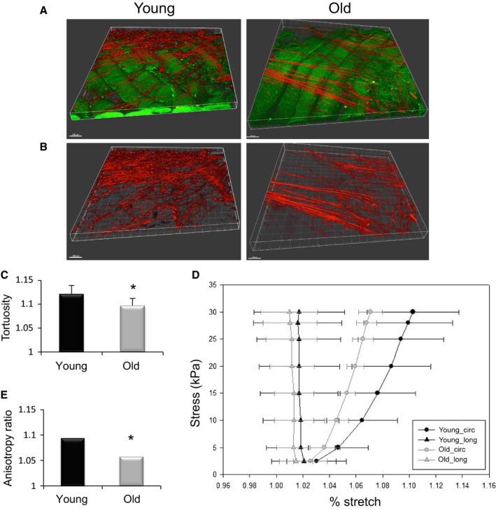
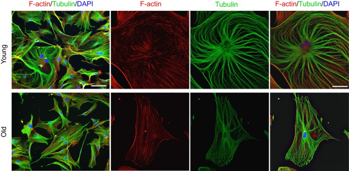
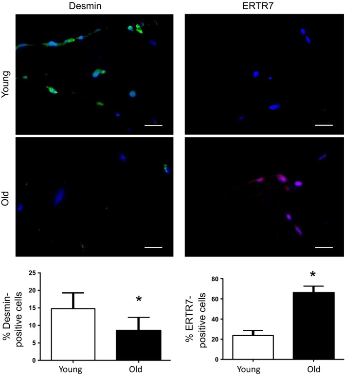
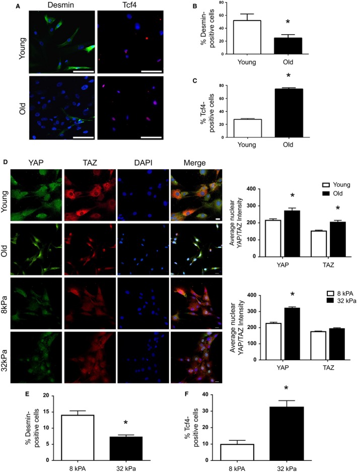
Verification
Claims the paper makes
The claims and statistics of this paper haven’t been checked yet.
Run the check — free, no account needed. It reads the open-access text and checks the sources it cites.
Claims other papers make about this one
Each is checked against this paper’s own text: does it say what the citing paper says it does?
In the citing paper
“Decreased collagen tortuosity and increased stiffness in the muscle niche are associated with aberrant MuSC differentiation82.”
From Ageing and rejuvenation of tissue stem cells and their niches (2023) · cites this paper as [82]
In the citing paper
“In muscle, quantitative proteomic studies also reveal striking remodelling in ECM during ageing66,82, and those changes can influence MuSC function.”
From Ageing and rejuvenation of tissue stem cells and their niches (2023) · cites this paper as [82]
In the citing paper
“In muscle, ECM stiffness also increases with age and is associated with decreased collagen tortuosity82. These ECM changes induce MuSCs to adopt a more fibrogenic fate, thereby diminishing their myogenic potential.”
From Ageing and rejuvenation of tissue stem cells and their niches (2023) · cites this paper as [82]
Self-check
Percentages, sums and reported differences re-computed from the paper’s own numbers.
The self-check needs the paper’s full text.
Citations
Cites
Matrix elasticity directs stem cell lineage specification
Engler, A. J., Sen, S., Sweeney, H. L., & Discher, D. E. (2006) · Cell · doi:10.1016/j.cell.2006.06.044 · Cited by 13,915
Role of YAP/TAZ in mechanotransduction
Dupont, S., Morsut, L., Aragona, M., Enzo, E., Giulitti, S., Cordenonsi, M., Zanconato, F., Le Digabel, J., Forcato, M., Bicciato, S., et al. (2011) · Nature · doi:10.1038/nature10137 · Cited by 5,979
SATELLITE CELL OF SKELETAL MUSCLE FIBERS
Mauro, A. (1961) · The Journal of Cell Biology · doi:10.1083/jcb.9.2.493 · Cited by 3,683
Rejuvenation of aged progenitor cells by exposure to a young systemic environment
Conboy, I. M., Conboy, M. J., Wagers, A. J., Girma, E. R., Weissman, I. L., & Rando, T. A. (2005) · Nature · doi:10.1038/nature03260 · Cited by 2,287
Self-citationMyotubes differentiate optimally on substrates with tissue-like stiffness
Engler, A. J., Griffin, M. A., Sen, S., Bönnemann, C. G., Sweeney, H. L., & Discher, D. E. (2004) · The Journal of Cell Biology · doi:10.1083/jcb.200405004 · Cited by 1,743
Aragona, M., Panciera, T., Manfrin, A., Giulitti, S., Michielin, F., Elvassore, N., Dupont, S., & Piccolo, S. (2013) · Cell · doi:10.1016/j.cell.2013.07.042 · Cited by 1,651
Cellular mechanotransduction: putting all the pieces together again
Ingber, D. E. (2006) · The FASEB Journal · doi:10.1096/fj.05-5424rev · Cited by 1,627
Substrate Elasticity Regulates Skeletal Muscle Stem Cell Self-Renewal in Culture
Gilbert, P. M., Havenstrite, K. L., Magnusson, K. E. G., Sacco, A., Leonardi, N. A., Kraft, P., Nguyen, N. K., Thrun, S., Lutolf, M. P., & Blau, H. M. (2010) · Science · doi:10.1126/science.1191035 · Cited by 1,587
Increased Wnt signaling during aging alters muscle stem cell fate and increases fibrosis
Brack, A. S., Conboy, M. J., Roy, S., Lee, M., Kuo, C. J., Keller, C., & Rando, T. A. (2007) · Science · doi:10.1126/science.1144090 · Cited by 1,564
Self-citationBrew, K., & Nagase, H. (2010) · Biochimica et Biophysica Acta (BBA) - Molecular Cell Research · doi:10.1016/j.bbamcr.2010.01.003 · Cited by 1,432
Cited by
Sorted by citations
Skeletal muscle: A review of molecular structure and function, in health and disease
Mukund, K., & Subramaniam, S. (2019) · WIREs Systems Biology and Medicine · doi:10.1002/wsbm.1462 · Cited by 789
Lin, H., Sohn, J., Shen, H., Langhans, M. T., & Tuan, R. S. (2018) · Biomaterials · doi:10.1016/j.biomaterials.2018.06.026 · Cited by 454
Self-citationAgeing and rejuvenation of tissue stem cells and their niches
Brunet, A., Goodell, M. A., & Rando, T. A. (2023) · Nature Reviews Molecular Cell Biology · doi:10.1038/s41580-022-00510-w · Cited by 407
Self-citationChemical strategies to engineer hydrogels for cell culture
Lou, J., & Mooney, D. J. (2022) · Nature Reviews Chemistry · doi:10.1038/s41570-022-00420-7 · Cited by 407
Bao, H., Cao, J., Chen, M., Min, C., Chen, W., Chen, X., Chen, Y., Chen, Y., Chen, Y., Chen, Z., et al. (2023) · Science China Life Sciences · doi:10.1007/s11427-023-2305-0 · Cited by 402
Skeletal muscle fibrosis: an overview
Mahdy, M. A. A. (2018) · Cell and Tissue Research · doi:10.1007/s00441-018-2955-2 · Cited by 320
Mechanoregulation of YAP and TAZ in Cellular Homeostasis and Disease Progression
Cai, X., Wang, K.-C., & Meng, Z. (2021) · Frontiers in Cell and Developmental Biology · doi:10.3389/fcell.2021.673599 · Cited by 333
Extracellular matrix: an important regulator of cell functions and skeletal muscle development
Zhang, W., Liu, Y., & Zhang, H. (2021) · Cell & Bioscience · doi:10.1186/s13578-021-00579-4 · Cited by 289
Mesenchymal Stem/Stromal Cell Senescence: Hallmarks, Mechanisms, and Combating Strategies
Weng, Z., Wang, Y., Ouchi, T., Liu, H., Qiao, X., Wu, C., Zhao, Z., Li, L., & Li, B. (2022) · Stem Cells Translational Medicine · doi:10.1093/stcltm/szac004 · Cited by 303
Cellular and epigenetic drivers of stem cell ageing
Ermolaeva, M., Neri, F., Ori, A., & Rudolph, K. L. (2018) · Nature Reviews Molecular Cell Biology · doi:10.1038/s41580-018-0020-3 · Cited by 272
Identity Noise and Adipogenic Traits Characterize Dermal Fibroblast Aging
Salzer, M. C., Lafzi, A., Berenguer, A., Youssif, C., Castellanos‐Martín, A., Solanas, G., Peixoto, F. O., Attolini, C. S., Prats, N., Aguilera, M., et al. (2018) · Cell · doi:10.1016/j.cell.2018.10.012 · Cited by 246
Mechanisms of Arterial Stiffening
Lacolley, P. J., Regnault, V., & Laurent, S. (2020) · Arteriosclerosis Thrombosis and Vascular Biology · doi:10.1161/atvbaha.119.313129 · Cited by 203
Perandini, L. A., Chimin, P., Lutkemeyer, D. D. S., & Câmara, N. O. S. (2018) · FEBS Journal · doi:10.1111/febs.14417 · Cited by 167
Vitamin D Promotes Skeletal Muscle Regeneration and Mitochondrial Health
Latham, C. M., Brightwell, C. R., Keeble, A. R., Munson, B. D., Thomas, N. T., Zagzoog, A. M., Fry, C. S., & Fry, J. L. (2021) · Frontiers in Physiology · doi:10.3389/fphys.2021.660498 · Cited by 171
Inflammaging: The ground for sarcopenia?
Liang, Z., Zhang, T., Liu, H., Li, Z., Peng, L., Wang, C., & Wang, T. (2022) · Experimental Gerontology · doi:10.1016/j.exger.2022.111931 · Cited by 156
The Muscle Stem Cell Niche in Health and Disease
Mashinchian, O., Pisconti, A., Moal, E. L., & Bentzinger, C. F. (2017) · Current topics in developmental biology/Current Topics in Developmental Biology · doi:10.1016/bs.ctdb.2017.08.003 · Cited by 139
In Vitro Tissue‐Engineered Skeletal Muscle Models for Studying Muscle Physiology and Disease
Khodabukus, A., Prabhu, N. K., Wang, J., & Bursac, N. (2018) · Advanced Healthcare Materials · doi:10.1002/adhm.201701498 · Cited by 136
Role of YAP/TAZ in Cell Lineage Fate Determination and Related Signaling Pathways
Heng, B. C., Zhang, X., Aubel, D., Bai, Y., Li, X., Yan, W., Fussenegger, M., & Deng, X. (2020) · Frontiers in Cell and Developmental Biology · doi:10.3389/fcell.2020.00735 · Cited by 133
Iijima, H., Gilmer, G. G., Wang, K., Bean, A., He, Y., Lin, H., Tang, W., Lamont, D. N., Tai, C., Ito, A., et al. (2023) · Nature Communications · doi:10.1038/s41467-022-35359-2 · Cited by 118
Self-citationPavan, P. G., Monti, E., Bondı̀, M., Fan, C., Stecco, C., Narici, M. V., Reggiani, C., & Marcucci, L. (2020) · International Journal of Molecular Sciences · doi:10.3390/ijms21113992 · Cited by 106
Shang, G., Han, L., Wang, Z., Liu, Y., Yan, S., Sai, W.-W., Wang, D., Li, Y., Zhang, W., & Zhong, M. (2020) · Journal of Cachexia Sarcopenia and Muscle · doi:10.1002/jcsm.12560 · Cited by 104
Loreti, M., & Sacco, A. (2022) · npj Regenerative Medicine · doi:10.1038/s41536-022-00204-z · Cited by 111
Decellularized tissues as platforms for in vitro modeling of healthy and diseased tissues
McCrary, M. W., Bousalis, D., Mobini, S., Song, Y. H., & Schmidt, C. E. (2020) · Acta Biomaterialia · doi:10.1016/j.actbio.2020.05.031 · Cited by 98
Ala, M., & Ala, M. (2021) · ACS Pharmacology & Translational Science · doi:10.1021/acsptsci.1c00167 · Cited by 97
Kramerova, I. A., Kumagai-Cresse, C., Ermolova, N. V., Mokhonova, E. I., Marinov, M., Capote, J. C., Becerra, D., Quattrocelli, M., Crosbie, R. H., Welch, E. M., et al. (2019) · Human Molecular Genetics · doi:10.1093/hmg/ddz181 · Cited by 97
Epigenetic regulation of satellite cell fate during skeletal muscle regeneration
Massenet, J., Gardner, E., Chazaud, B., & Dilworth, F. J. (2021) · Skeletal Muscle · doi:10.1186/s13395-020-00259-w · Cited by 99
Opportunities for organoids as new models of aging
Hu, J. L., Todhunter, M. E., LaBarge, M. A., & Gartner, Z. J. (2017) · The Journal of Cell Biology · doi:10.1083/jcb.201709054 · Cited by 92
Stochastic non-enzymatic modification of long-lived macromolecules - A missing hallmark of aging
Fedintsev, A., & Moskalev, A. (2020) · Ageing Research Reviews · doi:10.1016/j.arr.2020.101097 · Cited by 72
Tissue-Engineered Skeletal Muscle Models to Study Muscle Function, Plasticity, and Disease
Khodabukus, A. (2021) · Frontiers in Physiology · doi:10.3389/fphys.2021.619710 · Cited by 92
Passive stiffness of fibrotic skeletal muscle in mdx mice relates to collagen architecture
Brashear, S. E., Wohlgemuth, R. P., Gonzalez, G., & Smith, L. (2020) · The Journal of Physiology · doi:10.1113/jp280656 · Cited by 78
Extracellular matrix: Brick and mortar in the skeletal muscle stem cell niche
Schüler, S. C., Liu, Y., Dumontier, S., Grandbois, M., Moal, E. L., Cornelison, D., & Bentzinger, C. F. (2022) · Frontiers in Cell and Developmental Biology · doi:10.3389/fcell.2022.1056523 · Cited by 88
Wohlgemuth, R. P., Brashear, S. E., & Smith, L. (2023) · American Journal of Physiology-Cell Physiology · doi:10.1152/ajpcell.00287.2023 · Cited by 98
Tissue-specific parameters for the design of ECM-mimetic biomaterials
Tonti, O. R., Larson, H., Lipp, S. N., Luetkemeyer, C. M., Makam, M., Vargas, D., Wilcox, S. M., & Calve, S. (2021) · Acta Biomaterialia · doi:10.1016/j.actbio.2021.04.017 · Cited by 84
Forcina, L., Miano, C., Scicchitano, B. M., & Musarò, A. (2019) · Cells · doi:10.3390/cells8030232 · Cited by 82
Mitochondrial Dysfunction in Cancer Cachexia: Impact on Muscle Health and Regeneration
Beltrà, M., Pin, F., Ballarò, R., Costelli, P., & Penna, F. (2021) · Cells · doi:10.3390/cells10113150 · Cited by 78
Giroud, J., Bouriez, I., Paulus, H., Pourtier, A., Debacq‐Chainiaux, F., & Pluquet, O. (2023) · International Journal of Molecular Sciences · doi:10.3390/ijms241310788 · Cited by 87
Wang, D. M., Zhang, X., Huang, S., Liu, Y., Fu, S., Mak, K. K., Blocki, A. M., Yung, P. S., Tuan, R. S., & Ker, D. F. E. (2021) · Biomaterials · doi:10.1016/j.biomaterials.2021.120789 · Cited by 71
Self-citationGhosh, D., Peña, C. M., Quach, N. D., Xuan, B., Lee, A. H., & Dawson, M. (2020) · Journal of Cell Science · doi:10.1242/jcs.232470 · Cited by 70
Liu, X., Gao, Y., Long, X., Hayashi, T., Mizuno, K., Hattori, S., Fujisaki, H., Ogura, T., Wang, D. O., & Ikejima, T. (2019) · Food & Function · doi:10.1039/c9fo01346f · Cited by 70
Zullo, A., Fleckenstein, J., Schleip, R., Hoppe, K., Wearing, S. C., & Klingler, W. (2020) · Frontiers in Physiology · doi:10.3389/fphys.2020.00592 · Cited by 73
The cellular mechanobiology of aging: from biology to mechanics
Bajpai, A., Li, R., & Chen, W. (2020) · Annals of the New York Academy of Sciences · doi:10.1111/nyas.14529 · Cited by 71
Lipotoxicity, aging, and muscle contractility: does fiber type matter?
Carter, C. S., Justice, J. N., & Thompson, L. (2019) · GeroScience · doi:10.1007/s11357-019-00077-z · Cited by 66
Dysregulation of the Hippo pathway signaling in aging and cancer
Yeung, Y. T., Guerrero‐Castilla, A., Cano, M., Muñoz, M. F., Ayala, A., & Argüelles, S. (2019) · Pharmacological Research · doi:10.1016/j.phrs.2019.03.018 · Cited by 62
The Effects of Aging on the Intramuscular Connective Tissue
Fede, C., Fan, C., Pirri, C., Petrelli, L., Biz, C., Porzionato, A., Macchi, V., De, R., & Stecco, C. (2022) · International Journal of Molecular Sciences · doi:10.3390/ijms231911061 · Cited by 64
Advanced Glycation End-Products in Skeletal Muscle Aging
Olson, L. C., Redden, J. T., Schwartz, Z., Cohen, D. J., & McClure, M. J. (2021) · Bioengineering · doi:10.3390/bioengineering8110168 · Cited by 66
Rando, T. A., Brunet, A., & Goodell, M. A. (2025) · Cell stem cell · doi:10.1016/j.stem.2025.06.004 · Cited by 68
Self-citationAgeing of skeletal muscle extracellular matrix and mitochondria: finding a potential link
Cai, L., Shi, L., Peng, Z., Sun, Y., & Chen, J. (2023) · Annals of Medicine · doi:10.1080/07853890.2023.2240707 · Cited by 63
Mechanisms of muscle cells alterations and regeneration decline during aging
Chinvattanachot, G., Rivas, D., & Duque, G. (2024) · Ageing Research Reviews · doi:10.1016/j.arr.2024.102589 · Cited by 61
Stem Cell Aging in Lifespan and Disease: A State-of-the-Art Review
Sameri, S., Samadi, P., Dehghan, R., Salem, E., Hosseini, N. F., & Amini, R. (2020) · Current Stem Cell Research & Therapy · doi:10.2174/1574888x15666200213105155 · Cited by 57
Rahman, F. A., Angus, S. A., Stokes, K., Karpowicz, P. A., & Krause, M. P. (2020) · International Journal of Molecular Sciences · doi:10.3390/ijms21134575 · Cited by 57
Obstacles and challenges for tissue engineering and regenerative medicine: Australian nuances
Grounds, M. D. (2017) · Clinical and Experimental Pharmacology and Physiology · doi:10.1111/1440-1681.12899 · Cited by 54
Dietary epicatechin improves survival and delays skeletal muscle degeneration in aged mice
Si, H., Wang, X., Zhang, L., Parnell, L. D., Ahmed, B., LeRoith, T., Ansah, T.-A., Zhang, L., Li, J., Ordovás, J. M., et al. (2018) · The FASEB Journal · doi:10.1096/fj.201800554rr · Cited by 56
Abbott, C. B., Lawrence, M. M., Kobak, K. A., Lopes, E. B. P., Peelor, F. F., Donald, E. J., Remmen, H. V., Griffin, T. M., & Miller, B. F. (2021) · Function · doi:10.1093/function/zqab028 · Cited by 54
Sassoli, C., Pierucci, F., Zecchi‐Orlandini, S., & Meacci, E. (2019) · International Journal of Molecular Sciences · doi:10.3390/ijms20225545 · Cited by 55
Brightwell, C. R., Latham, C. M., Thomas, N. T., Keeble, A. R., Murach, K. A., & Fry, C. S. (2022) · American Journal of Physiology-Cell Physiology · doi:10.1152/ajpcell.00200.2022 · Cited by 52
Careccia, G., Mangiavini, L., & Cirillo, F. (2023) · International Journal of Molecular Sciences · doi:10.3390/ijms25010512 · Cited by 66
Zhou, J., So, K. K. H., Li, Y., Li, Y., Yuan, J., Ding, Y., Chen, F., Huang, Y., Liu, J., Lee, W., et al. (2019) · Aging Cell · doi:10.1111/acel.12996 · Cited by 52
Targeting the stem cell niche micro-environment as therapeutic strategies in aging
Farahzadi, R., Valipour, B., Montazersaheb, S., & Fathi, E. (2023) · Frontiers in Cell and Developmental Biology · doi:10.3389/fcell.2023.1162136 · Cited by 51
Anoikis in cell fate, physiopathology, and therapeutic interventions
Mei, J., Jiang, X., Tian, H., Rong, D., Song, J., Wang, L., Chen, Y., Wong, R. C. B., Guo, C., Wang, L., et al. (2024) · MedComm · doi:10.1002/mco2.718 · Cited by 59
Cellular Biomechanics in Skeletal Muscle Regeneration
Li, E. W., McKee-Muir, O. C., & Gilbert, P. M. (2017) · Current topics in developmental biology/Current Topics in Developmental Biology · doi:10.1016/bs.ctdb.2017.08.007 · Cited by 55
Fuqua, J. D., Lawrence, M. M., Hettinger, Z. R., Borowik, A., Brecheen, P., Szczygiel, M. M., Abbott, C. B., Peelor, F. F., Confides, A. L., Kinter, M. T., et al. (2023) · Journal of Cachexia Sarcopenia and Muscle · doi:10.1002/jcsm.13285 · Cited by 47
Hsp90β interacts with MDM2 to suppress p53‐dependent senescence during skeletal muscle regeneration
He, M. Y., Xu, S. B., Qu, Z., Guo, Y. M., Liu, X. C., Cong, X. X., Wang, J. F., Low, B. C., Li, L., Wu, Q., et al. (2019) · Aging Cell · doi:10.1111/acel.13003 · Cited by 48
Skeletal muscle as an experimental model of choice to study tissue aging and rejuvenation
Etienne, J., Liu, C., Skinner, C. M., Conboy, M. J., & Conboy, I. M. (2020) · Skeletal Muscle · doi:10.1186/s13395-020-0222-1 · Cited by 53
Liu, Y., Li, M., Lv, X., Bao, K., Tian, X. Y., He, L., Shi, L., Zhu, Y., & Ai, D. (2022) · Circulation Research · doi:10.1161/circresaha.121.320464 · Cited by 43
Changes in cardiac resident fibroblast physiology and phenotype in aging
Trial, J., & Cieslik, K. A. (2018) · American Journal of Physiology-Heart and Circulatory Physiology · doi:10.1152/ajpheart.00237.2018 · Cited by 46
Insight into muscle stem cell regeneration and mechanobiology
Pang, K. T., Loo, L. S. W., Chia, S., Ong, F. Y. T., Yu, H., & Walsh, I. (2023) · Stem Cell Research & Therapy · doi:10.1186/s13287-023-03363-y · Cited by 46
Differentiation reveals latent features of aging and an energy barrier in murine myogenesis
Kimmel, J. C., Yi, N., Roy, M., Hendrickson, D. G., & Kelley, D. R. (2021) · Cell Reports · doi:10.1016/j.celrep.2021.109046 · Cited by 42
Age-associated changes in the mechanical properties of human cadaveric pelvic floor muscles
Burnett, L. A., Cook, M. S., Shah, S. B., Wong, M., Kado, D. M., & Alperin, M. (2019) · Journal of Biomechanics · doi:10.1016/j.jbiomech.2019.109436 · Cited by 42
Available In Vitro Models for Human Satellite Cells from Skeletal Muscle
Romagnoli, C., Iantomasi, T., & Brandi, M. L. (2021) · International Journal of Molecular Sciences · doi:10.3390/ijms222413221 · Cited by 44
Age-Associated Differences in Recovery from Exercise-Induced Muscle Damage
Li, D. C. W., Rudloff, S., Langer, H. T., Norman, K., & Herpich, C. (2024) · Cells · doi:10.3390/cells13030255 · Cited by 45
Lyu, A., Zhu, S., Chen, J., Zhao, Y., Pu, D., Luo, C., Lyu, Q., Fan, Z., Sun, Y., Wu, J., et al. (2019) · Experimental Gerontology · doi:10.1016/j.exger.2019.04.008 · Cited by 40
Xu, J., Fu, S. N., & Hug, F. (2021) · BMC Musculoskeletal Disorders · doi:10.1186/s12891-021-04519-8 · Cited by 41
Age-Related Changes in the Matrisome of the Mouse Skeletal Muscle
Lofaro, F. D., Cisterna, B., Lacavalla, M. A., Boschi, F., Malatesta, M., Quaglino, D., Zancanaro, C., & Boraldi, F. (2021) · International Journal of Molecular Sciences · doi:10.3390/ijms221910564 · Cited by 40
Taetzsch, T., Brayman, V. L., & Valdez, G. (2018) · Biochimica et Biophysica Acta (BBA) - Molecular Basis of Disease · doi:10.1016/j.bbadis.2018.06.009 · Cited by 42
Thrown for a loop: fibro-adipogenic progenitors in skeletal muscle fibrosis
Loomis, T., & Smith, L. (2023) · American Journal of Physiology-Cell Physiology · doi:10.1152/ajpcell.00245.2023 · Cited by 39
Acun, A., Oganesyan, R., Uygun, K., Yeh, H., Yarmush, M. L., & Uygun, B. E. (2021) · Biomaterials · doi:10.1016/j.biomaterials.2021.120689 · Cited by 39
Myoblast mechanotransduction and myotube morphology is dependent on BAG3 regulation of YAP and TAZ
Günay, K. A., Silver, J. S., Chang, T.-L., Bednarski, O. J., Bannister, K. L., Rogowski, C. J., Olwin, B. B., & Anseth, K. S. (2021) · Biomaterials · doi:10.1016/j.biomaterials.2021.121097 · Cited by 42
Caloric restriction maintains stem cells through niche and regulates stem cell aging
Maharajan, N., Vijayakumar, K. A., Jang, C. H., & Cho, G. (2019) · Journal of Molecular Medicine · doi:10.1007/s00109-019-01846-1 · Cited by 38
Sosa, P., Alcalde‐Estévez, E., Asenjo‐Bueno, A., Plaza, P., Carrillo‐López, N., Olmos, G., López‐Ongil, S., & Ruiz‐Torres, M. P. (2021) · Journal of Cachexia Sarcopenia and Muscle · doi:10.1002/jcsm.12750 · Cited by 39
Giha, H. A., Sater, M. S., & Alamin, O. A. O. (2022) · Acta Diabetologica · doi:10.1007/s00592-022-01860-9 · Cited by 39
Alcalde-Estévez, E., Asenjo-Bueno, A., Sosa, P., Olmos, G., Plaza, P., Caballero-Mora, M. Á., Rodríguez-Puyol, D., Ruíz-Torres, M. P., & López-Ongil, S. (2020) · Aging · doi:10.18632/aging.103450 · Cited by 35
Extracellular matrix/mitochondria pathway: A novel potential target for sarcopenia
Melouane, A., Yoshioka, M., & St‐Amand, J. (2019) · Mitochondrion · doi:10.1016/j.mito.2019.10.007 · Cited by 35
Natural products and skeletal muscle health
Qu, Z., Zhou, S., Li, P., Liu, C., Yuan, B., Zhang, S., & Liu, A. (2021) · The Journal of Nutritional Biochemistry · doi:10.1016/j.jnutbio.2021.108619 · Cited by 35
Xiong, X., Li, W., Nam, J., Qu, M., Kay, S. A., & Ma, K. (2022) · Journal of Cell Science · doi:10.1242/jcs.260094 · Cited by 34
Age-Related Changes in Extracellular Matrix
Guvatova, Z. G., Borisov, P. V., Alekseev, A. A., & Moskalev, A. (2022) · Biochemistry (Moscow) · doi:10.1134/s0006297922120112 · Cited by 35
Decellularized Cell‐Secreted Extracellular Matrices as Biomaterials for Tissue Engineering
Ramos‐Rodriguez, D. H., & Leach, J. K. (2024) · Small Science · doi:10.1002/smsc.202400335 · Cited by 35
Narayanan, N., & Calve, S. (2020) · Connective Tissue Research · doi:10.1080/03008207.2020.1814263 · Cited by 35
Cellular enlargement - A new hallmark of aging?
Davies, D., van den Handel, K., Bharadwaj, S., & Lengefeld, J. (2022) · Frontiers in Cell and Developmental Biology · doi:10.3389/fcell.2022.1036602 · Cited by 32
Liao, J., Zheng, Y., Hu, M., Xu, P., Lin, L., Liu, X., Wu, Y., Huang, B., Ye, X., Li, S., et al. (2021) · Hypertension · doi:10.1161/hypertensionaha.121.18363 · Cited by 33
Odd skipped-related 1 controls the pro-regenerative response of fibro-adipogenic progenitors
Kotsaris, G., Qazi, T. H., Bucher, C. H., Zahid, H., Pöhle‐Kronawitter, S., Ugorets, V., Jarassier, W., Börno, S. T., Timmermann, B., Giesecke‐Thiel, C., et al. (2023) · npj Regenerative Medicine · doi:10.1038/s41536-023-00291-6 · Cited by 31
Kobak, K. A., Lawrence, M. M., Pharaoh, G. A., Borowik, A., Peelor, F. F., Shipman, P. D., Griffin, T. M., Remmen, H. V., & Miller, B. F. (2021) · Journal of Cachexia Sarcopenia and Muscle · doi:10.1002/jcsm.12772 · Cited by 31
Chen, S., Huang, L., Liu, B., Duan, H., Li, Z., Liu, Y., Li, H., Fu, X., Lin, J., Xu, Y., et al. (2023) · Science China Life Sciences · doi:10.1007/s11427-023-2400-3 · Cited by 33
Bechshøft, C. J. L., Jensen, S. M., Schjerling, P., Andersen, J. L., Svensson, R. B., Eriksen, C. S., Mkumbuzi, N. S., Kjær, M., & Mackey, A. L. (2019) · American Journal of Physiology-Cell Physiology · doi:10.1152/ajpcell.00049.2019 · Cited by 31
Skeletal muscle aging – Stem cells in the spotlight
Henze, H., Jung, M. J., Ahrens, H. E., Steiner, S., & von Maltzahn, J. (2020) · Mechanisms of Ageing and Development · doi:10.1016/j.mad.2020.111283 · Cited by 30
RhoA within myofibers controls satellite cell microenvironment to allow hypertrophic growth
Noviello, C., Kobon, K., Delivry, L., Guilbert, T., Britto, F. A., Julienne, F., Maire, P., Randrianarison-Huetz, V., & Sotiropoulos, A. (2021) · iScience · doi:10.1016/j.isci.2021.103616 · Cited by 29
Mechanical control of the mammalian circadian clock via YAP/TAZ and TEAD
Abenza, J. F., Rossetti, L., Mouelhi, M., Burgués, J., Andreu, I., Kennedy, K., Roca‐Cusachs, P., Marco, S., García‐Ojalvo, J., & Trepat, X. (2023) · The Journal of Cell Biology · doi:10.1083/jcb.202209120 · Cited by 29
Fischer, A. G., Elliott, E. M., Brittian, K. R., Garrett, L. T., Sadri, G., Aebersold, J., Singhal, R. A., Nong, Y., Leask, A., Jones, S. P., et al. (2024) · Matrix Biology · doi:10.1016/j.matbio.2024.08.001 · Cited by 30
Hunt, E. R., Confides, A. L., Abshire, S. M., Dupont‐Versteegden, E. E., & Butterfield, T. A. (2019) · Physiological Reports · doi:10.14814/phy2.14200 · Cited by 26
Sinha, U. S., Malis, V., Chen, J., Csapo, R., Kinugasa, R., Narici, M. V., & Sinha, S. (2020) · Frontiers in Physiology · doi:10.3389/fphys.2020.00626 · Cited by 27
Aparicio, P., Navarrete‐Villanueva, D., Gómez‐Cabello, A., López‐Royo, T., Santamaría, E., Fernández‐Irigoyen, J., Ausín, K., Arruebo, M., Sebastián, V., Vicente‐Rodríguez, G., et al. (2024) · Journal of Cachexia Sarcopenia and Muscle · doi:10.1002/jcsm.13539 · Cited by 27
Metadata
- Authors
- Stearns-Reider, K. M., D'Amore, A., Beezhold, K., Rothrauff, B., Cavalli, L., Wagner, W. R., Vorp, D. A., Tsamis, A., Shinde, S., Zhang, C., Barchowsky, A., Rando, T. A., Tuan, R. S., & Ambrosio, F.
- Affiliations
- University of Pittsburgh (US)
- Department of Surgery; University of Pittsburgh; 450 Technology Drive, Suite 300 Pittsburgh PA 15219 USA
- University of Leicester (GB)
- VA Palo Alto Health Care System (US)
- Corresponding author
- Ambrosio, Fabrisia
- ORCID
- Venue
- Aging Cell
- Publisher
- Wiley
- Published
- 2017-03-30
- Volume
- 16
- Issue
- 3
- Pages
- 518-528
- ISSN
- 1474-9718 (ISSN-L)
- PMC
- PMC5418187
- OpenAlex
- W2601343417
- Cited by
- 265
- References
- 52
- Citations by year
- 2026: 41 · 2025: 30 · 2024: 31 · 2023: 31 · 2022: 25 · 2021: 38 · 2020: 29 · 2019: 22 · 2018: 13 · 2017: 5
- FWCI
- 7.71
- Type
- journal article
- Language
- en
- Open access
- gold
- doi.org
- Locations
- Aging Cell (publishedVersion, cc-by)
- Europe PMC (PubMed Central) (publishedVersion, other-oa)
- PubMed Central (publishedVersion, cc-by)
- PubMed (publishedVersion)
- Figshare (submittedVersion, cc-by)
- Leicester Research Archive (University of Leicester) (submittedVersion, cc-by)
- Funders
- National Institutes of Health
- National Institute on Aging (K01AG039477)
- National Institute of Environmental Health Sciences (F32ES022134, R01ES023696, R01ES025529)
- University of Pittsburgh Medical Center Rehabilitation Institute
- Topics
- Life Sciences › Biochemistry, Genetics and Molecular Biology › Molecular Biology › Muscle Physiology and Disorders
- Health Sciences › Medicine › Surgery › Tissue Engineering and Regenerative Medicine
- Life Sciences › Biochemistry, Genetics and Molecular Biology › Cell Biology › Hippo pathway signaling and YAP/TAZ
- Keywords
- skeletal muscle aging, muscle stem cells, extracellular matrix, muscle fibrosis, muscle stiffness, matricellular proteins, muscle regeneration
- MeSH terms
- YAP-Signaling Proteins, Acyltransferases, Aging, Animals, Biomechanical Phenomena, Cell Differentiation, Extracellular Matrix, Fibroblasts, Fibrosis, Gene Expression Regulation, Male, Mice, Inbred C57BL, Phosphoproteins, Stem Cells, Transcription Factors, Muscle, Skeletal, Cell Cycle Proteins, Muscle Development, Myoblasts, Adaptor Proteins, Signal Transducing, Mice, Torsion, Mechanical, Primary Cell Culture
About these checks
Registry, retraction, Cites and statistics results are computed, not judged, with one exception: a printed reference that no index lists is read once by a model before it counts as a citation error. Claims are extracted by a model and kept only when their quote appears verbatim in the text; each cited source is then checked, with a model’s help, for whether it says what the claim relies on. Those verdicts can be wrong — read the claim’s quote and the cited source before relying on one.
Statistics and arithmetic are recomputed, not judged.
Confidence bands are shown only for fields calibrated on our annotated test set. None is calibrated yet, so no band is shown.
What counts as a claim ›How we measure confidence ›How we check this ›
Are you an author, or do you see a problem with this page? Request a review
