

The DOI matches its Crossref record; 25 of the 27 references it cites were found in a registry or an index.
Transplantation of macroencapsulated human islets within the bioartificial pancreas βAir to patients with type 1 diabetes mellitus
Open accessAbstract
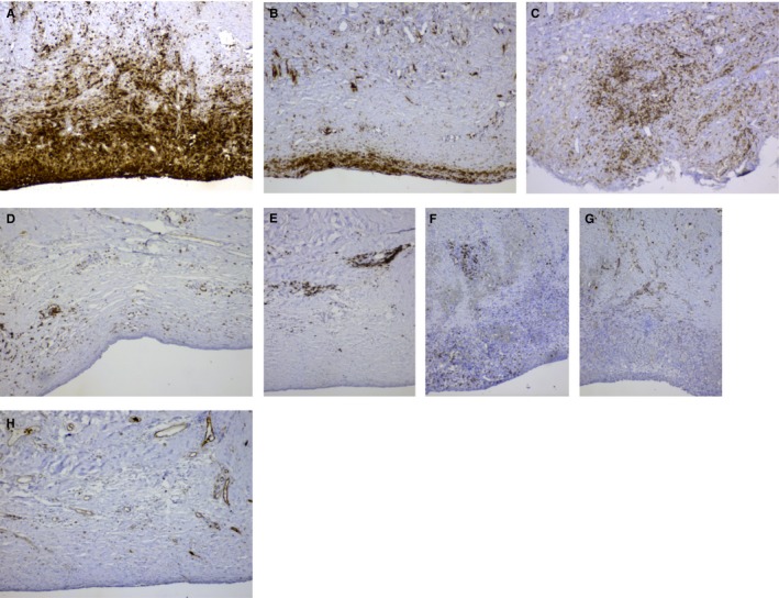
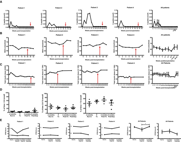
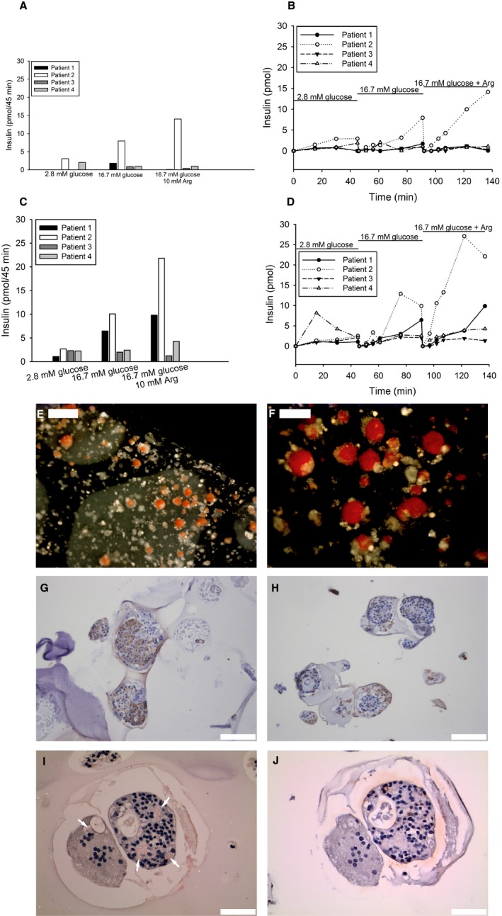
Macroencapsulation devices provide the dual possibility of immunoprotecting transplanted cells while also being retrievable, the latter bearing importance for safety in future trials with stem cell-derived cells. However, macroencapsulation entails a problem with oxygen supply to the encapsulated cells. The βAir device solves this with an incorporated refillable oxygen tank. This phase 1 study evaluated the safety and efficacy of implanting the βAir device containing allogeneic human pancreatic islets into patients with type 1 diabetes. Four patients were transplanted with 1-2 βAir devices, each containing 155 000-180 000 islet equivalents (ie, 1800-4600 islet equivalents per kg body weight), and monitored for 3-6 months, followed by the recovery of devices. Implantation of the βAir device was safe and successfully prevented immunization and rejection of the transplanted tissue. However, although beta cells survived in the device, only minute levels of circulating C-peptide were observed with no impact on metabolic control. Fibrotic tissue with immune cells was formed in capsule surroundings. Recovered devices displayed a blunted glucose-stimulated insulin response, and amyloid formation in the endocrine tissue. We conclude that the βAir device is safe and can support survival of allogeneic islets for several months, although the function of the transplanted cells was limited (Clinicaltrials.gov: NCT02064309).
Overview
The claims and statistics of this paper haven’t been checked yet.
Run the check — free, no account needed. It reads the open-access text and checks the sources it cites.
Integrity signals
Registry
Cites
Statistics
Figures
Verification
Claims the paper makes
The claims and statistics of this paper haven’t been checked yet.
Run the check — free, no account needed. It reads the open-access text and checks the sources it cites.
Claims other papers make about this one
Each is checked against this paper’s own text: does it say what the citing paper says it does?
The source backs part of the claim, not all of it — or backs it for a different population, condition or comparison.
In the citing paper
“these macroencapsulation devices can also promote fibrosis around the implant, further preventing nutrient diffusion.”
From Developments in stem cell-derived islet replacement therapy for treating type 1 diabetes (2023) · cites this paper as [92]
Experimental — accuracy not yet measured
Self-check
Percentages, sums and reported differences re-computed from the paper’s own numbers.
The self-check needs the paper’s full text.
Citations
Cites
The rand 36‐item health survey 1.0
Hays, R. D., Sherbourne, C. D., & Mazel, R. M. (1993) · Health Economics · doi:10.1002/hec.4730020305 · Cited by 2,971
Hering, B. J., Clarke, W. R., Bridges, N. D., Eggerman, T. L., Alejandro, R., Bellin, M. D., Chaloner, K., Czarniecki, C. W., Goldstein, J. S., Hunsicker, L. G., et al. (2016) · Diabetes Care · doi:10.2337/dc15-1988 · Cited by 645 · cited 1×
“22-24 The problems encountered could be attributed to (1) the significant contribution of glucose effectiveness for glucose metabolism in small animals,25 (2) limited islet survival due to diffusion problems of oxygen and nutrients occurring when the device has been upscaled from harboring a few hundred islets to enco…”
Self-citationLive encapsulated porcine islets from a type 1 diabetic patient 9.5 yr after xenotransplantation
Elliott, R. B., Escobar, L., Tan, P., Muzina, M., Zwain, S., & Buchanan, C. M. (2007) · Xenotransplantation · doi:10.1111/j.1399-3089.2007.00384.x · Cited by 409
Bradley, C., & Lewis, K. S. (1990) · Diabetic Medicine · doi:10.1111/j.1464-5491.1990.tb01421.x · Cited by 380
Microencapsulated Pancreatic Islet Allografts Into Nonimmunosuppressed Patients With Type 1 Diabetes
Calafiore, R., Basta, G., Luca, G., Lemmi, A., Montanucci, M. P., Calabrese, G., Racanicchi, L., Mancuso, F., & Brunetti, P. (2006) · Diabetes Care · doi:10.2337/diacare.29.01.06.dc05-1270 · Cited by 342
Safety and Viability of Microencapsulated Human Islets Transplanted Into Diabetic Humans
Tuch, B., Keogh, G., Williams, L., Wu, W., Foster, J., Vaithilingam, V., & Philips, R. (2009) · Diabetes Care · doi:10.2337/dc09-0744 · Cited by 329
Transplantation of human islets without immunosuppression
Ludwig, B., Reichel, A., Steffen, A., Zimerman, B., Schally, A. V., Block, N. L., Colton, C. K., Ludwig, S., Kersting, S., Bonifacio, E. E., et al. (2013) · Proceedings of the National Academy of Sciences · doi:10.1073/pnas.1317561110 · Cited by 286 · cited 3×
“Even so, the kinetics of glucosestimulated insulin release during an intravenous glucose tolerance test showed a significant delay in secreted Cpeptide over a 4- hour period also at the preperitoneal space.(7) Given the physiological differences between different tissue compartments, especially regarding vascularity,…”
InfluentialSelf-citationStaining methods for identification of amyloid in tissue.
Westermark, G., Johnson, K., & Westermark, P. (1999) · Methods in Enzymology · doi:10.1016/s0076-6879(99)09003-5 · Cited by 266 · cited 1×
“Alginateembedded formalinfixed slabs and islets from the pancreas donors were stained with Congo red according to Puchtler and Sweat,(12) and the presence of amyloid was determined in polarized light.(13) Biopsies from the tissue surrounding the device were obtained at explantation and fixed in formalin.”
Self-citationGoto, M., Eich, T., Felldin, M., Foss, A., Källén, R., Salmela, K., Tibell, A., Tufveson, G., Fujimori, K., Engkvist, M., et al. (2004) · Transplantation · doi:10.1097/01.tp.0000140882.53773.dc · Cited by 235 · cited 1×
“Human pancreatic islets were isolated from pancreases procured donation after brain death (DBD) donors in GMP facilities according to clinical routine.(8) Descriptive data for each islet donor are provided in Table S1.”
Self-citationWhy do microencapsulated islet grafts fail in the absence of fibrotic overgrowth?
de Vos, P., van Straaten, J. F. M., Nieuwenhuizen, A., de Groot, M., Ploeg, R., de Haan, B. D., & van Schilfgaarde, R. (1999) · Diabetes · doi:10.2337/diabetes.48.7.1381 · Cited by 206
Research works printed but not linked: 2
- Ekberg, 2007, C-Peptide replacement therapy and sensory nerve function in type 1 diabetic neuropathy, Diabetes Care., 30, 71, 10.2337/dc06-1274 · DOI
- Leu, 1992, Microencapsulated pancreatic islets: a pathologic study, J Formos Med Assoc., 91, 849
Cited by
Sorted by citations
Pancreatic Islet Transplantation in Humans: Recent Progress and Future Directions
Rickels, M. R., & Robertson, R. P. (2019) · Endocrine Reviews · doi:10.1210/er.2018-00154 · Cited by 329
Biotechnology and Bioengineering: Volume 117, Number 1, January 2020
Anane, E., García, Á. C., Haby, B., Hans, S., Krausch, N., Krewinkel, M., Hauptmann, P., Neubauer, P., & Bournazou, M. N. C. (2019) · Biotechnology and Bioengineering · doi:10.1002/bit.26757 · Cited by 246
Developments in stem cell-derived islet replacement therapy for treating type 1 diabetes
Hogrebe, N. J., Ishahak, M., & Millman, J. R. (2023) · Cell Stem Cell · doi:10.1016/j.stem.2023.04.002 · Cited by 210
Cell encapsulation: Overcoming barriers in cell transplantation in diabetes and beyond
Farina, M., Alexander, J. F., Thekkedath, U. R., Mauro Ferrari, B., & Grattoni, A. (2018) · Advanced Drug Delivery Reviews · doi:10.1016/j.addr.2018.04.018 · Cited by 194
Engineering islets from stem cells for advanced therapies of diabetes
Siehler, J., Blöchinger, A. K., Meier, M., & Lickert, H. (2021) · Nature Reviews Drug Discovery · doi:10.1038/s41573-021-00262-w · Cited by 125
Therapies for Type 1 Diabetes: Current Scenario and Future Perspectives
Pathak, V., Pathak, N., O’Neill, C. L., Guduric‐Fuchs, J., & Medina, R. J. (2019) · Clinical Medicine Insights Endocrinology and Diabetes · doi:10.1177/1179551419844521 · Cited by 144
3D Bioprinting of Functional Islets of Langerhans in an Alginate/Methylcellulose Hydrogel Blend
Duin, S., Schütz, K., Ahlfeld, T., Lehmann, S., Lode, A., Ludwig, B., & Gelinsky, M. (2019) · Advanced Healthcare Materials · doi:10.1002/adhm.201801631 · Cited by 113
Strategies for durable β cell replacement in type 1 diabetes
Brusko, T. M., Russ, H. A., & Stabler, C. L. (2021) · Science · doi:10.1126/science.abh1657 · Cited by 119
Engineering immunomodulatory biomaterials for type 1 diabetes
Stabler, C. L., Li, Y., Stewart, J. M., & Keselowsky, B. G. (2019) · Nature Reviews Materials · doi:10.1038/s41578-019-0112-5 · Cited by 128
Grattoni, A., Korbutt, G., Tomei, A. A., García, A. J., Pepper, A. R., Stabler, C., Brehm, M., Papas, K., Citro, A., Shirwan, H., et al. (2025) · Nature Reviews Endocrinology · doi:10.1038/s41574-024-01029-0 · Cited by 92
“Furthermore, immune rejection requires lifelong systemic immunosuppressive enabling on-target cell types to be purified 22,25,26 , which might improve the functional potency and safety of the stem cell-derived islet drug product, but this approach might be difficult to scale up for manufacturing.”
Hu, X., Gattis, C., Olroyd, A. G., Friera, A. M., White, K. A., Young, C., Basco, R., Lamba, M., Wells, F., Ankala, R., et al. (2023) · Science Translational Medicine · doi:10.1126/scitranslmed.adg5794 · Cited by 104
Coronel, M. M., Liang, J.-P., Li, Y., & Stabler, C. L. (2019) · Biomaterials · doi:10.1016/j.biomaterials.2019.04.017 · Cited by 85
“Given that aggressive foreign body responses to an implant can lead to significant impairment of clinical function [58], the benign response of the host is encouraging.”
BackgroundClinical Translation of Pluripotent Stem Cell Therapies: Challenges and Considerations
Desgres, M., & Menasché, P. (2019) · Cell stem cell · doi:10.1016/j.stem.2019.10.001 · Cited by 88
Domingo‐Lopez, D. A., Lattanzi, G., Schreiber, L. H. J., Wallace, E. J., Wylie, R., O’Sullivan, J., Dolan, E. B., & Duffy, G. P. (2022) · Advanced Drug Delivery Reviews · doi:10.1016/j.addr.2022.114280 · Cited by 109
Oxygenation strategies for encapsulated islet and beta cell transplants
Papas, K. K., León, H. D., Suszynski, T. M., & Johnson, R. C. (2019) · Advanced Drug Delivery Reviews · doi:10.1016/j.addr.2019.05.002 · Cited by 88
An inverse-breathing encapsulation system for cell delivery
Wang, L.-H., Ernst, A. U., Flanders, J. A., Liu, W., Wang, X., Datta, A. K., Epel, B., Kotecha, M., Papas, K. K., & Ma, M. (2021) · Science Advances · doi:10.1126/sciadv.abd5835 · Cited by 73
Espona‐Noguera, A., Ciriza, J., Cañibano-Hernández, A., Orive, G., Hernández, R. M., del Burgo, L. S., & Pedraz, J. L. (2019) · Pharmaceutics · doi:10.3390/pharmaceutics11110597 · Cited by 88
“In addition, in the mentioned clinical trial using the β-Air device [94], results showed that such device prevented immunization and rejection of the transplanted islets.”
BackgroundEngineering a Clinically Translatable Bioartificial Pancreas to Treat Type I Diabetes
Orive, G., Emerich, D. F., Khademhosseini, A., Matsumoto, S., Hernández, R. M., Pedraz, J. L., Desai, T. A., Calafiore, R., & de Vos, P. (2018) · Trends in biotechnology · doi:10.1016/j.tibtech.2018.01.007 · Cited by 78
Current state and future evolution of pancreatic islet transplantation
Anazawa, T., Okajima, H., Masui, T., & Üemoto, S. (2018) · Annals of Gastroenterological Surgery · doi:10.1002/ags3.12214 · Cited by 76
Transplant strategies for type 1 diabetes: whole pancreas, islet and porcine beta cell therapies
Bellin, M. D., & Dunn, T. B. (2020) · Diabetologia · doi:10.1007/s00125-020-05184-7 · Cited by 76
“However, these devices were not sufficient to maintain long-term islet viability and no long-term glycaemic benefit was observed [30].”
BackgroundAdvances in Encapsulation and Delivery Strategies for Islet Transplantation
Wu, S., Wang, L., Fang, Y., Huang, H. W., You, X., & Wu, J. (2021) · Advanced Healthcare Materials · doi:10.1002/adhm.202100965 · Cited by 80
Nanotechnology in cell replacement therapies for type 1 diabetes
Ernst, A. U., Bowers, D. T., Wang, L., Shariati, K., Plesser, M. D., Brown, N. K., Mehrabyan, T., & Ma, M. (2019) · Advanced Drug Delivery Reviews · doi:10.1016/j.addr.2019.01.013 · Cited by 77
“During clinical trials, nonsignificant reduction of HbA1c levels and transient circulating C-peptide levels were observed [253].”
BackgroundYang, K., O’Cearbhaill, E. D., Liu, S. S., Zhou, A., Chitnis, G. D., Hamilos, A. E., Xu, J., Verma, M. K. S., Giraldo, J. A., Kudo, Y., et al. (2021) · Proceedings of the National Academy of Sciences · doi:10.1073/pnas.2101258118 · Cited by 58
“The remaining cells were neither capable of restoring euglycemia in rodents (1,000 to ∼ 2,000 IEQ required) nor sustaining a therapeutic dosage needed for humans ( ∼ 500,000 islets) in a reasonably sized device (13, 24 – 26).”
Safley, S. A., Kenyon, N. S., Berman, D. M., Barber, G. F., Willman, M. A., Duncanson, S., Iwakoshi, N. N., Holdcraft, R. W., Gazda, L. S., THOMPSON, P. W., et al. (2018) · Xenotransplantation · doi:10.1111/xen.12450 · Cited by 57
Paez‐Mayorga, J., Campa‐Carranza, J. N., Capuani, S., Hernandez, N., Liu, H., Chua, C. Y. X., Pons-Faudoa, F. P., Malgir, G., Alvarez, B., Niles, J. A., et al. (2022) · Nature Communications · doi:10.1038/s41467-022-35629-z · Cited by 64
Liang, J.-P., Accolla, R. P., Soundirarajan, M., Emerson, A. E., Coronel, M. M., & Stabler, C. (2021) · Acta Biomaterialia · doi:10.1016/j.actbio.2021.05.028 · Cited by 52
Progress and challenges in developing allogeneic cell therapies
Deuse, T., & Schrepfer, S. (2025) · Cell Stem Cell · doi:10.1016/j.stem.2025.03.004 · Cited by 51
Boscari, F., & Avogaro, A. (2021) · Reviews in Endocrine and Metabolic Disorders · doi:10.1007/s11154-021-09635-3 · Cited by 70
“Results were not satisfactory in terms of c peptide production and metabolic control but lead to a new strategy to protect transplanted cells [155].”
BackgroundPaez‐Mayorga, J., Capuani, S., Hernandez, N., Farina, M., Chua, C. Y. X., Blanchard, R., Sizovs, A., Liu, H., Fraga, D. W., Niles, J. A., et al. (2020) · Biomaterials · doi:10.1016/j.biomaterials.2020.120232 · Cited by 54
From Mesenchymal Stromal/Stem Cells to Insulin-Producing Cells: Progress and Challenges
Ghoneim, M. A., Refaie, A. F., Elbassiouny, B. L., Gabr, M. M., & Zakaria, M. M. (2020) · Stem Cell Reviews and Reports · doi:10.1007/s12015-020-10036-3 · Cited by 62
“Carlsson and associates reported their clinical experience with the β-Air device in a phase 1 study [146].”
BackgroundPaving the way for successful islet encapsulation
Dimitrioglou, N. G., Kanelli, M., Papageorgiou, E. G., Karatzas, T., & Hatziavramidis, D. (2019) · Drug Discovery Today · doi:10.1016/j.drudis.2019.01.020 · Cited by 56
Hydrogel-Encapsulated Pancreatic Islet Cells as a Promising Strategy for Diabetic Cell Therapy
Huan, Z., Li, J., Luo, Z., Yu, Y., & Li, L. (2024) · Research · doi:10.34133/research.0403 · Cited by 42
“Nevertheless, there was only a small amount of circulating C-peptide in the subjects, and metabolic control was not improved in the end [90].”
BackgroundTransplantation of Macroencapsulated Insulin-Producing Cells
Hwa, A. J., & Weir, G. C. (2018) · Current Diabetes Reports · doi:10.1007/s11892-018-1028-y · Cited by 49
Samojlik, M. M., & Stabler, C. L. (2021) · Acta Biomaterialia · doi:10.1016/j.actbio.2021.05.039 · Cited by 38
Jiang, L., Shen, Y., Liu, Y., Zhang, L. J., & Jiang, W. (2022) · Theranostics · doi:10.7150/thno.66670 · Cited by 46
“In this device, oxygen is supplied by the oxygen tank compartment which is fed with oxygen periodically [180].”
BackgroundImmune Protection of Stem Cell-Derived Islet Cell Therapy for Treating Diabetes
Tahbaz, M., & Yoshihara, E. (2021) · Frontiers in Endocrinology · doi:10.3389/fendo.2021.716625 · Cited by 43
Krishnan, S. R., Liu, C., Bochenek, M. A., Bose, S., Khatib, N., Walters, B., O’Keeffe, L., Facklam, A., Langer, R., & Anderson, D. G. (2023) · Proceedings of the National Academy of Sciences of the United States of America · doi:10.1073/pnas.2311707120 · Cited by 36
“Encapsulation has the potential to support a broad range of cell types, ranging from primary cells (4) to stem - cell - derived products (5) and immortalized, engineered cell lines (3, 6).”
Directed self-assembly of a xenogeneic vascularized endocrine pancreas for type 1 diabetes
Citro, A., Neroni, A., Pignatelli, C., Campo, F., Policardi, M., Monieri, M., Pellegrini, S., Dugnani, E., Manenti, F., Maffia, M. C., et al. (2023) · Nature Communications · doi:10.1038/s41467-023-36582-1 · Cited by 41
Emerging strategies for beta cell transplantation to treat diabetes
Paez‐Mayorga, J., Lukin, I., Emerich, D. F., de Vos, P., Orive, G., & Grattoni, A. (2021) · Trends in Pharmacological Sciences · doi:10.1016/j.tips.2021.11.007 · Cited by 43
Islet Transplantation: Current Limitations and Challenges for Successful Outcomes
Langlois, A., Pinget, M., Kessler, L., & Bouzakri, K. (2024) · Cells · doi:10.3390/cells13211783 · Cited by 42
Type 1 diabetes and engineering enhanced islet transplantation
Jeyagaran, A., Lu, C., Zbinden, A., Birkenfeld, A. L., Brucker, S. Y., & Layland, S. L. (2022) · Advanced Drug Delivery Reviews · doi:10.1016/j.addr.2022.114481 · Cited by 42
Li, Y., Frei, A. W., Yang, E. Y., Labrada-Miravet, I., Sun, C., Rong, Y., Samojlik, M. M., Bayer, A., & Stabler, C. (2019) · bioRxiv · doi:10.1101/2019.12.11.872978 · Cited by 33
Polymer scaffolds for pancreatic islet transplantation — Progress and challenges
Smink, A., de Haan, B. D., Lakey, J., & de Vos, P. (2018) · American Journal of Transplantation · doi:10.1111/ajt.14942 · Cited by 33
Tondin, A. R., & Lanzoni, G. (2025) · BioDrugs · doi:10.1007/s40259-025-00703-7 · Cited by 35
Encapsulated islet transplantation
Liu, S. S., Shim, S., Kudo, Y., Stabler, C. L., O’Cearbhaill, E. D., Karp, J. M., & Yang, K. (2024) · Nature Reviews Bioengineering · doi:10.1038/s44222-024-00238-6 · Cited by 42
“In a healthy pancreas, β cells may last for several decades 7 ; this is reduced to only a few years or months after the cells are placed into encapsulation devices 8–10 .”
InfluentialWang, L., Marfil‐Garza, B. A., Ernst, A. U., Pawlick, R. L., Pepper, A. R., Okada, K., Epel, B., Viswakarma, N., Kotecha, M., Flanders, J. A., et al. (2023) · Nature Biomedical Engineering · doi:10.1038/s41551-023-01145-8 · Cited by 38
“Many strategies have been developed to provide exogenous O 2 supply in cell encapsulation devices and sustain early engraftment and long-term cell survival, including O 2 -generating 34,35 and O 2 -filling 36–39 systems.”
Macroencapsulation Devices for Cell Therapy
Liu, W., Wang, Y., Wang, J., Lanier, O. L., Wechsler, M. E., Peppas, N. A., & Gu, Z. (2022) · Engineering · doi:10.1016/j.eng.2021.10.021 · Cited by 38
Qin, T., Smink, A. M., & de Vos, P. (2023) · Acta Biomaterialia · doi:10.1016/j.actbio.2023.06.038 · Cited by 35
Paez-Mayorga, J., Capuani, S., Farina, M., Lotito, M., Niles, J. A., Salazar, H. F., Rhudy, J., Esnaola, L., Chua, C. Y., Taraballi, F., et al. (2020) · Advanced Healthcare Materials · doi:10.1002/adhm.202000670 · Cited by 31
Encapsulation and immune protection for type 1 diabetes cell therapy
Kioulaphides, S., & Garcı́a, A. J. (2024) · Advanced Drug Delivery Reviews · doi:10.1016/j.addr.2024.115205 · Cited by 37
Tissue engineering and 3D printing of bioartificial pancreas for regenerative medicine in diabetes
Soetedjo, A. A. P., Lee, J. M., Lau, H. H., Goh, G. L., An, J., Koh, Y., Yeong, W. Y., & Teo, A. K. K. (2021) · Trends in Endocrinology and Metabolism · doi:10.1016/j.tem.2021.05.007 · Cited by 35
Advances and challenges of the cell-based therapies among diabetic patients
Raoufinia, R., Rahimi, H. R., Saburi, E., & Moghbeli, M. (2024) · Journal of Translational Medicine · doi:10.1186/s12967-024-05226-3 · Cited by 29
“Although, low insulin levels were produced for up to eight weeks, there was not any reduction in the required exogenous insulin [37].”
BackgroundHo, B. X., Teo, A. K. K., & Ng, N. H. J. (2024) · Frontiers in Immunology · doi:10.3389/fimmu.2024.1375177 · Cited by 32
Pignatelli, C., Campo, F., Neroni, A., Piemonti, L., & Citro, A. (2022) · Transplant International · doi:10.3389/ti.2022.10555 · Cited by 33
“A clinical study evaluated the β-Air bio-artificial pancreas, which had a daily refillable oxygen chamber between two layers of alginate encapsulating the islet to maintain an adequate oxygen supply (NCT02064309) (98,112,122). β-Air improved cell viability and supported graft function, which were detected for 10 month…”
BackgroundInfluentialCell delivery systems: Toward the next generation of cell therapies for type 1 diabetes
Dang, H. P., Chen, H., Dargaville, T. R., & Tuch, B. E. (2022) · Journal of Cellular and Molecular Medicine · doi:10.1111/jcmm.17499 · Cited by 29
Campa‐Carranza, J. N., Paez‐Mayorga, J., Chua, C. Y. X., Nichols, J. E., & Grattoni, A. (2022) · Expert Opinion on Drug Delivery · doi:10.1080/17425247.2022.2076834 · Cited by 29
“However, lack of direct cell apposition to blood vessels impair mass transport critical for metabolic activity, thus affecting graft viability and function [32,33].”
BackgroundFrom islet of Langerhans transplantation to the bioartificial pancreas
Berney, T., Wassmer, C., Lebreton, F., Bellofatto, K., Fonseca, L. M., Bignard, J., Hanna, R., Peloso, A., & Berishvili, E. (2022) · La Presse Médicale · doi:10.1016/j.lpm.2022.104139 · Cited by 26
Prevascularized Retrievable Hybrid Implant to Enhance Function of Subcutaneous Encapsulated Islets
Mridha, A. R., Dargaville, T. R., Dalton, P. D., Carroll, L. M., Morris, M. B., Vaithilingam, V., & Tuch, B. E. (2020) · Tissue Engineering Part A · doi:10.1089/ten.tea.2020.0179 · Cited by 30
Pancreatic islet transplantation: toward definitive treatment for diabetes mellitus
Takaki, T., & Shimoda, M. (2020) · Global Health & Medicine · doi:10.35772/ghm.2020.01057 · Cited by 33
“Amino acid supplementation during transplantation and devices that can supply oxygen are promising avenues of research (96,97).”
BackgroundKong, D.-F., Xu, H.-M., Chen, M., Yu, Y.-P., Qian, Y.-B., Qin, T., Tong, Y., Xia, Q., & Hang, H.-L. (2020) · Stem cell research & therapeutics · doi:10.1186/s13287-020-01962-7 · Cited by 23
A steady decline in pancreas transplantation rates
Benjamens, S., Leemkuil, M., Margreiter, C., Huurman, V. A. L., Leuvenink, H. G. D., & Pol, R. A. (2018) · Pancreatology · doi:10.1016/j.pan.2018.11.003 · Cited by 28
Jeang, W., Bochenek, M. A., Bose, S., Zhao, Y.-C., Wong, B. M., Yang, J.-W., Jiang, A. L., Langer, R., & Anderson, D. G. (2024) · Science Advances · doi:10.1126/sciadv.adk5949 · Cited by 22
“Strategies to improve the oxygenation of devices include developing antifibrotic materials ( 29 , 39 , 41 , 42 ), incorporating particles that chemically decompose to release oxygen ( 43 – 46 ), or supplementing oxygen delivery through transcutaneous gas ports ( 47 , 48 ).”
Tan, L. S., Chen, J., Lim, L., & Teo, A. (2022) · Cell Proliferation · doi:10.1111/cpr.13232 · Cited by 22
Ajima, K., Tsuda, N., Takaki, T., Furusako, S., Matsumoto, S., Shinohara, K., Yamashita, Y., Amano, S., Oyama, C., & Shimoda, M. (2022) · Cell Reports Methods · doi:10.1016/j.crmeth.2022.100370 · Cited by 22
Krentz, N. A. J., Shea, L. D., Huising, M. O., & Shaw, J. (2021) · The Lancet Diabetes & Endocrinology · doi:10.1016/s2213-8587(21)00170-4 · Cited by 39
Development of a Bioartificial Vascular Pancreas
Han, E. X., Wang, J., Kural, M. H., Jiang, B., Leiby, K. L., Chowdhury, N., Tellides, G., Kibbey, R. G., Lawson, J. H., & Niklason, L. E. (2021) · Journal of Tissue Engineering · doi:10.1177/20417314211027714 · Cited by 22
Dinnyés, A., Schnur, A., Muenthaisong, S., Bartenstein, P., Burcez, C.-T., Burton, N., Cyran, C., Gianello, P., Kemter, E., Németh, G., et al. (2020) · Cell Proliferation · doi:10.1111/cpr.12785 · Cited by 18
Safety and function of a new pre-vascularized bioartificial pancreas in an allogeneic rat model
Magisson, J., Sassi, A., Xhema, D., Kobalyan, A., Gianello, P., Mourer, B., Tran, N. H., Burcez, C.-T., Bouaoun, R., & Sigrist, S. (2020) · Journal of Tissue Engineering · doi:10.1177/2041731420924818 · Cited by 18
Naumova, N., & Iop, L. (2021) · Frontiers in Bioengineering and Biotechnology · doi:10.3389/fbioe.2021.673477 · Cited by 19
GPR44 as a Target for Imaging Pancreatic Beta-Cell Mass
Eriksson, O. (2019) · Current Diabetes Reports · doi:10.1007/s11892-019-1164-z · Cited by 18
Self-citationRecent advances in endocrine organoids for therapeutic application
Jeon, S., Lee, Y.-S., Oh, S. R., Jeong, J., Lee, D., So, K., & Hwang, N. S.-Y. (2023) · Advanced Drug Delivery Reviews · doi:10.1016/j.addr.2023.114959 · Cited by 20
3D Bioprinting and Translation of Beta Cell Replacement Therapies for Type 1 Diabetes
Gurlin, R. E., Giraldo, J. A., & Latres, E. (2020) · Tissue Engineering Part B Reviews · doi:10.1089/ten.teb.2020.0192 · Cited by 23
“36 43 While localized delivery of oxygen and angiogenic factors promote cell viability and vascularization, respectively, measures to protect the cells from rejection such as nanoencapsulation or microencapsulation, localized immunomodulation through controlled drug delivery, or immune engineering…”
Generation of Human Stem Cell-Derived Pancreatic Organoids (POs) for Regenerative Medicine
Navarro‐Tableros, V., Gomez, Y., Brizzi, M. F., & Camussi, G. (2019) · Advances in experimental medicine and biology · doi:10.1007/5584_2019_340 · Cited by 17
Artificial Pancreas or Novel Beta-Cell Replacement Therapies: a Race for Optimal Glycemic Control?
Nijhoff, M. F., & de Koning, E. J. P. (2018) · Current Diabetes Reports · doi:10.1007/s11892-018-1073-6 · Cited by 19
Clinical Applications of Cell Encapsulation Technology
Santos‐Vizcaíno, E., Orive, G., Pedraz, J. L., & Hernández, R. M. (2020) · Methods in molecular biology · doi:10.1007/978-1-0716-0215-7_32 · Cited by 18
Wong, J. M., & Pepper, A. R. (2024) · Frontiers in Transplantation · doi:10.3389/frtra.2024.1485444 · Cited by 19
Triolo, T. M., & Bellin, M. D. (2021) · Frontiers in Endocrinology · doi:10.3389/fendo.2021.636824 · Cited by 19
Applying Immunomodulation to Promote Longevity of Immunoisolated Pancreatic Islet Grafts
Kuwabara, R., Hu, S., Smink, A. M., Orive, G., Lakey, J. R. T., & de Vos, P. (2021) · Tissue Engineering Part B Reviews · doi:10.1089/ten.teb.2020.0326 · Cited by 17
“Four T1D patients received one or two bAir devices with human islets in the subcutaneous site and were monitored for 3–6 months.(38) Although also in this study the safety of the bAir device was demonstrated and islet grafts survived in the devices, only transient levels of circulating C-peptide were observed, while n…”
Sthijns, M. M. J. P. E., Jetten, M. J., Mohammed, S. G., Claessen, S. M. H., de Vries, R. H. W., Stell, A. L., de Bont, D. F. A., Engelse, M. A., Mumcuoglu, D., van Blitterswijk, C., et al. (2020) · Biomaterials · doi:10.1016/j.biomaterials.2020.120449 · Cited by 18
Current State and Evidence of Cellular Encapsulation Strategies in Type 1 Diabetes
Marfil‐Garza, B. A., Polishevska, K., Pepper, A. R., & Korbutt, G. S. (2020) · Comprehensive physiology · doi:10.1002/cphy.c190033 · Cited by 23
Karaoğlu, İ. C., Duymaz, D., Rashid, M. M., & Kızılel, S. (2025) · Frontiers in Immunology · doi:10.3389/fimmu.2025.1618086 · Cited by 15
Advancements and Challenges in Immune Protection Strategies for Islet Transplantation
Wang, X., Zeng, Z., Li, D., Wang, K., Zhang, W., Yu, Y., & Wang, X. (2025) · Journal of Diabetes · doi:10.1111/1753-0407.70048 · Cited by 18
Tissue Engineering Strategies for Improving Beta Cell Transplantation Outcome
Abadpour, S., Wang, C., Niemi, E. M., & Scholz, H. (2021) · Current Transplantation Reports · doi:10.1007/s40472-021-00333-2 · Cited by 21
Stephens, C. H., Morrison, R. A., McLaughlin, M. R., Orr, K. S., Tersey, S. A., Scott‐Moncrieff, J. C., Mirmira, R. G., Considine, R. V., & Voytik‐Harbin, S. L. (2020) · American Journal of Physiology-Endocrinology and Metabolism · doi:10.1152/ajpendo.00066.2020 · Cited by 16
Stem cell-derived pancreatic beta cells: a step closer to functional diabetes treatment?
Ogieuhi, I. J., Agbo, C. E., Ajekiigbe, V. O., Chidera Stanley Anthony, M., Onyehalu, J. C., Nwankwo, C. K., Agu, M. C., Lema, K. F., Adewole, O. A., Asade, O. O., et al. (2025) · BMC Endocrine Disorders · doi:10.1186/s12902-025-01997-y · Cited by 14
Mu-u-min, R., Diané, A., Allouch, A., & Al-Siddiqi, H. (2025) · Biomedicines · doi:10.3390/biomedicines13020383 · Cited by 14
“An improved variant of the Beta-air device is being generated for hPSC-derived β -cells [51,97].”
β cell replacement therapy for the cure of diabetes
Lee, J., & Yoon, K. (2022) · Journal of Diabetes Investigation · doi:10.1111/jdi.13884 · Cited by 15
The Current Status of Bioartificial Pancreas Devices
Photiadis, S. J., Gologorsky, R. C., & Sarode, D. N. (2020) · ASAIO Journal · doi:10.1097/mat.0000000000001252 · Cited by 17
Engineering a Porous Hydrogel-Based Device for Cell Transplantation
Naficy, S., Dehghani, F., Chew, Y. V., Hawthorne, W. J., & Le, T. Y. L. (2020) · ACS Applied Bio Materials · doi:10.1021/acsabm.9b01144 · Cited by 14
Three-dimensional bioprinting of functional β-islet-like constructs
Parvaneh, S., Kemény, L. V., Ghaffarinia, A., Yarani, R., & Veréb, Z. (2023) · International Journal of Bioprinting · doi:10.18063/ijb.v9i2.665 · Cited by 18
Mooranian, A., Jones, M., Ionescu, C. M., Walker, D., Wagle, S. R., Kovačević, B., Chester, J., Foster, T., Johnston, E., Kuthubutheen, J., et al. (2021) · Journal of Functional Biomaterials · doi:10.3390/jfb12040068 · Cited by 12
Monitoring the macrophage response towards biomaterial implants using label-free imaging
Lu, C., Levey, R. E., Ghersi, G., Schueller, N., Liebscher, S., Layland, S. L., Schenke‐Layland, K., Duffy, G. P., & Marzi, J. (2023) · Materials Today Bio · doi:10.1016/j.mtbio.2023.100696 · Cited by 18
Moeun, B. N., Lemaire, F., Smink, A. M., Orimi, H. E., Leask, R. L., de Vos, P., & Hoesli, C. A. (2025) · Journal of Tissue Engineering · doi:10.1177/20417314241284826 · Cited by 10
Oppler, S. H., Stone, L. H., Leishman, D. J., Janecek, J. L., Moore, M. E. G., Rangarajan, P., Willenberg, B. J., O’Brien, T. D., Modiano, J. F., Pheil, N., et al. (2024) · Science Advances · doi:10.1126/sciadv.adi4919 · Cited by 11
“The β air Bio-Artificial Pancreas increases local oxygen availability by directly supplying oxygen via daily injection through a subcutaneous port to the housed islets ( 67 , 68 ).”
BackgroundEinstein, S. A., Steyn, L. V., Weegman, B. P., Suszynski, T. M., Sambanis, A., O’Brien, T. D., Avgoustiniatos, E. S., Firpo, M. T., Graham, M. L., Janecek, J. L., et al. (2023) · Frontiers in Transplantation · doi:10.3389/frtra.2023.1257029 · Cited by 12
Integration of Islet/Beta-Cell Transplants with Host Tissue Using Biomaterial Platforms
Clough, D., King, J. L., Li, F., & Shea, L. D. (2020) · Endocrinology · doi:10.1210/endocr/bqaa156 · Cited by 16
Preservation of β-Cells as a Therapeutic Strategy for Diabetes
Taneera, J., & Saber-Ayad, M. M. (2024) · Hormone and Metabolic Research · doi:10.1055/a-2239-2668 · Cited by 11
“However, this strategy requires ensuring optimal oxygen and nutrient supply to the encapsulated islets, maintaining the viability and function of the encapsulated islets over time [24].”
BackgroundAdvances in single-cell nanoencapsulation and applications in diseases
Xue, Z., Mei, D., & Zhang, L. (2022) · Journal of Microencapsulation · doi:10.1080/02652048.2022.2111472 · Cited by 9
Alternative transplantation sites for islet transplantation
Pellegrini, S. (2020) · doi:10.1016/b978-0-12-814833-4.00065-4 · Cited by 9
Tokito, F., Shinohara, M., Maruyama, M., Inamura, K., Nishikawa, M., & Sakai, Y. (2020) · Journal of Bioscience and Bioengineering · doi:10.1016/j.jbiosc.2020.12.009 · Cited by 8
Metadata
- Authors
- Carlsson, P.-O., Espes, D., Sedigh, A., Rotem, A., Zimerman, B., Grinberg, H., Goldman, T., Barkai, U., Avni, Y., Westermark, G. T., Carlbom, L., Ahlström, H., Eriksson, O., Olerud, J., & Korsgren, O.
- Affiliations
- Uppsala University (SE)
- Beta-O2 Technologies (Israel) (IL)
- Corresponding author
- Carlsson, Per-Ola
- ORCID
- Venue
- American Journal of Transplantation
- Publisher
- Elsevier BV
- Published
- 2018-07
- Volume
- 18
- Issue
- 7
- Pages
- 1735-1744
- ISSN
- 1600-6135 (ISSN-L)
- PMC
- PMC6055594
- OpenAlex
- W2777721344
- Cited by
- 205
- References
- 27
- Citations by year
- 2026: 22 · 2025: 15 · 2024: 21 · 2023: 22 · 2022: 23 · 2021: 31 · 2020: 32 · 2019: 26 · 2018: 12
- FWCI
- 19.90
- Type
- journal article
- Language
- en
- Open access
- hybrid
- doi.org
- Licence
- Locations
- American Journal of Transplantation (publishedVersion, cc-by-nc)
- Europe PMC (PubMed Central) (publishedVersion, cc-by-nc)
- PubMed Central (publishedVersion, cc-by-nc)
- PubMed (publishedVersion)
- KTH Publication Database DiVA (KTH Royal Institute of Technology) (submittedVersion, other-oa)
- Funders
- Juvenile Diabetes Research Foundation International
- Diabetes Research & Wellness Foundation
- Stiftelsen Familjen Ernfors Fond
- Diabetesfonden
- Vetenskapsrådet (921-2014-7054, K2013-55X-15043, K2015-54X-12219-19-4, K2016-01040, K2016-GTW)
- Novo Nordisk
- European Research Council (F4-2013-602889, 646075-ELASTISLET)
- Leona M. and Harry B. Helmsley Charitable Trust
- Barndiabetesfonden
- EXODIAB
- Topics
- Health Sciences › Medicine › Endocrinology, Diabetes and Metabolism › Pancreatic function and diabetes
- Health Sciences › Medicine › Endocrinology, Diabetes and Metabolism › Diabetes Management and Research
- Life Sciences › Biochemistry, Genetics and Molecular Biology › Genetics › Diabetes and associated disorders
- Keywords
- macroencapsulation, bioartificial pancreas, type 1 diabetes, islet transplantation, oxygen supply, immunoprotection, islet survival, C-peptide, amyloid fibril formation, capsule fibrosis, device retrieval, treatment efficacy and safety
- MeSH terms
- Adolescent, Blood Glucose, Capsules, Child, Child, Preschool, Diabetes Mellitus, Type 1, Female, Follow-Up Studies, Humans, Islets of Langerhans, Male, Monitoring, Physiologic, Prognosis, Islets of Langerhans Transplantation, Pancreas, Artificial, Bioartificial Organs
About these checks
Registry, retraction, Cites and statistics results are computed, not judged, with one exception: a printed reference that no index lists is read once by a model before it counts as a citation error. Claims are extracted by a model and kept only when their quote appears verbatim in the text; each cited source is then checked, with a model’s help, for whether it says what the claim relies on. Those verdicts can be wrong — read the claim’s quote and the cited source before relying on one.
Statistics and arithmetic are recomputed, not judged.
Confidence bands are shown only for fields calibrated on our annotated test set. None is calibrated yet, so no band is shown.
What counts as a claim ›How we measure confidence ›How we check this ›
Are you an author, or do you see a problem with this page? Request a review
