

The DOI matches its Crossref record; 54 of the 54 references it cites were found in a registry or an index; none of the 37 sources its claims cite is checked for support yet.
p62/SQSTM1 cooperates with Parkin for perinuclear clustering of depolarized mitochondria
Open accessAbstract
PINK1 and Parkin were first identified as the causal genes responsible for familial forms of early‐onset Parkinson’s disease (PD), a prevalent neurodegenerative disorder. PINK1 encodes a mitochondrial serine/threonine protein kinase, whereas Parkin encodes an ubiquitin‐protein ligase. PINK1 and Parkin cooperate to maintain mitochondrial integrity; however, the detailed molecular mechanism of how Parkin‐catalyzed ubiquitylation results in mitochondrial integrity remains an enigma. In this study, we show that Parkin‐catalyzed K63‐linked polyubiquitylation of depolarized mitochondria resulted in ubiquitylated mitochondria being transported along microtubules to cluster in the perinuclear region, which was interfered by pathogenic mutations of Parkin. In addition, p62/SQSTM1 (hereafter referred to as p62) was recruited to depolarized mitochondria after Parkin‐directed ubiquitylation. Intriguingly, deletion of p62 in mouse embryonic fibroblasts resulted in a gross loss of mitochondrial perinuclear clustering but did not hinder mitochondrial degradation. Thus, p62 is required for ubiquitylation‐dependent clustering of damaged mitochondria, which resembles p62‐mediated ‘aggresome’ formation of misfolded/unfolded proteins after ubiquitylation.
Overview
AI snapshot
Written from the paper’s full text.
TL;DR
Parkin-catalyzed K63-linked ubiquitylation of depolarized mitochondria drives their microtubule-dependent perinuclear clustering, and p62/SQSTM1 is required for this clustering but not for mitochondrial degradation in MEFs. The study links Parkin, p62, and aggresome-like mitochondrial quarantine, while leaving the role of p62 in mitophagy unresolved.
Key findings
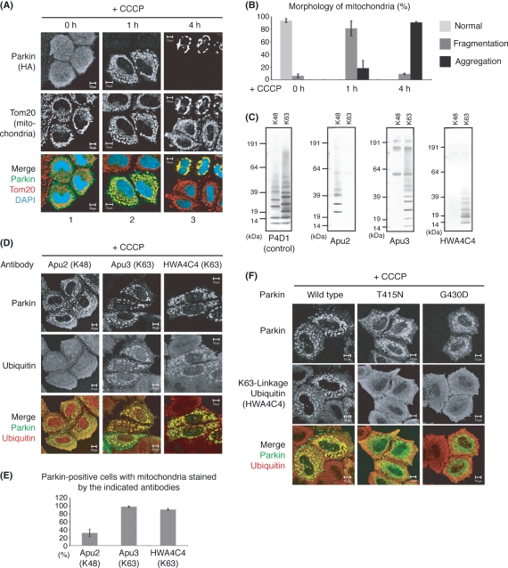
- Parkin rapidly moved to CCCP-depolarized mitochondria and ubiquitylated them by t = 1 h; mitochondria later concentrated in the perinuclear region at t > 4 h in Parkin-expressing HeLa cells, and this clustering was Parkin dependent.
- Linkage-specific antibodies showed that Parkin-localized mitochondria were stained by K63-linked ubiquitin antibodies Apu3 and HWA4C4, but not by K48-linked Apu2; signals disappeared with E3-inactivated Parkin mutations T415N and G430D.
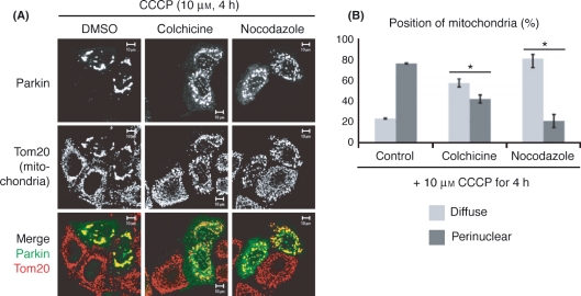
- Microtubule inhibitors nocodazole (10 μg/mL) and colchicine (50 μg/mL) dissipated perinuclear mitochondrial clusters, whereas latrunculin A/B had no effect.
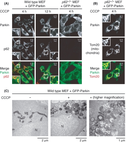
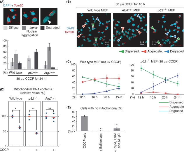
- p62 was recruited to depolarized mitochondria after Parkin-directed ubiquitylation; p62 deletion in MEFs caused gross loss of perinuclear clustering but did not hinder degradation. mtDNA 12s rRNA after CCCP fell to 62 ± 4% in wild-type MEFs, 95 ± 13% in Atg7−/− MEFs, and 43 ± 2% in p62−/− MEFs.
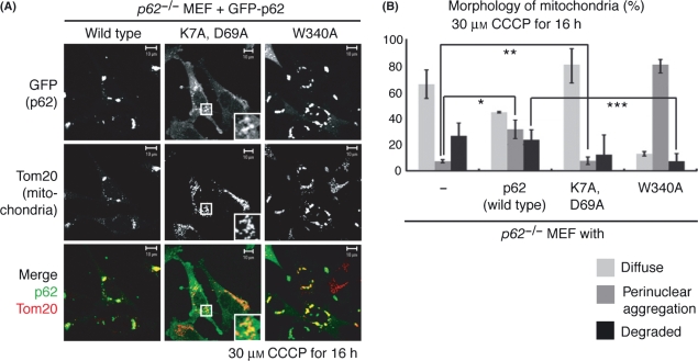
- p62 PB1 mutant K7A/D69A failed to restore clustering in p62−/− MEFs, whereas W340A restored clustering but delayed mitochondrial degradation.
Methods
- HeLa cells expressing HA-Parkin or pathogenic Parkin mutants were treated with 10 μM CCCP; MEFs (wild-type, p62−/−, Atg7−/−) expressing HA- or GFP-Parkin were treated with 30 μM CCCP and analyzed by immunocytochemistry.
- Mitochondrial morphology was scored in more than 100 cells per condition across at least three experiments, with Welch’s t-test or Student’s t-test where reported; linkage-specific anti-ubiquitin antibodies were validated by immunoblotting against K48- or K63-linked polyubiquitin chains.
- Additional assays included COX electron microscopy, real-time quantitative PCR for mitochondrial 12s ribosomal RNA normalized to GAPDH, and inhibitor treatments with bafilomycin A1 or E64d/pepstatin A/NH4Cl.
Limitations
- The paper states that its results “do not rule out completely the involvement of p62 in Parkin-mediated mitophagy” and that a role for p62 in degradation is substitutable in MEFs, possibly due to functional redundancy with proteins such as NBR1, Alfy, and BAG3.
- The authors note their p62 result conflicts with Geisler et al. (2010), who reported p62 is required for Parkin-dependent mitophagy; they attribute the discrepancy to methodological differences (p62 knockout MEFs versus p62 siRNA in HeLa cells) and state further analysis is needed.
- Evident from the text: the study relies on cultured HeLa cells and MEFs with exogenous Parkin and CCCP-induced depolarization, not in vivo neurons; MEFs required higher CCCP concentrations and longer incubation than HeLa cells, and the p62−/− phenotype was interpreted with possible redundant factors.
Model-written summary. Check it against the paper before relying on it.
Integrity signals
Registry
Cites
Statistics
Figures
Verification
Claims the paper makes
30 Own result · 23 Not checked yet · 17 Own interpretation · 6 Own method · 3 No basis given
None of the sources cited for this claim have been checked yet.
In the paper
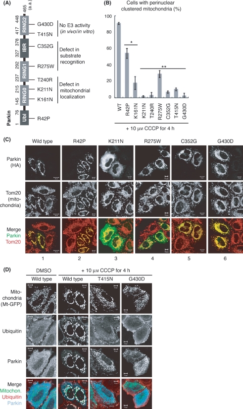
“The K161N, K211N and T240R mutations, which severely compromised the mitochondrial localization of Parkin (Geisler et al. 2010; Matsuda et al. 2010; Narendra et al. 2010), inhibited the mitochondrial clustering.”
§ p62/SQSTM1 cooperates with Parkin for perinuclear clustering of depolarized mitochondria > Results > Various pathogenic mutations of Parkin impede mitochondrial clustering
plaindirect- Asserted: passes — The authors put it forward as true.
- Checkable: passes — It can be checked against evidence.
- Load-bearing: passes — The paper’s conclusions depend on it.
- Based: passes — The paper gives a reason why it holds.
A result the paper reports from its own data — it cites no other source for it, so there is none to check. The quote shows it is in the paper.
In the paper
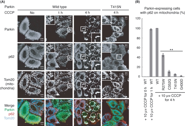
“Under steady-state conditions in HeLa cells, endogenous p62 was mainly localized throughout the cytosol irrespective of the presence or absence of Parkin, whereas p62 was rapidly recruited to the mitochondria only in Parkin-expressing cells after CCCP treatment for 1 h (Fig. 4A), as also reported by Springer et al. recently (Geisler et al. 2010).”
§ p62/SQSTM1 cooperates with Parkin for perinuclear clustering of depolarized mitochondria > Results > p62/SQSTM1 is not essential for mitochondrial degradation
plain- Asserted: passes — The authors put it forward as true.
- Checkable: passes — It can be checked against evidence.
- Load-bearing: unclear — It is unclear whether the paper’s conclusions depend on it.
- Based: passes — The paper gives a reason why it holds.
A result the paper reports from its own data — it cites no other source for it, so there is none to check. The quote shows it is in the paper.
In the paper
“In this study, we show that Parkin-catalyzed K63-linked polyubiquitylation of depolarized mitochondria resulted in ubiquitylated mitochondria being transported along microtubules to cluster in the perinuclear region, which was interfered by pathogenic mutations of Parkin.”
§ p62/SQSTM1 cooperates with Parkin for perinuclear clustering of depolarized mitochondria > Abstract
boosteddirect- Asserted: passes — The authors put it forward as true.
- Checkable: passes — It can be checked against evidence.
- Load-bearing: passes — The paper’s conclusions depend on it.
- Based: passes — The paper gives a reason why it holds.
A result the paper reports from its own data — it cites no other source for it, so there is none to check. The quote shows it is in the paper.
In the paper
“In addition, p62/SQSTM1 (hereafter referred to as p62) was recruited to depolarized mitochondria after Parkin-directed ubiquitylation.”
- Also stated in: § p62/SQSTM1 is not essential for mitochondrial degradation · plain
“whereas p62 was rapidly recruited to the mitochondria only in Parkin-expressing cells after CCCP treatment for 1 h (Fig. 4A)”
§ p62/SQSTM1 cooperates with Parkin for perinuclear clustering of depolarized mitochondria > Abstract
plaindirect- Asserted: passes — The authors put it forward as true.
- Checkable: passes — It can be checked against evidence.
- Load-bearing: passes — The paper’s conclusions depend on it.
- Based: passes — The paper gives a reason why it holds.
- Also stated in: § p62/SQSTM1 is not essential for mitochondrial degradation · plain
A result the paper reports from its own data — it cites no other source for it, so there is none to check. The quote shows it is in the paper.
In the paper
“which was interfered by pathogenic mutations of Parkin.”
- Also stated in: § Various pathogenic mutations of Parkin impede mitochondrial clustering · plain
“Various pathogenic mutations of Parkin impede mitochondrial clustering”
§ p62/SQSTM1 cooperates with Parkin for perinuclear clustering of depolarized mitochondria > Abstract
plaindirect- Asserted: passes — The authors put it forward as true.
- Checkable: passes — It can be checked against evidence.
- Load-bearing: passes — The paper’s conclusions depend on it.
- Based: passes — The paper gives a reason why it holds.
- Also stated in: § Various pathogenic mutations of Parkin impede mitochondrial clustering · plain
A result the paper reports from its own data — it cites no other source for it, so there is none to check. The quote shows it is in the paper.
In the paper
“Intriguingly, deletion of p62 in mouse embryonic fibroblasts resulted in a gross loss of mitochondrial perinuclear clustering but did not hinder mitochondrial degradation.”
- Also stated in: § p62/SQSTM1 is necessary for perinuclear clustering of damaged mitochondria · plain
“we noticed that the perinuclear clustering of depolarized mitochondria was almost completely inhibited in p62 deficient MEFs (Fig. 6A–C).”
§ p62/SQSTM1 cooperates with Parkin for perinuclear clustering of depolarized mitochondria > Abstract
plaindirect- Asserted: passes — The authors put it forward as true.
- Checkable: passes — It can be checked against evidence.
- Load-bearing: passes — The paper’s conclusions depend on it.
- Based: passes — The paper gives a reason why it holds.
- Also stated in: § p62/SQSTM1 is necessary for perinuclear clustering of damaged mitochondria · plain
A result the paper reports from its own data — it cites no other source for it, so there is none to check. The quote shows it is in the paper.
In the paper
“After treatment with nocodazole (10 μg/mL) or colchicine (50 μg/mL), the perinuclear mitochondria clusters dissipated (Fig. 2A).”
§ p62/SQSTM1 cooperates with Parkin for perinuclear clustering of depolarized mitochondria > Results > Mitochondrial clustering is microtubule dependent
plaindirect- Asserted: passes — The authors put it forward as true.
- Checkable: passes — It can be checked against evidence.
- Load-bearing: passes — The paper’s conclusions depend on it.
- Based: passes — The paper gives a reason why it holds.
A result the paper reports from its own data — it cites no other source for it, so there is none to check. The quote shows it is in the paper.
In the paper
“Interestingly, mitochondria later concentrated to the perinuclear region at t > 4 h (Fig. 1A,B) and finally degraded as reported (Narendra et al. 2008) (not shown).”
§ p62/SQSTM1 cooperates with Parkin for perinuclear clustering of depolarized mitochondria > Results > Parkin-catalyzed K63-linked polyubiquitylation promotes mitochondrial clustering
plain- Asserted: passes — The authors put it forward as true.
- Checkable: passes — It can be checked against evidence.
- Load-bearing: passes — The paper’s conclusions depend on it.
- Based: passes — The paper gives a reason why it holds.
A result the paper reports from its own data — it cites no other source for it, so there is none to check. The quote shows it is in the paper.
In the paper
“but did not hinder mitochondrial degradation.”
§ p62/SQSTM1 cooperates with Parkin for perinuclear clustering of depolarized mitochondria > Abstract
plain- Asserted: passes — The authors put it forward as true.
- Checkable: passes — It can be checked against evidence.
- Load-bearing: passes — The paper’s conclusions depend on it.
- Based: passes — The paper gives a reason why it holds.
A result the paper reports from its own data — it cites no other source for it, so there is none to check. The quote shows it is in the paper.
In the paper
“These results suggest that Parkin ubiquitylates the damaged mitochondria mainly via K63-linked polyubiquitylation and are consistent, in part, with the recent report by Geisler et al. (2010).”
§ p62/SQSTM1 cooperates with Parkin for perinuclear clustering of depolarized mitochondria > Results > Parkin-catalyzed K63-linked polyubiquitylation promotes mitochondrial clustering
hedgeddirect- Asserted: passes — The authors put it forward as true.
- Checkable: passes — It can be checked against evidence.
- Load-bearing: passes — The paper’s conclusions depend on it.
- Based: passes — The paper gives a reason why it holds.
- It may state two separate claims as one. 14
- Probably not a substantial claim. 17
A result the paper reports from its own data — it cites no other source for it, so there is none to check. The quote shows it is in the paper.
In the paper
“In the R275W and C352G mutations, Parkin associated with depolarized mitochondria, but mitochondrial ubiquitylation and juxtanuclear clustering were severely inhibited (Fig. 3C and not shown).”
§ p62/SQSTM1 cooperates with Parkin for perinuclear clustering of depolarized mitochondria > Results > Various pathogenic mutations of Parkin impede mitochondrial clustering
plaindirect- Asserted: passes — The authors put it forward as true.
- Checkable: passes — It can be checked against evidence.
- Load-bearing: passes — The paper’s conclusions depend on it.
- Based: passes — The paper gives a reason why it holds.
It may state two separate claims as one.
None of the sources cited for this claim have been checked yet.
In the paper
“This perinuclear transport and clustering of mitochondria are Parkin dependent, because this phenomenon was only observed in Parkin-expressing HeLa cells that lack an endogenous Parkin gene (Denison et al. 2003) (Fig. 1A, panel 3).”
§ p62/SQSTM1 cooperates with Parkin for perinuclear clustering of depolarized mitochondria > Results > Parkin-catalyzed K63-linked polyubiquitylation promotes mitochondrial clustering
plaindirect- Asserted: passes — The authors put it forward as true.
- Checkable: passes — It can be checked against evidence.
- Load-bearing: passes — The paper’s conclusions depend on it.
- Based: passes — The paper gives a reason why it holds.
It may state two separate claims as one.
A result the paper reports from its own data — it cites no other source for it, so there is none to check. The quote shows it is in the paper.
In the paper
“In mitochondria fragmented with the mitochondrial uncoupler, carbonyl cyanide m-chlorophenylhydrazone (CCCP), Parkin quickly moved to depolarized mitochondria and ubiquitylated them (t = 1 h).”
§ p62/SQSTM1 cooperates with Parkin for perinuclear clustering of depolarized mitochondria > Results > Parkin-catalyzed K63-linked polyubiquitylation promotes mitochondrial clustering
plaindirect- Asserted: passes — The authors put it forward as true.
- Checkable: passes — It can be checked against evidence.
- Load-bearing: passes — The paper’s conclusions depend on it.
- Based: passes — The paper gives a reason why it holds.
It may state two separate claims as one.
A result the paper reports from its own data — it cites no other source for it, so there is none to check. The quote shows it is in the paper.
In the paper
“In wild-type MEFs, p62 was again selectively recruited to the mitochondria after CCCP treatment at t = 4 h, although the transport efficiency was lower than that of HeLa cells (data not shown), and depolarized mitochondria were later clustered in the perinuclear region similar to HeLa cells at t > 12 h (Fig. 5A).”
§ p62/SQSTM1 cooperates with Parkin for perinuclear clustering of depolarized mitochondria > Results > p62/SQSTM1 is not essential for mitochondrial degradation
plaincorrelational- Asserted: passes — The authors put it forward as true.
- Checkable: passes — It can be checked against evidence.
- Load-bearing: unclear — It is unclear whether the paper’s conclusions depend on it.
- Based: passes — The paper gives a reason why it holds.
It may state two separate claims as one.
A result the paper reports from its own data — it cites no other source for it, so there is none to check. The quote shows it is in the paper.
In the paper
“Mitochondrial localization of p62 was only observed in Parkin-expressing cells and disappeared when Parkin mutants deficient in substrate recognition or E3 activity (R275W, C352G, T415N and G430D; Fig. 3A) were introduced (Fig. 4), suggesting that ubiquitylation-catalyzed Parkin is required for the recruitment of p62 onto depolarized mitochondria.”
§ p62/SQSTM1 cooperates with Parkin for perinuclear clustering of depolarized mitochondria > Results > p62/SQSTM1 is not essential for mitochondrial degradation
hedgeddirect- Asserted: passes — The authors put it forward as true.
- Checkable: passes — It can be checked against evidence.
- Load-bearing: passes — The paper’s conclusions depend on it.
- Based: passes — The paper gives a reason why it holds.
It may state two separate claims as one.
A result the paper reports from its own data — it cites no other source for it, so there is none to check. The quote shows it is in the paper.
In the paper
“Electron microscopic analysis showed that juxtanuclear-clustered mitochondria aggregated in grape-like clusters and not fused to each other to make single large mitochondrion (Fig. 5C).”
§ p62/SQSTM1 cooperates with Parkin for perinuclear clustering of depolarized mitochondria > Results > p62/SQSTM1 is not essential for mitochondrial degradation
plain- Asserted: passes — The authors put it forward as true.
- Checkable: passes — It can be checked against evidence.
- Load-bearing: unclear — It is unclear whether the paper’s conclusions depend on it.
- Based: passes — The paper gives a reason why it holds.
It may state two separate claims as one.
None of the sources cited for this claim have been checked yet.
In the paper
“PINK1 then recruits Parkin from the cytoplasm to depolarized mitochondria that then undergo Parkin-catalyzed ubiquitylation, meaning that PINK1 and Parkin cooperate to ubiquitylate mitochondria with low membrane potentials (Narendra et al. 2008, 2010; Geisler et al. 2010; Matsuda et al. 2010; Vives-Bauza et al. 2010).”
§ p62/SQSTM1 cooperates with Parkin for perinuclear clustering of depolarized mitochondria > Introduction
What the sources say
- Not checked yetcites Narendra et al. 2008, 2010
- Not checked yetcites Geisler et al. 2010
- Not checked yetcites Matsuda et al. 2010
plaindirect- Asserted: passes — The authors put it forward as true.
- Checkable: passes — It can be checked against evidence.
- Load-bearing: passes — The paper’s conclusions depend on it.
- Based: passes — The paper gives a reason why it holds.
Probably not a substantial claim. It may state two separate claims as one.
None of the sources cited for this claim have been checked yet.
In the paper
“PINK1 then recruits Parkin from the cytoplasm to depolarized mitochondria that then undergo Parkin-catalyzed ubiquitylation, meaning that PINK1 and Parkin cooperate to ubiquitylate mitochondria with low membrane potentials (Narendra et al. 2008, 2010; Geisler et al. 2010; Matsuda et al. 2010; Vives-Bauza et al. 2010).”
§ p62/SQSTM1 cooperates with Parkin for perinuclear clustering of depolarized mitochondria > Introduction
What the sources say
- Not checked yetcites Narendra et al. 2008, 2010
- Not checked yetcites Geisler et al. 2010
- Not checked yetcites Matsuda et al. 2010
plaindirect- Asserted: passes — The authors put it forward as true.
- Checkable: passes — It can be checked against evidence.
- Load-bearing: passes — The paper’s conclusions depend on it.
- Based: passes — The paper gives a reason why it holds.
Probably not a substantial claim.
None of the sources cited for this claim have been checked yet.
In the paper
“Parkin has the potential to catalyze several types of ubiquitylation: multiple monoubiquitylation, Lys-48-linked polyubiquitylation and Lys-63-linked polyubiquitylation (Lim 2007; Matsuda & Tanaka 2010).”
§ p62/SQSTM1 cooperates with Parkin for perinuclear clustering of depolarized mitochondria > Results > Parkin-catalyzed K63-linked polyubiquitylation promotes mitochondrial clustering
What the sources say
- Not checked yetcites Lim 2007
- Not checked yetcites Matsuda & Tanaka 2010
hedgedconditional- Asserted: passes — The authors put it forward as true.
- Checkable: passes — It can be checked against evidence.
- Load-bearing: fails — The paper’s conclusions may not depend on it.
- Based: passes — The paper gives a reason why it holds.
Probably not a substantial claim. The paper’s conclusions may not depend on it. It may state two separate claims as one.
What the paper did — it cites no source for this, so there is none to check. The quote shows it is in the paper.
In the paper
“Staining with single antibodies alone indicated that the merged data described earlier were not derived from channel crosstalk (Fig. S1 in Supporting Information).”
§ p62/SQSTM1 cooperates with Parkin for perinuclear clustering of depolarized mitochondria > Results > p62/SQSTM1 is not essential for mitochondrial degradation
hedged- Asserted: passes — The authors put it forward as true.
- Checkable: passes — It can be checked against evidence.
- Load-bearing: fails — The paper’s conclusions may not depend on it.
- Based: passes — The paper gives a reason why it holds.
Probably not a substantial claim. The paper’s conclusions may not depend on it.
None of the sources cited for this claim have been checked yet.
In the paper
“p62 can interact with both ubiquitin and the autophagic machinery LC-3 (Bjorkoy et al. 2005; Komatsu et al. 2007; Pankiv et al. 2007; Ichimura et al. 2008; Noda et al. 2008; Shvets et al. 2008) and thus p62 is a plausible factor that links Parkin-catalyzed ubiquitylation with mitochondrial degradation via autophagy.”
§ p62/SQSTM1 cooperates with Parkin for perinuclear clustering of depolarized mitochondria > Results > p62/SQSTM1 is not essential for mitochondrial degradation
What the sources say
- Not checked yetcites Bjorkoy et al. 2005
- Not checked yetcites Komatsu et al. 2007
- Not checked yetcites Pankiv et al. 2007
plaincorrelational- Asserted: passes — The authors put it forward as true.
- Checkable: passes — It can be checked against evidence.
- Load-bearing: passes — The paper’s conclusions depend on it.
- Based: passes — The paper gives a reason why it holds.
It may state two separate claims as one.
In the paper
“bafilomycin A1 (an inhibitor of vacuolar-type H+-ATPase that prevents autophagosome-lysosome fusion and/or intralysosomal degradation)”
§ p62/SQSTM1 cooperates with Parkin for perinuclear clustering of depolarized mitochondria > Results > p62/SQSTM1 is not essential for mitochondrial degradation
What the sources say
- Not checked yetcites Mizushima et al. 2010
plaindirect- Asserted: passes — The authors put it forward as true.
- Checkable: passes — It can be checked against evidence.
- Load-bearing: passes — The paper’s conclusions depend on it.
- Based: passes — The paper gives a reason why it holds.
Probably not a substantial claim. It may state two separate claims as one.
What the paper did — it cites no source for this, so there is none to check. The quote shows it is in the paper.
In the paper
“Immunoblotting against K48- and K63-linked polyubiquitin chains, whose linkage was directly confirmed by MALDI-TOF mass spectrometry (data not shown), showed the high specificity of the antibodies (Fig. 1C).”
§ p62/SQSTM1 cooperates with Parkin for perinuclear clustering of depolarized mitochondria > Results > Parkin-catalyzed K63-linked polyubiquitylation promotes mitochondrial clustering
plain- Asserted: passes — The authors put it forward as true.
- Checkable: passes — It can be checked against evidence.
- Load-bearing: passes — The paper’s conclusions depend on it.
- Based: passes — The paper gives a reason why it holds.
It may state two separate claims as one.
None of the sources cited for this claim have been checked yet.
In the paper
“However, overproduction of mutant ubiquitin can change the ubiquitylation linkage, and thus careful interpretation of observed results is required (Pickart & Raasi 2005; Saeki et al. 2009).”
§ p62/SQSTM1 cooperates with Parkin for perinuclear clustering of depolarized mitochondria > Results > Parkin-catalyzed K63-linked polyubiquitylation promotes mitochondrial clustering
What the sources say
- Not checked yetcites Pickart & Raasi 2005
- Not checked yetcites Saeki et al. 2009
plaindirect- Asserted: passes — The authors put it forward as true.
- Checkable: passes — It can be checked against evidence.
- Load-bearing: passes — The paper’s conclusions depend on it.
- Based: passes — The paper gives a reason why it holds.
Probably not a substantial claim.
None of the sources cited for this claim have been checked yet.
In the paper
“Moreover, PINK1 functions upstream of Parkin, because forced overexpression of Parkin rescues loss-of-function phenotypes of PINK1 but not vice versa (Clark et al. 2006; Park et al. 2006; Yang et al. 2006).”
§ p62/SQSTM1 cooperates with Parkin for perinuclear clustering of depolarized mitochondria > Introduction
What the sources say
- Not checked yetcites Clark et al. 2006
- Not checked yetcites Park et al. 2006
- Not checked yetcites Yang et al. 2006
plaindirect- Asserted: passes — The authors put it forward as true.
- Checkable: passes — It can be checked against evidence.
- Load-bearing: fails — The paper’s conclusions may not depend on it.
- Based: passes — The paper gives a reason why it holds.
Probably not a substantial claim. The paper’s conclusions may not depend on it.
What the paper did — it cites no source for this, so there is none to check. The quote shows it is in the paper.
In the paper
“To check the specificity of linkage-specific anti-ubiquitin antibodies, Lys 48- or Lys 63-linked polyubiquitin chain (Enzo Life Sciences) was used as a positive control and subjected to immunoblotting.”
§ p62/SQSTM1 cooperates with Parkin for perinuclear clustering of depolarized mitochondria > Experimental procedures > Immunocytochemistry
plain- Asserted: passes — The authors put it forward as true.
- Checkable: passes — It can be checked against evidence.
- Load-bearing: fails — The paper’s conclusions may not depend on it.
- Based: passes — The paper gives a reason why it holds.
Probably not a substantial claim. The paper’s conclusions may not depend on it.
In the paper
“mitochondria are delivered to areas of the axon where metabolic demand is high, such as synapses, active growth cones and branches (Hollenbeck & Saxton 2005).”
§ p62/SQSTM1 cooperates with Parkin for perinuclear clustering of depolarized mitochondria > Discussion > Function of depolarized mitochondria juxtanuclear clustering
What the sources say
- Not checked yetcites Hollenbeck & Saxton 2005
plaindirect- Asserted: passes — The authors put it forward as true.
- Checkable: passes — It can be checked against evidence.
- Load-bearing: fails — The paper’s conclusions may not depend on it.
- Based: passes — The paper gives a reason why it holds.
Probably not a substantial claim. The paper’s conclusions may not depend on it.
What the paper did — it cites no source for this, so there is none to check. The quote shows it is in the paper.
In the paper
“To prevent carry-over contamination, 0.5 unit of Uracil DNA Glycosylase (Invitrogen) was added during PCR”
§ p62/SQSTM1 cooperates with Parkin for perinuclear clustering of depolarized mitochondria > Experimental procedures > Measurement of mitochondrial DNA contents
plaindirect- Asserted: passes — The authors put it forward as true.
- Checkable: passes — It can be checked against evidence.
- Load-bearing: fails — The paper’s conclusions may not depend on it.
- Based: passes — The paper gives a reason why it holds.
Probably not a substantial claim. The paper’s conclusions may not depend on it.
A conclusion the paper draws from its own results — it cites no other source for it, so there is none to check. The quote shows it is in the paper.
In the paper
“(note that complete disruption of p62 cannot cause this type of titration and thus cannot confer the dominant effect).”
§ p62/SQSTM1 cooperates with Parkin for perinuclear clustering of depolarized mitochondria > Discussion > Function of p62 in the degradation of depolarized mitochondria
plaindirect- Asserted: passes — The authors put it forward as true.
- Checkable: passes — It can be checked against evidence.
- Load-bearing: passes — The paper’s conclusions depend on it.
- Based: passes — The paper gives a reason why it holds.
It may state two separate claims as one.
- Rests on an assumption: Complete disruption of p62 cannot cause this type of titration and thus cannot confer the dominant effect.
A conclusion the paper draws from its own results — it cites no other source for it, so there is none to check. The quote shows it is in the paper.
In the paper
“and thus mitochondrial clustering might be involved in this process.”
§ p62/SQSTM1 cooperates with Parkin for perinuclear clustering of depolarized mitochondria > Discussion > Function of depolarized mitochondria juxtanuclear clustering
hedgedconditional- Asserted: passes — The authors put it forward as true.
- Checkable: passes — It can be checked against evidence.
- Load-bearing: fails — The paper’s conclusions may not depend on it.
- Based: passes — The paper gives a reason why it holds.
Probably not a substantial claim. The paper’s conclusions may not depend on it.
None of the sources cited for this claim have been checked yet.
In the paper
“with mitochondria-dedicated selective autophagy (termed mitophagy) considered to be essential for this clearance pathway (Narendra et al. 2008; Twig et al. 2008).”
§ p62/SQSTM1 cooperates with Parkin for perinuclear clustering of depolarized mitochondria > Introduction
What the sources say
- Not checked yetcites Narendra et al. 2008
- Not checked yetcites Twig et al. 2008
plaindirect- Asserted: passes — The authors put it forward as true.
- Checkable: passes — It can be checked against evidence.
- Load-bearing: passes — The paper’s conclusions depend on it.
- Based: passes — The paper gives a reason why it holds.
Probably not a substantial claim.
What the paper did — it cites no source for this, so there is none to check. The quote shows it is in the paper.
In the paper
“samples were prepared in triplicate for each condition.”
§ p62/SQSTM1 cooperates with Parkin for perinuclear clustering of depolarized mitochondria > Experimental procedures > Measurement of mitochondrial DNA contents
plain- Asserted: passes — The authors put it forward as true.
- Checkable: passes — It can be checked against evidence.
- Load-bearing: fails — The paper’s conclusions may not depend on it.
- Based: passes — The paper gives a reason why it holds.
Probably not a substantial claim. The paper’s conclusions may not depend on it.
In the paper
“These results suggest that oligomerization of p62 but not interaction with LC3 is essential for the mitochondrial clustering triggered by Parkin.”
§ p62/SQSTM1 cooperates with Parkin for perinuclear clustering of depolarized mitochondria > Results > p62/SQSTM1 is necessary for perinuclear clustering of damaged mitochondria
What the sources say
- Not checked yetcites Lamark et al. 2003
hedgeddirect- Asserted: passes — The authors put it forward as true.
- Checkable: passes — It can be checked against evidence.
- Load-bearing: passes — The paper’s conclusions depend on it.
- Based: passes — The paper gives a reason why it holds.
It may state two separate claims as one.
- Rests on an assumption: The K7A/D69A double mutation in the PB1 domain abolishes p62 oligomerization.
In the paper
“The defect in p62−/− MEFs, however, is concealed by other functionally redundant protein(s) (see Discussion).”
§ p62/SQSTM1 cooperates with Parkin for perinuclear clustering of depolarized mitochondria > Results > p62/SQSTM1 is necessary for perinuclear clustering of damaged mitochondria
What the sources say
- Not checked yetcites Kirkin et al. 2009
plaindirect- Asserted: passes — The authors put it forward as true.
- Checkable: passes — It can be checked against evidence.
- Load-bearing: fails — The paper’s conclusions may not depend on it.
- Based: passes — The paper gives a reason why it holds.
Probably not a substantial claim. The paper’s conclusions may not depend on it.
In the paper
“methodological differences (i.e. we used p62 knockout MEFs whereas they used p62 siRNA in HeLa cells) may account for the conflicting observations”
§ p62/SQSTM1 cooperates with Parkin for perinuclear clustering of depolarized mitochondria > Discussion > Function of p62 in the degradation of depolarized mitochondria
What the sources say
- Not checked yetcites Geisler et al. (2010)
hedgedconditional- Asserted: passes — The authors put it forward as true.
- Checkable: passes — It can be checked against evidence.
- Load-bearing: fails — The paper’s conclusions may not depend on it.
- Based: passes — The paper gives a reason why it holds.
Probably not a substantial claim. The paper’s conclusions may not depend on it.
A conclusion the paper draws from its own results — it cites no other source for it, so there is none to check. The quote shows it is in the paper.
In the paper
“The most plausible explanation for this result is that the p62 W340A mutant competes for certain common factor(s) with functionally redundant protein(s), and thus has a dominant effect”
§ p62/SQSTM1 cooperates with Parkin for perinuclear clustering of depolarized mitochondria > Discussion > Function of p62 in the degradation of depolarized mitochondria
hedgeddirectSingle case- Asserted: passes — The authors put it forward as true.
- Checkable: passes — It can be checked against evidence.
- Load-bearing: passes — The paper’s conclusions depend on it.
- Based: passes — The paper gives a reason why it holds.
It may state two separate claims as one.
- Rests on an assumption: The p62 W340A mutant cannot interact with LC3.
A conclusion the paper draws from its own results — it cites no other source for it, so there is none to check. The quote shows it is in the paper.
In the paper
“The similarity between aggresome and mitochondrial clustering, although superficial at the moment, suggests that the two ubiquitin-mediated cytoprotective events have something in common.”
§ p62/SQSTM1 cooperates with Parkin for perinuclear clustering of depolarized mitochondria > Discussion > Function of depolarized mitochondria juxtanuclear clustering
hedgedcorrelational- Asserted: passes — The authors put it forward as true.
- Checkable: fails — It may be too vague to check against evidence.
- Load-bearing: fails — The paper’s conclusions may not depend on it.
- Based: passes — The paper gives a reason why it holds.
Probably not a substantial claim. It may be too vague to check against evidence. The paper’s conclusions may not depend on it.
A conclusion the paper draws from its own results — it cites no other source for it, so there is none to check. The quote shows it is in the paper.
In the paper
“This series of events is reminiscent of aggresome formation”
§ p62/SQSTM1 cooperates with Parkin for perinuclear clustering of depolarized mitochondria > Discussion > Function of depolarized mitochondria juxtanuclear clustering
plaincorrelational- Asserted: passes — The authors put it forward as true.
- Checkable: passes — It can be checked against evidence.
- Load-bearing: fails — The paper’s conclusions may not depend on it.
- Based: passes — The paper gives a reason why it holds.
Probably not a substantial claim. The paper’s conclusions may not depend on it.
The paper relies on this without support.
Endogenous Parkin is undetectable in mouse embryonic fibroblasts.
“Because endogenous Parkin is undetectable in MEFs, HA- or GFP-Parkin was introduced into control (p62+/+) and p62 KO (p62−/−) MEFs (Komatsu et al. 2007) by retro-viral transfection (Kitamura et al. 2003).”
The paper relies on this without support.
MEFs are less sensitive than HeLa cells to CCCP, requiring higher concentrations and longer incubation to observe the phenotype.
“Because MEFs are less sensitive than HeLa cells to CCCP, higher concentrations of CCCP (30 μm) and longer incubation are required to observe the phenotype.”
The paper relies on this without support.
The p62 W340A mutant cannot interact with LC3.
“the p62 W340A mutant, which cannot interact with LC3 (Fig. 7B)”
The paper relies on this without support.
Complete disruption of p62 cannot cause this type of titration and thus cannot confer the dominant effect.
“(note that complete disruption of p62 cannot cause this type of titration and thus cannot confer the dominant effect).”
The paper relies on this without support.
The K7A/D69A double mutation in the PB1 domain abolishes p62 oligomerization.
“we introduced a K7A/D69A double mutation in the PB1 domain”
Claims other papers make about this one
Each is checked against this paper’s own text: does it say what the citing paper says it does?
The cited source doesn’t support the claim as written.
In the citing paper
“This results in the preferential degradation of mitochondrial outer membrane proteins by the proteasome, while inner membrane proteins and mitochondrial DNA 558 remain intact.”
From Guidelines for the use and interpretation of assays for monitoring autophagy (2012) · cites this paper as [558]
Checked by a reasoning model — may be wrong.
Experimental — accuracy not yet measuredIn the citing paper
“During mitochondrial degradation in cultured cells, depolarized mitochondria cluster with p6224.”
From Loss of autophagy in dopaminergic neurons causes Lewy pathology and motor dysfunction in aged mice (2018) · cites this paper as [24]
In the citing paper
“During mitochondrial degradation in cultured cells, depolarized mitochondria cluster with p6224. Depolarized mitochondria are ubiquitylated by Parkin, recognized by p62, transported via microtubules to aggregates, and degraded by autophagy.”
From Loss of autophagy in dopaminergic neurons causes Lewy pathology and motor dysfunction in aged mice (2018) · cites this paper as [24]
Self-check
Percentages, sums and reported differences re-computed from the paper’s own numbers.
Each contradiction between two passages listed here was confirmed by an independent check.
Rated minor: it would not touch a result — a value no result rests on, a p-value that stays on the same side of the significance level, or a conflict that is clerical, resolved elsewhere in the paper, or outside its own findings.
In the paper
§ p62/SQSTM1 cooperates with Parkin for perinuclear clustering of depolarized mitochondria > Discussion > Function of p62 in the degradation of depolarized mitochondria
“They showed that knockdown of p62 in HeLa cells dramatically inhibited the final clearance of damaged mitochondria”
§ p62/SQSTM1 cooperates with Parkin for perinuclear clustering of depolarized mitochondria > Discussion > Function of p62 in the degradation of depolarized mitochondria
“cells with uncleared mitochondria decreased from 85% to 20% with p62 siRNA treatment.”
Rated minor: it would not touch a result — a value no result rests on, a p-value that stays on the same side of the significance level, or a conflict that is clerical, resolved elsewhere in the paper, or outside its own findings.
In the paper
§ p62/SQSTM1 cooperates with Parkin for perinuclear clustering of depolarized mitochondria > Results > Various pathogenic mutations of Parkin impede mitochondrial clustering
“HA-Parkin mutants harboring one of eight pathogenic mutations (R42P, K161N, T240R, R275W, C352G, T415N and G430D; Fig. 3A)”
§ p62/SQSTM1 cooperates with Parkin for perinuclear clustering of depolarized mitochondria > Results > Various pathogenic mutations of Parkin impede mitochondrial clustering
“The K161N, K211N and T240R mutations, which severely compromised the mitochondrial localization of Parkin (Geisler et al. 2010; Matsuda et al. 2010; Narendra et al. 2010), inhibited the mitochondrial clustering.”
Citations
Cites
Mutations in the parkin gene cause autosomal recessive juvenile parkinsonism
Kitada, T., Asakawa, S., Hattori, N., Matsumine, H., Yamamura, Y., Minoshima, S., Yokochi, M., Mizuno, Y., & Shimizu, N. (1998) · Nature · doi:10.1038/33416 · Cited by 5,227 · cited 1×
“One familial form of the PD gene causing autosomal recessive juvenile Parkinsonism (AR-JP) is Parkin (also known as PARK2), which encodes an E3 ubiquitin ligase, a substrate recognition member of the ubiquitylation pathway (Kitada et al. 1998; Shimura et al. 2000).”
BackgroundSelf-citationPankiv, S., Clausen, T. H., Lamark, T., Brech, A., Bruun, J.-A., Outzen, H., Øvervatn, A., Bjørkøy, G., & Johansen, T. (2007) · Journal of Biological Chemistry · doi:10.1074/jbc.m702824200 · Cited by 4,578
Methods in Mammalian Autophagy Research
Mizushima, N., Yoshimori, T., & Levine, B. (2010) · Cell · doi:10.1016/j.cell.2010.01.028 · Cited by 4,497 · cited 2×
“Moreover, treatment with bafilomycin A1 (an inhibitor of vacuolar-type H-ATPase that prevents autophagosome-lysosome fusion and ⁄ or intralysosomal degradation) or a cocktail of lysosomal inhibitors (E64d, pepstatin A and ammonium chloride) (Mizushima et al. 2010) inhibited the mitochondrial degradation of p62 ⁄ ) MEF…”
BackgroundParkin is recruited selectively to impaired mitochondria and promotes their autophagy
Narendra, D., Tanaka, A., Suen, D.-F., & Youle, R. J. (2008) · The Journal of Cell Biology · doi:10.1083/jcb.200809125 · Cited by 4,026 · cited 5×
“…endogenous and exogenous stresses, such as unnecessary generation of reactive oxygen species (ROS) and mtDNA mutations, is critical, with mitochondria-dedicated selective autophagy (termed mitophagy) considered to be essential for this clearance pathway (Narendra et al. 2008; Twig et al. 2008).”
BackgroundInfluentialHereditary Early-Onset Parkinson's Disease Caused by Mutations in PINK1
Valente, E. M., Abou-Sleiman, P. M., Caputo, V., Muqit, M. M. K., Harvey, K., Gispert, S., Ali, Z., Del Turco, D., Bentivoglio, A. R., Healy, D. G., et al. (2004) · Science · doi:10.1126/science.1096284 · Cited by 3,481 · cited 2×
“The other early-onset PD gene encodes a mitochondria-targeted serine-threonine kinase termed PTEN induced putative kinase 1 (PINK1) (Valente et al. 2004).”
BackgroundBjørkøy, G., Lamark, T., Brech, A., Outzen, H., Perander, M., Øvervatn, A., Stenmark, H., & Johansen, T. (2005) · The Journal of Cell Biology · doi:10.1083/jcb.200507002 · Cited by 3,276
Fission and selective fusion govern mitochondrial segregation and elimination by autophagy
Twig, G., Elorza, A., Molina, A. J. A., Mohamed, H., Wikstrom, J. D., Walzer, G., Stiles, L., Haigh, S. E., Katz, S., Las, G., et al. (2008) · The EMBO Journal · doi:10.1038/sj.emboj.7601963 · Cited by 3,082 · cited 2×
“…endogenous and exogenous stresses, such as unnecessary generation of reactive oxygen species (ROS) and mtDNA mutations, is critical, with mitochondria-dedicated selective autophagy (termed mitophagy) considered to be essential for this clearance pathway (Narendra et al. 2008; Twig et al. 2008).”
BackgroundPINK1 Is Selectively Stabilized on Impaired Mitochondria to Activate Parkin
Narendra, D. P., Jin, S. M., Tanaka, A., Suen, D.-F., Gautier, C. A., Shen, J., Cookson, M. R., & Youle, R. J. (2010) · PLoS Biology · doi:10.1371/journal.pbio.1000298 · Cited by 2,944 · cited 5×
“…by likely inhibiting an as-yet-unknown processing protease that catalyzes conversion of the mature PINK1 (approximately 60 kDa) to an intermediate short form (approximately 50 kDa), which is further exclusively degraded by the proteasome pathway (Matsuda et al. 2010; Narendra et al. 2010).”
BackgroundInfluentialPINK1/Parkin-mediated mitophagy is dependent on VDAC1 and p62/SQSTM1
Geisler, S., Holmström, K. M., Skujat, D., Fiesel, F. C., Rothfuss, O. C., Kahle, P. J., & Springer, W. (2010) · Nature Cell Biology · doi:10.1038/ncb2012 · Cited by 2,835 · cited 10×
“PINK1 then recruits Parkin from the cytoplasm to depolarized mitochondria that then undergo Parkin-catalyzed ubiquitylation, meaning that PINK1 and Parkin cooperate to ubiquitylate mitochondria with low membrane potentials (Narendra et al. 2008, 2010; Geisler et al. 2010; Matsuda et al. 2010; Vives-Bauza et al. 2010).”
BackgroundMethodInfluentialImpairment of starvation-induced and constitutive autophagy in Atg7 -deficient mice
Komatsu, M., Waguri, S., Ueno, T., Iwata, J., Murata, S., Tanida, I., Ezaki, J., Mizushima, N., Ohsumi, Y., Uchiyama, Y., et al. (2005) · The Journal of Cell Biology · doi:10.1083/jcb.200412022 · Cited by 2,331 · cited 1×
“When Atg7 (an essential gene for autophagy) KO MEFs (Komatsu et al. 2005) were used as a positive control, the clearance of depolarized mitochondria was considerably impeded after CCCP treatment for 24 h [Fig.”
MethodSelf-citationAggresomes: A Cellular Response to Misfolded Proteins
Johnston, J. A., Ward, C. L., & Kopito, R. R. (1998) · The Journal of Cell Biology · doi:10.1083/jcb.143.7.1883 · Cited by 2,148 · cited 2×
“…concentrated into proteinacious inclusion bodies and ⁄ or recognized by HDAC6 to be transported along microtubles, organized into a perinuclear aggregation (an aggresome) and sometimes degraded by autophagy (Johnston et al. 1998; Garcia-Mata et al. 1999; Olzmann et al. 2007; Kirkin et al. 2009).”
BackgroundHomeostatic levels of p62 control cytoplasmic inclusion body formation in autophagy-deficient mice.
Komatsu, M., Waguri, S., Koike, M., Sou, Y.-S., Ueno, T., Hara, T., Mizushima, N., Iwata, J.-I., Ezaki, J., Murata, S., et al. (2007) · Cell · doi:10.1016/j.cell.2007.10.035 · Cited by 2,124 · cited 5×
“…of p62 in Parkin-mediated mitochondrial degradation. p62 can interact with both ubiquitin and the autophagic machinery LC-3 (Bjorkoy et al. 2005; Komatsu et al. 2007; Pankiv et al. 2007; Ichimura et al. 2008; Noda et al. 2008; Shvets et al. 2008) and thus p62 is a plausible factor that links…”
BackgroundInfluentialSelf-citationFamilial Parkinson disease gene product, parkin, is a ubiquitin-protein ligase
Shimura, H., Hattori, N., Kubo, S.-I., Mizuno, Y., Asakawa, S., Minoshima, S., Shimizu, N., Iwai, K., Chiba, T., Tanaka, K., et al. (2000) · Nature Genetics · doi:10.1038/77060 · Cited by 2,024 · cited 1×
“One familial form of the PD gene causing autosomal recessive juvenile Parkinsonism (AR-JP) is Parkin (also known as PARK2), which encodes an E3 ubiquitin ligase, a substrate recognition member of the ubiquitylation pathway (Kitada et al. 1998; Shimura et al. 2000).”
BackgroundSelf-citationMatsuda, N., Sato, S., Shiba, K., Okatsu, K., Saisho, K., Gautier, C. A., Sou, Y.-S., Saiki, S., Kawajiri, S., Sato, F., et al. (2010) · Journal of Cell Biology · doi:10.1083/jcb.200910140 · Cited by 1,969
Self-citationDrosophila pink1 is required for mitochondrial function and interacts genetically with parkin
Clark, I. E., Dodson, M. W., Jiang, C., Cao, J. H., Huh, J. R., Seol, J. H., Yoo, S. J., Hay, B. A., & Guo, M. (2006) · Nature · doi:10.1038/nature04779 · Cited by 1,788 · cited 1×
“Moreover, PINK1 functions upstream of Parkin, because forced overexpression of Parkin rescues loss-of-function phenotypes of PINK1 but not vice versa (Clark et al. 2006; Park et al. 2006; Yang et al. 2006).”
BackgroundMitochondrial dysfunction in Drosophila PINK1 mutants is complemented by parkin
Park, J., Lee, S. B., Lee, S., Kim, Y., Song, S., Kim, S., Bae, E., Kim, J., Shong, M., Kim, J.-M., et al. (2006) · Nature · doi:10.1038/nature04788 · Cited by 1,705 · cited 1×
“Moreover, PINK1 functions upstream of Parkin, because forced overexpression of Parkin rescues loss-of-function phenotypes of PINK1 but not vice versa (Clark et al. 2006; Park et al. 2006; Yang et al. 2006).”
BackgroundKinesin and Dynein Superfamily Proteins and the Mechanism of Organelle Transport
Hirokawa, N. (1998) · Science · doi:10.1126/science.279.5350.519 · Cited by 1,689 · cited 2×
“Mitochondrial clustering is microtubule dependent Within the cell, many organelles including mitochondria are transported along microtubules, which function as intracellular railways (Hirokawa 1998).”
BackgroundPINK1-dependent recruitment of Parkin to mitochondria in mitophagy
Vives-Bauza, C., Zhou, C., Huang, Y., Cui, M., de Vries, R. L. A., Kim, J., May, J., Tocilescu, M. A., Liu, W., Ko, H. S., et al. (2010) · Proceedings of the National Academy of Sciences · doi:10.1073/pnas.0911187107 · Cited by 1,616 · cited 5×
“PINK1 then recruits Parkin from the cytoplasm to depolarized mitochondria that then undergo Parkin-catalyzed ubiquitylation, meaning that PINK1 and Parkin cooperate to ubiquitylate mitochondria with low membrane potentials (Narendra et al. 2008, 2010; Geisler et al. 2010; Matsuda et al. 2010; Vives-Bauza et al. 2010).”
BackgroundResultInfluentialThemes and variations on ubiquitylation
Weissman, A. M. (2001) · Nature Reviews Molecular Cell Biology · doi:10.1038/35056563 · Cited by 1,507
A Role for Ubiquitin in Selective Autophagy
Kirkin, V., McEwan, D. G., Novak, I., & Đikić, I. (2009) · Molecular Cell · doi:10.1016/j.molcel.2009.04.026 · Cited by 1,268 · cited 12×
“7), confirming that the observed defects were caused by the loss of p62. p62 possesses an N-terminal Phox and Bem1 (PB1) domain that mediates hetero- and homo-oligomerization of p62 (Lamark et al. 2003) and the LC3 recognition sequence (LRS) ⁄LC3 interacting region (LIR) identified in murine (Ichimura et al. 2008) and…”
BackgroundInfluentialThe axonal transport of mitochondria
Saxton, W. M., & Hollenbeck, P. J. (2012) · Journal of Cell Science · doi:10.1242/jcs.053850 · Cited by 1,218 · cited 1×
“Indeed, mitochondria are delivered to areas of the axon where metabolic demand is high, such as synapses, active growth cones and branches (Hollenbeck & Saxton 2005).”
BackgroundSeligman, A. M., Karnovsky, M. J., Wasserkrug, H. L., & Hanker, J. S. (1968) · The Journal of Cell Biology · doi:10.1083/jcb.38.1.1 · Cited by 883 · cited 2×
“The fixed samples were rinsed with 0.05 M phosphate buffer (pH 7.4) at room temperature and stained for COX activity (Seligman et al. 1968).”
MethodInfluentialStructural Basis for Sorting Mechanism of p62 in Selective Autophagy
Ichimura, Y., Kumanomidou, T., Sou, Y.-S., Mizushima, T., Ezaki, J., Ueno, T., Kominami, E., Yamane, T., Tanaka, K., & Komatsu, M. (2008) · Journal of Biological Chemistry · doi:10.1074/jbc.m802182200 · Cited by 803 · cited 9×
“…degradation. p62 can interact with both ubiquitin and the autophagic machinery LC-3 (Bjorkoy et al. 2005; Komatsu et al. 2007; Pankiv et al. 2007; Ichimura et al. 2008; Noda et al. 2008; Shvets et al. 2008) and thus p62 is a plausible factor that links Parkin-catalyzed ubiquitylation with…”
BackgroundMethodInfluentialSelf-citationYang, Y., Gehrke, S., Imai, Y., Huang, Z., Ouyang, Y., Wang, J.-W., Yang, L., Beal, M. F., Vogel, H., & Lu, B. (2006) · Proceedings of the National Academy of Sciences · doi:10.1073/pnas.0602493103 · Cited by 798 · cited 1×
“Moreover, PINK1 functions upstream of Parkin, because forced overexpression of Parkin rescues loss-of-function phenotypes of PINK1 but not vice versa (Clark et al. 2006; Park et al. 2006; Yang et al. 2006).”
BackgroundThe axonal transport of mitochondria
Hollenbeck, P. J., & Saxton, W. M. (2005) · Journal of Cell Science · doi:10.1242/jcs.02745 · Cited by 771
Retrovirus-mediated gene transfer and expression cloning: powerful tools in functional genomics
Kitamura, T., Koshino, Y., Shibata, F., Oki, T., Nakajima, H., Nosaka, T., & Kumagai, H. (2003) · Experimental Hematology · doi:10.1016/j.exphem.2003.07.005 · Cited by 738 · cited 6×
“Retrovirus packaging cells, PLAT-E were provided by Dr T. Kitamura from the University of Tokyo (Kitamura et al. 2003) were transfected with the aforementioned vectors and were cultured at 37 C for 24 h.”
BackgroundMethodInfluentialDrosophila Parkin requires PINK1 for mitochondrial translocation and ubiquitinates Mitofusin
Ziviani, E., Tao, R. N., & Whitworth, A. J. (2010) · Proceedings of the National Academy of Sciences · doi:10.1073/pnas.0913485107 · Cited by 735 · cited 2×
“…which ubiquitylates the outer membrane protein(s) of mitochondria with no membrane potential with the ubiquitylated mitochondria destined for final degradation in part via mitophagy (Narendra et al. 2008, 2010; Geisler et al. 2010; Matsuda et al. 2010; Vives-Bauza et al. 2010; Ziviani et al. 2010).”
BackgroundSekiguchi, M. (1997) · Trends in Biochemical Sciences · doi:10.1016/s0968-0004(97)83119-2 · Cited by 668
Retrovirus-mediated gene transfer and expression cloning: powerful tools in functional genomics
Kitamura, T., KOSHINO, Y., Shibata, F., et al. (2003) · Experimental Hematology · doi:10.1016/s0301-472x(03)00260-1 · Cited by 642
What is the role of protein aggregation in neurodegeneration?
Ross, C. A., & Poirier, M. A. (2005) · Nature Reviews Molecular Cell Biology · doi:10.1038/nrm1742 · Cited by 623 · cited 1×
“Such inclusion bodies have been suggested to be pathogenic; however, growing lines of evidence indicate that they are not the main cause of toxicity but a consequence of a protective cellular response (Ross & Poirier 2005).”
BackgroundThe Adaptor Protein p62/SQSTM1 Targets Invading Bacteria to the Autophagy Pathway
Zheng, Y. T., Shahnazari, S., Brech, A., Lamark, T., Johansen, T., & Brumell, J. H. (2009) · The Journal of Immunology · doi:10.4049/jimmunol.0900441 · Cited by 616 · cited 1×
“This idea is based on the preceding paper regarding p62-mediated pexophagy and xenophagy i.e., a selective autophagy for cytoplasm-invading bacteria and superfluous peroxisomes (Kim et al. 2008; Yoshikawa et al. 2009; Zheng et al. 2009).”
BackgroundCharacterization and Dynamics of Aggresome Formation by a Cytosolic Gfp-Chimera✪
García‐Mata, R., Bebők, Z., Sorscher, E. J., & Sztul, E. S. (1999) · The Journal of Cell Biology · doi:10.1083/jcb.146.6.1239 · Cited by 597 · cited 2×
“…concentrated into proteinacious inclusion bodies and ⁄ or recognized by HDAC6 to be transported along microtubles, organized into a perinuclear aggregation (an aggresome) and sometimes degraded by autophagy (Johnston et al. 1998; Garcia-Mata et al. 1999; Olzmann et al. 2007; Kirkin et al. 2009).”
BackgroundUbiquitin Chain Editing Revealed by Polyubiquitin Linkage-Specific Antibodies
Newton, K., Matsumoto, M. L., Wertz, I. E., Kirkpatrick, D. S., Lill, J. R., Tan, J., Dugger, D. L., Gordon, N. C., Sidhu, S. S., Fellouse, F. A., et al. (2008) · Cell · doi:10.1016/j.cell.2008.07.039 · Cited by 562 · cited 4×
“In immunocytochemistry experiments, the Apu3 and HWA4C4 antibodies, which exclusively react with ubiquitin chain linked at K63, clearly stained Parkin-localized mitochondria, whereas Apu2, which specifically recognizes K48-linkage polyubiquitylation, did not stain Parkin-localized mitochondria as well (Fig.”
MethodInfluentialUbiquitin signals autophagic degradation of cytosolic proteins and peroxisomes
Kim, P. K., Hailey, D. W., Mullen, R. T., & Lippincott‐Schwartz, J. (2008) · Proceedings of the National Academy of Sciences · doi:10.1073/pnas.0810611105 · Cited by 556 · cited 2×
“This idea is based on the preceding paper regarding p62-mediated pexophagy and xenophagy i.e., a selective autophagy for cytoplasm-invading bacteria and superfluous peroxisomes (Kim et al. 2008; Yoshikawa et al. 2009; Zheng et al. 2009).”
BackgroundLee, J., Nagano, Y., Taylor, J. P., Lim, K. L., & Yao, T. (2010) · The Journal of Cell Biology · doi:10.1083/jcb.201001039 · Cited by 545 · cited 1×
“After our manuscript had been accepted, Lee et al. (2010) independently published results that are related to those described herein.”
ResultPhysiological significance of selective degradation of p62 by autophagy
Komatsu, M., & Ichimura, Y. (2010) · FEBS Letters · doi:10.1016/j.febslet.2010.02.017 · Cited by 524 · cited 1×
“…lines of evidence indicate that p62, a selective substrate of autophagy, acts as a shuttle protein that transports ubiquitylated proteins to the autophagosome via interactions with both the ubiquitin and autophagic component LC3 (Kirkin et al. 2009; Komatsu & Ichimura 2010).”
BackgroundSelf-citationListeria monocytogenes ActA-mediated escape from autophagic recognition
Yoshikawa, Y., Ogawa, M., Hain, T., Yoshida, M., Fukumatsu, M., Kim, M., Mimuro, H., Nakagawa, I., Yanagawa, T., Ishii, T., et al. (2009) · Nature Cell Biology · doi:10.1038/ncb1967 · Cited by 469
Structural basis of target recognition by Atg8/LC3 during selective autophagy
Noda, N. N., Kumeta, H., Nakatogawa, H., Satoo, K., Adachi, W., Ishii, J., Fujioka, Y., Ohsumi, Y., & Inagaki, F. (2008) · Genes to Cells · doi:10.1111/j.1365-2443.2008.01238.x · Cited by 437 · cited 3×
“p62 can interact with both ubiquitin and the autophagic machinery LC-3 (Bjorkoy et al. 2005; Komatsu et al. 2007; Pankiv et al. 2007; Ichimura et al. 2008; Noda et al. 2008; Shvets et al. 2008) and thus p62 is a plausible factor that links Parkin-catalyzed ubiquitylation with mitochondrial degradation via autophagy.”
BackgroundNakada, K., Inoue, K., Ono, T., Isobe, K., Ogura, A., Goto, Y., Nonaka, I., & Hayashi, J. (2001) · Nature Medicine · doi:10.1038/90976 · Cited by 430 · cited 1×
“COX electronmicrographs were carried out as reported previously (Nonaka et al. 1989; Nakada et al. 2001).”
MethodSelf-citationInteraction Codes within the Family of Mammalian Phox and Bem1p Domain-containing Proteins*
Lamark, T., Perander, M., Outzen, H., Kristiansen, K., Øvervatn, A., Michaelsen, E., Bjørkøy, G., & Johansen, T. (2003) · Journal of Biological Chemistry · doi:10.1074/jbc.m303221200 · Cited by 409 · cited 2×
“p62 possesses an N-terminal Phox and Bem1 (PB1) domain that mediates hetero- and homo-oligomerization of p62 (Lamark et al. 2003) and the LC3 recognition sequence (LRS) ⁄LC3 interacting region (LIR) identified in murine (Ichimura et al.”
BackgroundControl of mitochondrial transport and localization in neurons
MacAskill, A. F., & Kittler, J. T. (2009) · Trends in Cell Biology · doi:10.1016/j.tcb.2009.11.002 · Cited by 372 · cited 1×
“In axons, microtubules are oriented with their plus ends toward the periphery, and damaged mitochondria are subjected to retrograde transport along microtubules toward the cell body, which is the same direction of aggresome formation (MacAskill & Kittler 2010).”
BackgroundOlzmann, J. A., Li, L., Chudaev, M. V., Chen, J., Perez, F. A., Palmiter, R. D., & Chin, L. (2007) · The Journal of Cell Biology · doi:10.1083/jcb.200611128 · Cited by 343 · cited 2×
“…concentrated into proteinacious inclusion bodies and ⁄ or recognized by HDAC6 to be transported along microtubles, organized into a perinuclear aggregation (an aggresome) and sometimes degraded by autophagy (Johnston et al. 1998; Garcia-Mata et al. 1999; Olzmann et al. 2007; Kirkin et al. 2009).”
BackgroundBedford, L., Hay, D., Devoy, A., Paine, S., Powe, D. G., Seth, R., Gray, T., Topham, I., Fone, K., Rezvani, N., et al. (2008) · The Journal of Neuroscience · doi:10.1523/jneurosci.2218-08.2008 · Cited by 327 · cited 2×
“(2000) showed that many mitochondria are concentrated in the early forms of Lewy bodies and Bedford et al. (2008) also demonstrated deposits of mitochondria in the early form of the Lewy body (pale body) in patients with Parkinson’s disease.”
BackgroundLysine 63‐linked polyubiquitin chain may serve as a targeting signal for the 26S proteasome
Saeki, Y., Kudo, T., Sone, T., Kikuchi, Y., Yokosawa, H., Toh‐e, A., & Tanaka, K. (2009) · The EMBO Journal · doi:10.1038/emboj.2008.305 · Cited by 273 · cited 1×
“However, overproduction of mutant ubiquitin can change the ubiquitylation linkage, and thus careful interpretation of observed results is required (Pickart & Raasi 2005; Saeki et al. 2009).”
BackgroundSelf-citationGai, W. P., Yuan, H. X., Li, X. Q., Power, J. T. H., Blumbergs, P. C., & Jensen, P. H. (2000) · Experimental Neurology · doi:10.1006/exnr.2000.7527 · Cited by 272 · cited 1×
“Interestingly, electron microscopic analysis by Gai et al. (2000) showed that many mitochondria are concentrated in the early forms of Lewy bodies and Bedford et al. (2008) also demonstrated deposits of mitochondria in the early form of the Lewy body (pale body) in patients with Parkinson’s disease.”
BackgroundControlled Synthesis of Polyubiquitin Chains
Pickart, C. M., & Raasi, S. (2005) · Methods in enzymology on CD-ROM/Methods in enzymology · doi:10.1016/s0076-6879(05)99002-2 · Cited by 259 · cited 2×
“However, overproduction of mutant ubiquitin can change the ubiquitylation linkage, and thus careful interpretation of observed results is required (Pickart & Raasi 2005; Saeki et al. 2009).”
BackgroundAlterations in the common fragile site gene Parkin in ovarian and other cancers
Denison, S. R., Wang, F., Becker, N. A., Schüle, B., Kock, N., Phillips, L. A., Klein, C., & Smith, D. I. (2003) · Oncogene · doi:10.1038/sj.onc.1207072 · Cited by 224 · cited 1×
“This perinuclear transport and clustering of mitochondria are Parkin dependent, because this phenomenon was only observed in Parkin-expressing HeLa cells that lack an endogenous Parkin gene (Denison et al. 2003) (Fig.”
BackgroundHampe, C., Ardila-Osorio, H., Fournier, M., Brice, A., & Corti, O. (2006) · Human Molecular Genetics · doi:10.1093/hmg/ddl131 · Cited by 222 · cited 2×
“…(i.e., the MBP moiety of the MBP-fused Parkin in vitro and the GFP moiety of the GFP-Parkin in cell) was intact in these Parkin mutations (Hampe et al. 2006; Matsuda et al. Genes to Cells (2010) 15, 887–900 2010 The Authors Journal compilation 2010 by the Molecular Biology Society of…”
BackgroundDiverse Effects of Pathogenic Mutations of Parkin That Catalyze Multiple Monoubiquitylation in Vitro
Matsuda, N., Kitami, T., Suzuki, T., Mizuno, Y., Hattori, N., & Tanaka, K. (2006) · Journal of Biological Chemistry · doi:10.1074/jbc.m510393200 · Cited by 167 · cited 1×
“Furthermore, mutations in the RING2 catalytic domain (T415N and G430D), which abolished the E3 activity of Parkin in vitro (Hampe et al. 2006; Matsuda et al. 2006), impeded the mitochondrial clustering (Fig.”
BackgroundSelf-citationThe N-terminus and Phe52 residue of LC3 recruit p62/SQSTM1 into autophagosomes
Shvets, E., Fass, E., Scherz‐Shouval, R., & Elazar, Z. (2008) · Journal of Cell Science · doi:10.1242/jcs.026005 · Cited by 166 · cited 3×
“p62 can interact with both ubiquitin and the autophagic machinery LC-3 (Bjorkoy et al. 2005; Komatsu et al. 2007; Pankiv et al. 2007; Ichimura et al. 2008; Noda et al. 2008; Shvets et al. 2008) and thus p62 is a plausible factor that links Parkin-catalyzed ubiquitylation with mitochondrial degradation via autophagy.”
BackgroundMatsuda, N., & Tanaka, K. (2010) · Journal of Alzheimer s Disease · doi:10.3233/jad-2010-1231 · Cited by 88 · cited 1×
“Parkin has the potential to catalyze several types of ubiquitylation: multiple monoubiquitylation, Lys-48-linked polyubiquitylation and Lys-63-linked polyubiquitylation (Lim 2007; Matsuda & Tanaka 2010).”
BackgroundSelf-citationUbiquitin–proteasome system dysfunction in Parkinson’s disease: current evidence and controversies
Lim, K. (2007) · Expert Review of Proteomics · doi:10.1586/14789450.4.6.769 · Cited by 72 · cited 1×
“Parkin has the potential to catalyze several types of ubiquitylation: multiple monoubiquitylation, Lys-48-linked polyubiquitylation and Lys-63-linked polyubiquitylation (Lim 2007; Matsuda & Tanaka 2010).”
BackgroundWang, H., Matsuzawa, A., Brown, S. A., Zhou, J., Guy, C. S., Tseng, P., Forbes, K., Nicholson, T. P., Sheppard, P. W., Häcker, H., et al. (2008) · Proceedings of the National Academy of Sciences · doi:10.1073/pnas.0810461105 · Cited by 68 · cited 4×
“In immunocytochemistry experiments, the Apu3 and HWA4C4 antibodies, which exclusively react with ubiquitin chain linked at K63, clearly stained Parkin-localized mitochondria, whereas Apu2, which specifically recognizes K48-linkage polyubiquitylation, did not stain Parkin-localized mitochondria as well (Fig.”
MethodInfluentialTissue specificity in cytochrome c oxidase deficient myopathy
Nonaka, I., Koga, Y., Ohtaki, E., & Yamamoto, M. (1989) · Journal of the Neurological Sciences · doi:10.1016/0022-510x(89)90136-6 · Cited by 33 · cited 1×
“COX electronmicrographs were carried out as reported previously (Nonaka et al. 1989; Nakada et al. 2001).”
Method
Cited by
Sorted by citations
Autophagy: Renovation of Cells and Tissues
Mizushima, N., & Komatsu, M. (2011) · Cell · doi:10.1016/j.cell.2011.10.026 · Cited by 6,615
“occurs normally in p62-deficient cells (Narendra et al., 2010b; Okatsu et al., 2010).”
BackgroundSelf-citationGuidelines for the use and interpretation of assays for monitoring autophagy (3rd edition).
Klionsky, D. J., Abdelmohsen, K., Abe, A., Abedin, M. J., Abeliovich, H., Acevedo Arozena, A., Adachi, H., Adams, C. M., Adams, P. D., Adeli, K., et al. (2016) · Autophagy · doi:10.1080/15548627.2015.1100356 · Cited by 5,894
Self-citationGuidelines for the use and interpretation of assays for monitoring autophagy
Klionsky, D. J., Abdalla, F. C., Abeliovich, H., Abraham, R. T., Acevedo-Arozena, A., Adeli, K., Agholme, L., Agnello, M., Agostinis, P., Aguirre-Ghiso, J. A., et al. (2012) · Autophagy · doi:10.4161/auto.19496 · Cited by 3,996
Self-citationYoule, R. J., & Narendra, D. P. (2011) · Nature Reviews Molecular Cell Biology · doi:10.1038/nrm3028 · Cited by 3,273
Guidelines for the use and interpretation of assays for monitoring autophagy (4th edition) 1
Klionsky, D. J., Abdel-Aziz, A. K., Abdelfatah, S., Abdellatif, M., Abdoli, A., Abel, S., Abeliovich, H., Abildgaard, M. H., Abudu, Y. P., Acevedo-Arozena, A., et al. (2021) · Autophagy · doi:10.1080/15548627.2020.1797280 · Cited by 2,897
Self-citation
Metadata
- Authors
- Okatsu, K., Saisho, K., Shimanuki, M., Nakada, K., Shitara, H., Sou, Y., Kimura, M., Sato, S., Hattori, N., Komatsu, M., Tanaka, K., & Matsuda, N.
- Affiliations
- Tokyo Metropolitan Institute of Medical Science (JP)
- University of Tsukuba (JP)
- Juntendo University (JP)
- Japan Science and Technology Agency (JP)
- Corresponding author
- Tanaka, Keiji
- Matsuda, Noriyuki
- ORCID
- Venue
- Genes to Cells
- Publisher
- Wiley
- Published
- 2010-07-17
- Volume
- 15
- Issue
- 8
- Pages
- 887-900
- ISSN
- 1356-9597 (ISSN-L)
- 1365-2443
- PMC
- PMC2970908
- OpenAlex
- W1904531393
- Semantic Scholar
- 0960aff7e7bc308a5932acbd9c26a685cf83635d
- Cited by
- 397
- References
- 54
- Citations by year
- 2026: 8 · 2025: 14 · 2024: 9 · 2023: 9 · 2022: 11 · 2021: 22 · 2020: 27 · 2019: 24 · 2018: 33 · 2017: 25 · 2016: 27 · 2015: 40 · 2014: 43 · 2013: 30 · 2012: 43
- FWCI
- 20.04
- Type
- journal article
- Language
- en
- Open access
- bronze
- doi.org
- Locations
- Genes to Cells (publishedVersion)
- PubMed Central (publishedVersion, cc-by)
- PubMed (publishedVersion)
- Funders
- Ministry of Education, Culture, Sports, Science and Technology
- Japan Society for the Promotion of Science (22650091)
- Japan Science and Technology Agency
- Topics
- Health Sciences › Medicine › Clinical Neurology › Parkinson's Disease Mechanisms and Treatments
- Life Sciences › Biochemistry, Genetics and Molecular Biology › Cell Biology › Autophagy in Disease and Therapy
- Life Sciences › Biochemistry, Genetics and Molecular Biology › Molecular Biology › Ubiquitin and proteasome pathways
- Keywords
- p62, Parkin, PINK1, mitochondrial depolarization, ubiquitin-protein ligase, microtubule transport, mitochondrial quality control, mitochondrial degradation, aggresomes, Parkinson's disease, parkin mutations
- MeSH terms
- Sequestosome-1 Protein, Cell Nucleus, Cells, Cultured, DNA, Mitochondrial, HeLa Cells, Humans, Mitochondria, Protein Folding, Ubiquitin, Ubiquitin-Protein Ligases, Adaptor Proteins, Signal Transducing, Ubiquitination, Biocatalysis
About these checks
Registry, retraction, Cites and statistics results are computed, not judged, with one exception: a printed reference that no index lists is read once by a model before it counts as a citation error. Claims are extracted by a model and kept only when their quote appears verbatim in the text; each cited source is then checked, with a model’s help, for whether it says what the claim relies on. Those verdicts can be wrong — read the claim’s quote and the cited source before relying on one.
A language model drew the claims from the paper on 2026-10-10, under version 3 of the claim definition (model deepseek-v4.1-flash, prompt extract-v3).
A second model labelled each claim on 2026-10-10 (model jev-1.13-20260917, definition revision gold-guideline-v0.2).
Statistics and arithmetic are recomputed, not judged.
Confidence bands are shown only for fields calibrated on our annotated test set. None is calibrated yet, so no band is shown.
What counts as a claim ›How we measure confidence ›How we check this ›
Are you an author, or do you see a problem with this page? Request a review
