

The DOI matches its Crossref record; 39 of the 39 references it cites were found in a registry or an index.
Skin-interfaced microfluidic system with personalized sweating rate and sweat chloride analytics for sports science applications
Open accessAbstract
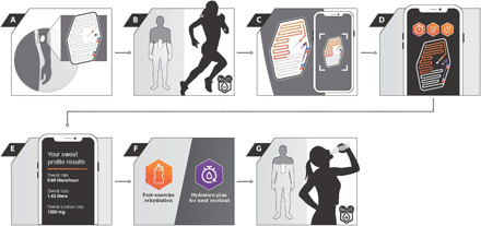
Wearable microfluidic system enables remote analysis of sweat rate and chloride loss for assessing personalized fluid needs.
Overview
The claims and statistics of this paper haven’t been checked yet.
Free: it reads the open-access text and checks the first 5 claims that cite a source we can check. Create a free account to check every claim.
Integrity signals
Registry
Cites
Statistics
Figures


Verification
Claims the paper makes
The claims and statistics of this paper haven’t been checked yet.
Free: it reads the open-access text and checks the first 5 claims that cite a source we can check. Create a free account to check every claim.
Claims other papers make about this one
Each is checked against this paper’s own text: does it say what the citing paper says it does?
The source backs part of the claim, not all of it — or backs it for a different population, condition or comparison.
In the citing paper
“Sweat detection can respond in real-time to changes in the volume and rate of sweat loss,[241,242] as well as the concentration of various ions and metabolites in the body.[45,243–245]”
From Flexible Electronics and Devices as Human–Machine Interfaces for Medical Robotics (2022) · cites this paper as [244]
Experimental — accuracy not yet measured
Self-check
Percentages, sums and reported differences re-computed from the paper’s own numbers.
The self-check needs the paper’s full text.
Citations
Cites
A concordance correlation coefficient to evaluate reproducibility.
Lin, L. I.-K. (1989) · Biometrics · doi:10.2307/2532051 · Cited by 8,785
Fully integrated wearable sensor arrays for multiplexed in situ perspiration analysis
Gao, W., Emaminejad, S., Nyein, H. Y. Y., Challa, S., Chen, K., Peck, A., Fahad, H. M., Ota, H., Shiraki, H., Kiriya, D., et al. (2016) · Nature · doi:10.1038/nature16521 · Cited by 5,087
Wearable biosensors for healthcare monitoring
Kim, J., Campbell, A. S., de Ávila, B. E.-F., & Wang, J. (2019) · Nature Biotechnology · doi:10.1038/s41587-019-0045-y · Cited by 3,373
ACSM Position Stand: Exercise and Fluid Replacement
Convertino, V. A., Armstrong, L. E., Coyle, E. F., Mack, G. W., Sawka, M. N., Senay, L. C., & Sherman, W. M. (1996) · Medicine & Science in Sports & Exercise · doi:10.1097/00005768-199610000-00045 · Cited by 1,965
Bio-Integrated Wearable Systems: A Comprehensive Review
Ray, T. R., Choi, J., Bandodkar, A. J., Krishnan, S., Gutruf, P., Tian, L., Ghaffari, R., & Rogers, J. A. (2019) · Chemical Reviews · doi:10.1021/acs.chemrev.8b00573 · Cited by 1,350
Self-citationA soft, wearable microfluidic device for the capture, storage, and colorimetric sensing of sweat
Koh, A., Kang, D., Xue, Y., Lee, S., Pielak, R. M., Kim, J., Hwang, T., Min, S., Banks, A., Bastien, P., et al. (2016) · Science Translational Medicine · doi:10.1126/scitranslmed.aaf2593 · Cited by 1,308
Self-citationLee, H., Song, C., Hong, Y. S., Kim, M., Cho, H. R., Kang, T., Shin, K., Choi, S. H., Hyeon, T., & Kim, D.-H. (2017) · Science Advances · doi:10.1126/sciadv.1601314 · Cited by 1,118
Method agreement analysis: A review of correct methodology
Watson, P. F., & Petrie, A. (2010) · Theriogenology · doi:10.1016/j.theriogenology.2010.01.003 · Cited by 881
Bandodkar, A. J., Gutruf, P., Choi, J., Lee, K., Sekine, Y., Reeder, J. T., Jeang, W. J., Aranyosi, A. J., Lee, S. P., Model, J. B., et al. (2019) · Science Advances · doi:10.1126/sciadv.aav3294 · Cited by 761
Self-citationAccessing analytes in biofluids for peripheral biochemical monitoring
Heikenfeld, J., Jajack, A., Feldman, B., Granger, S. W., Gaitonde, S., Begtrup, G., & Katchman, B. A. (2019) · Nature Biotechnology · doi:10.1038/s41587-019-0040-3 · Cited by 758
Research works printed but not linked: 2
- ACSM ACSM’s Guidelines for Exercise Testing and Prescription (Lippincott Williams & Wilkins ed. 9 2014) 456 pp.
- J. R. Thomas J. K. Nelson Measuring research variables in Research Methods in Physical Activity J. R. Thomas J. K. Nelson Eds. (Human Kinetics ed. 4 2001) pp. 181–200.
Cited by
Technology Roadmap for Flexible Sensors
Luo, Y., Abidian, M. R., Ahn, J.-H., Akinwande, D., Andrews, A. M., Antonietti, M., Bao, Z., Berggren, M., Berkey, C. A., Bettinger, C. J., et al. (2023) · ACS Nano · doi:10.1021/acsnano.2c12606 · Cited by 1,383
Self-citationEnd-to-end design of wearable sensors
Ates, H. C., Nguyen, P. Q., Gonzalez-Macia, L., Morales-Narváez, E., Güder, F., Collins, J. J., & Dincer, C. (2022) · Nature Reviews Materials · doi:10.1038/s41578-022-00460-x · Cited by 1,306
Skin-Interfaced Wearable Sweat Sensors for Precision Medicine
Min, J., Tu, J., Xu, C., Lukas, H., Shin, S., Yang, Y., Solomon, S. A., Mukasa, D., & Gao, W. (2023) · Chemical Reviews · doi:10.1021/acs.chemrev.2c00823 · Cited by 712
Tehrani, F., Teymourian, H., Wuerstle, B., Kavner, J., Patel, R., Furmidge, A., Aghavali, R., Hosseini-Toudeshki, H., Brown, C., Zhang, F., et al. (2022) · Nature Biomedical Engineering · doi:10.1038/s41551-022-00887-1 · Cited by 625
Flexible Electronics and Devices as Human–Machine Interfaces for Medical Robotics
Heng, W., Solomon, S., & Gao, W. (2022) · Advanced Materials · doi:10.1002/adma.202107902 · Cited by 609
Metadata
- Authors
- Baker, L. B., Model, J. B., Barnes, K. A., Anderson, M. L., Lee, S. P., Lee, K. A., Brown, S. D., Reimel, A. J., Roberts, T. J., Nuccio, R. P., Bonsignore, J. L., Ungaro, C. T., Carter, J. M., Li, W., Seib, M. S., Reeder, J. T., Aranyosi, A. J., Rogers, J. A., & Ghaffari, R.
- Affiliations
- Gatorade Sports Science Institute (US)
- Northwestern University (US)
- PepsiCo (United Kingdom) (GB)
- Epicore Biosystems Inc, Cambridge, MA 02139, USA.
- Corresponding author
- Baker, Lindsay B.
- Rogers, John A.
- Ghaffari, Roozbeh
- ORCID
- Venue
- Science Advances
- Published
- 2020-12-11
- Volume
- 6
- Issue
- 50
- ISSN
- 2375-2548 (ISSN-L)
- PMC
- PMC7732194
- OpenAlex
- W3111366224
- Cited by
- 243
- References
- 39
- Citations by year
- 2026: 48 · 2025: 47 · 2024: 52 · 2023: 41 · 2022: 34 · 2021: 21
- FWCI
- 7.52
- Type
- journal article
- Language
- en
- Open access
- gold
- doi.org
- Licence
- Locations
- Science Advances (publishedVersion, cc-by)
- PubMed Central (publishedVersion, cc-by)
- PubMed (publishedVersion)
- Funders
- Gatorade Sports Science Institute (NA)
- Querrey Simpson Institute for Bioelectronics at Northwestern University
- Topics
- Physical Sciences › Engineering › Biomedical Engineering › Advanced Sensor and Energy Harvesting Materials
- Physical Sciences › Engineering › Biomedical Engineering › Microfluidic and Capillary Electrophoresis Applications
- Health Sciences › Medicine › Physiology (medical) › Thermoregulation and physiological responses
- Keywords
- sweat analysis, sweat chloride, wearable sensors, sports science, low-cost sensing
About these checks
Registry, retraction, Cites and statistics results are computed, not judged, with one exception: a printed reference that no index lists is read once by a model before it counts as a citation error. Claims are extracted by a model and kept only when their quote appears verbatim in the text; each cited source is then checked, with a model’s help, for whether it says what the claim relies on. Those verdicts can be wrong — read the claim’s quote and the cited source before relying on one.
Statistics and arithmetic are recomputed, not judged.
Confidence bands are shown only for fields calibrated on our annotated test set. None is calibrated yet, so no band is shown.
What counts as a claim ›How we measure confidence ›How we check this ›
Are you an author, or do you see a problem with this page? Request a review
How to cite
Cite CoChat for the checks on this page, and the paper for its own findings.
- This page
- CoChat. (2026). Citation check: Skin-interfaced microfluidic system with personalized sweating rate and sweat chloride analytics for sports science applications. https://app.cochat.ai/papers/10.1126/sciadv.abe3929
- The paper
- Baker, L. B., Model, J. B., Barnes, K. A., Anderson, M. L., Lee, S. P., Lee, K. A., Brown, S. D., Reimel, A. J., Roberts, T. J., Nuccio, R. P., Bonsignore, J. L., Ungaro, C. T., Carter, J. M., Li, W., Seib, M. S., Reeder, J. T., Aranyosi, A. J., Rogers, J. A., & Ghaffari, R. (2020). Skin-interfaced microfluidic system with personalized sweating rate and sweat chloride analytics for sports science applications. Science Advances. https://doi.org/10.1126/sciadv.abe3929
Data sources
Bibliographic data: Semantic Scholar, OpenAlex, Crossref.
Contains information from the Semantic Scholar Academic Graph Datasets, made available under the ODC Attribution License (ODC-BY 1.0).
Semantic Scholar’s data is described in: Kinney, R., et al. (2023). The Semantic Scholar Open Data Platform. arXiv:2301.10140.
