Cited by 81

The DOI matches its Crossref record; 13 of the 13 references it cites were found in a registry or an index.

Cortical wiring by synapse type–specific control of local protein synthesis

Bernard, Clémence, Exposito-Alonso, David, Selten, Martijn, Sanalidou, Stella, Hanusz-Godoy, Alicia, Aguilera, Alfonso, Hamid, Fursham, Oozeer, Fazal, Maeso, Patricia, Allison, Leanne, Russell, Matthew, Fleck, Roland A., Rico, Beatriz, Marín, Oscar·2022·Science

Published version of a preprint

Abstract

Neurons use local protein synthesis to support their morphological complexity, which requires independent control across multiple subcellular compartments up to the level of individual synapses. We identify a signaling pathway that regulates the local synthesis of proteins required to form excitatory synapses on parvalbumin-expressing (PV + ) interneurons in the mouse cerebral cortex. This process involves regulation of the TSC subunit 2 (Tsc2) by the Erb-B2 receptor tyrosine kinase 4 (ErbB4), which enables local control of messenger RNA {mRNA} translation in a cell type–specific and synapse type–specific manner. Ribosome-associated mRNA profiling reveals a molecular program of synaptic proteins downstream of ErbB4 signaling required to form excitatory inputs on PV + interneurons. Thus, specific connections use local protein synthesis to control synapse formation in the nervous system.

Overview

The claims and statistics of this paper haven’t been checked yet.

Run the check — free, no account needed. It reads the open-access text and checks the sources it cites.

A few free checks a day · papers already verified open instantly · results are public

Integrity signals

Registry

Crossref · 10.1126/science.abm7466

Cites

13 of 13 references verified0 citation errors · 0 not checkable

Statistics

Figures

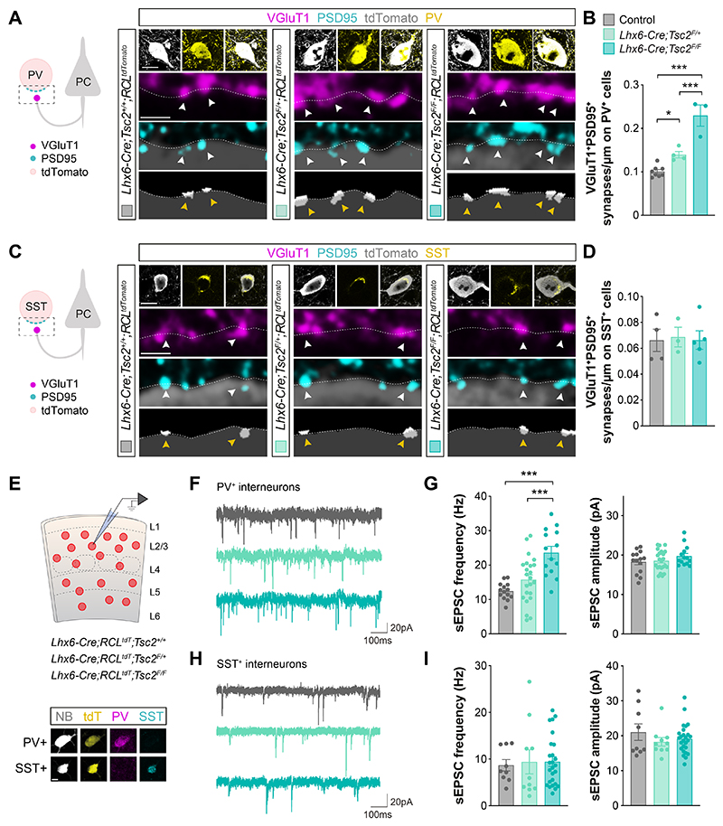
Fig. 1 Differential contribution of Tsc2 to synapse development. (A) Schematic of synaptic markers analyzed (left). Confocal images (top) and binary images (bottom) illustrating presynaptic VGluT1+ puncta (magenta) and postsynaptic PSD95+ clusters (cyan) in PV+ (yellow) tdTomato+ (grey) interneurons from P18-21 control, heterozygous and homozygous conditional Tsc2 mutants. (B) Quantification of the density of VGluT1+PSD95+ synapses contacting PV+ interneurons (control, n =111 cells from 8 mice; heterozygous, n = 70 cells from 4 mice; homozygous, n = 46 cells from 3 mice). (C) Schematic of synaptic markers analyzed (left). Confocal images (top) and binary images (bottom) illustrating presynaptic VGluT1+ puncta (magenta) and postsynaptic PSD95+ clusters (cyan) in SST+ (yellow) tdTomato+ (grey) interneurons from P18-21 control, heterozygous and homozygous conditional Tsc2 mutants. (D) Quantification of the density of VGluT1+PSD95+ synapses contacting SST+ interneurons (control, n = 67 cells from 4 mice; heterozygous, n = 41 cells from 3 mice; homozygous, n = 60 cells from 5 mice). (E) Schematic of experimental design (top) and post-recording labelling of neurobiotin (NB, grey) -filled tdTomato+ (yellow) cells with PV (magenta) and SST (cyan) (bottom). (F) Example traces of sEPSCs recorded from PV+ interneurons from P18-21 control, heterozygous and homozygous conditional Tsc2 mutants. (G) Quantification of the frequency (left) and amplitude (right) of sEPSCs from PV+ interneurons (control n =14 cells from 5 mice, Lhx6-Cre;Tsc2F/+ n=23 cells from 8 mice, Lhx6-Cre;Tsc2F/F n =14 cells from 6 mice). (H) Example traces of sEPSCs recorded from SST+ interneurons from P18-21 control, heterozygous and homozygous conditional Tsc2 mutants. (I) Quantification of the frequency (left) and amplitude (right) of sEPSCs from SST+ interneurons (control, n = 9 cells from 5 mice; heterozygous, n = 10 cells from 9 mice; homozygous, n = 26 cells from 7 mice). One-way ANOVA followed by Tukey’s multiple comparisons test or Kruskal-Wallis followed by Dunn’s multiple comparisons test: *P < 0.05, **P < 0.01, ***P < 0.001. The dashed lines in the images shown in A and C outline the surface of the cells. Data are mean ± s.e.m. Scale bar, 10 μm and 1 μm (high magnification) (A, C), and 10 μm (E).
Fig. 1 Differential contribution of Tsc2 to synapse development. (A) Schematic of synaptic markers analyzed (left). Confocal images (top) and binary images (bottom) illustrating presynaptic VGluT1+ puncta (magenta) and postsynaptic PSD95+ clusters (cyan) in PV+ (yellow) tdTomato+ (grey) interneurons from P18-21 control, heterozygous and homozygous conditional Tsc2 mutants. (B) Quantification of the density of VGluT1+PSD95+ synapses contacting PV+ interneurons (control, n =111 cells from 8 mice; heterozygous, n = 70 cells from 4 mice; homozygous, n = 46 cells from 3 mice). (C) Schematic of synaptic markers analyzed (left). Confocal images (top) and binary images (bottom) illustrating presynaptic VGluT1+ puncta (magenta) and postsynaptic PSD95+ clusters (cyan) in SST+ (yellow) tdTomato+ (grey) interneurons from P18-21 control, heterozygous and homozygous conditional Tsc2 mutants. (D) Quantification of the density of VGluT1+PSD95+ synapses contacting SST+ interneurons (control, n = 67 cells from 4 mice; heterozygous, n = 41 cells from 3 mice; homozygous, n = 60 cells from 5 mice). (E) Schematic of experimental design (top) and post-recording labelling of neurobiotin (NB, grey) -filled tdTomato+ (yellow) cells with PV (magenta) and SST (cyan) (bottom). (F) Example traces of sEPSCs recorded from PV+ interneurons from P18-21 control, heterozygous and homozygous conditional Tsc2 mutants. (G) Quantification of the frequency (left) and amplitude (right) of sEPSCs from PV+ interneurons (control n =14 cells from 5 mice, Lhx6-Cre;Tsc2F/+ n=23 cells from 8 mice, Lhx6-Cre;Tsc2F/F n =14 cells from 6 mice). (H) Example traces of sEPSCs recorded from SST+ interneurons from P18-21 control, heterozygous and homozygous conditional Tsc2 mutants. (I) Quantification of the frequency (left) and amplitude (right) of sEPSCs from SST+ interneurons (control, n = 9 cells from 5 mice; heterozygous, n = 10 cells from 9 mice; homozygous, n = 26 cells from 7 mice). One-way ANOVA followed by Tukey’s multiple comparisons test or Kruskal-Wallis followed by Dunn’s multiple comparisons test: *P < 0.05, **P < 0.01, ***P < 0.001. The dashed lines in the images shown in A and C outline the surface of the cells. Data are mean ± s.e.m. Scale bar, 10 μm and 1 μm (high magnification) (A, C), and 10 μm (E).CC BY · PubMed Central
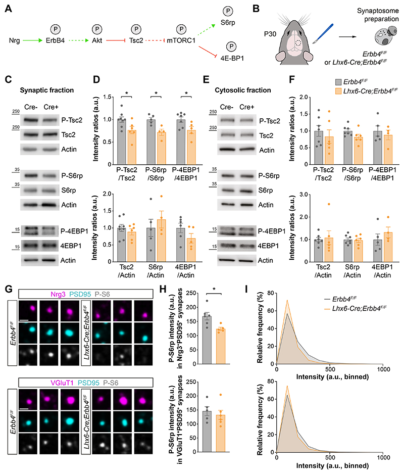
Fig. 2 ErbB4 regulates mTOR at excitatory synapses contacting PV+ interneurons. (A) Hypothetical signaling pathway. P indicates phosphorylation; green arrow activation; red arrow inhibition; dotted arrows indicate indirect regulation. (B) Schematic of experimental design. (C) Phosphorylation and protein expression of Tsc2, S6rp, 4EBP1 and actin assessed by Western blot of cortical synaptic fractions from P30 homozygous conditional Erbb4 mice and their control littermates. (D) Quantification of phosphorylation of Tsc2, S6rp and 4EBP1 normalized to the total expression of the corresponding protein (top). Quantification of expression levels of Tsc2, S6rp and 4EBP1 normalized to actin (bottom) (Tsc2: control, n = 8 mice, homozygous, n = 6 mice; S6rp: control, n = 5 mice, homozygous, n = 4 mice; 4EBP1: control, n = 7 mice, homozygous, n = 5 mice). (E) Phosphorylation and protein expression of Tsc2, S6rp, 4EBP1 and actin assessed by Western blot of cortical cytosolic fractions from P30 homozygous conditional Erbb4 mice and their control littermates. (F) Quantification of phosphorylation of Tsc2, S6rp and 4EBP1 normalized to the total expression of the corresponding protein (top). Quantification of expression levels of Tsc2, S6rp and 4EBP1 normalized to actin (bottom) (Tsc2 and S6rp: control, n = 7 mice, homozygous, n = 6 mice; 4EBP1: control, n = 5 mice, homozygous, n = 4 mice). (G) Confocal images illustrating phosphorylation of S6rp (P-S6rp, grey) in Nrg3+ (magenta) PSD95+ (cyan) synaptosomes (top) and in VGluT1+ (magenta) PSD95+ (cyan) synaptosomes (bottom) from P21 homozygous conditional Erbb4 mice and their control littermates. (H) Quantification of P-S6rp staining intensity in Nrg3+PSD95+ (top) and VGluT1+PSD95+ synaptosomes (bottom). (I) Relative frequency distribution of P-S6rp staining intensity in Nrg3+PSD95+ synaptosomes (top) and in VGluT1+PSD95+ synaptosomes (bottom) (Nrg3+PSD95+: control, n = 22,063 synaptosomes from 6 mice, homozygous, n = 17,523 synaptosomes from 5 mice; VGluT1+PSD95+: control, n = 39,395 synaptosomes from 5 mice, homozygous, n = 48,057 synaptosomes from 6 mice). Two-tailed Student’s unpaired t-tests: *P < 0.05. Data are mean ± s.e.m. Scale bar, 1 μm.
Fig. 2 ErbB4 regulates mTOR at excitatory synapses contacting PV+ interneurons. (A) Hypothetical signaling pathway. P indicates phosphorylation; green arrow activation; red arrow inhibition; dotted arrows indicate indirect regulation. (B) Schematic of experimental design. (C) Phosphorylation and protein expression of Tsc2, S6rp, 4EBP1 and actin assessed by Western blot of cortical synaptic fractions from P30 homozygous conditional Erbb4 mice and their control littermates. (D) Quantification of phosphorylation of Tsc2, S6rp and 4EBP1 normalized to the total expression of the corresponding protein (top). Quantification of expression levels of Tsc2, S6rp and 4EBP1 normalized to actin (bottom) (Tsc2: control, n = 8 mice, homozygous, n = 6 mice; S6rp: control, n = 5 mice, homozygous, n = 4 mice; 4EBP1: control, n = 7 mice, homozygous, n = 5 mice). (E) Phosphorylation and protein expression of Tsc2, S6rp, 4EBP1 and actin assessed by Western blot of cortical cytosolic fractions from P30 homozygous conditional Erbb4 mice and their control littermates. (F) Quantification of phosphorylation of Tsc2, S6rp and 4EBP1 normalized to the total expression of the corresponding protein (top). Quantification of expression levels of Tsc2, S6rp and 4EBP1 normalized to actin (bottom) (Tsc2 and S6rp: control, n = 7 mice, homozygous, n = 6 mice; 4EBP1: control, n = 5 mice, homozygous, n = 4 mice). (G) Confocal images illustrating phosphorylation of S6rp (P-S6rp, grey) in Nrg3+ (magenta) PSD95+ (cyan) synaptosomes (top) and in VGluT1+ (magenta) PSD95+ (cyan) synaptosomes (bottom) from P21 homozygous conditional Erbb4 mice and their control littermates. (H) Quantification of P-S6rp staining intensity in Nrg3+PSD95+ (top) and VGluT1+PSD95+ synaptosomes (bottom). (I) Relative frequency distribution of P-S6rp staining intensity in Nrg3+PSD95+ synaptosomes (top) and in VGluT1+PSD95+ synaptosomes (bottom) (Nrg3+PSD95+: control, n = 22,063 synaptosomes from 6 mice, homozygous, n = 17,523 synaptosomes from 5 mice; VGluT1+PSD95+: control, n = 39,395 synaptosomes from 5 mice, homozygous, n = 48,057 synaptosomes from 6 mice). Two-tailed Student’s unpaired t-tests: *P < 0.05. Data are mean ± s.e.m. Scale bar, 1 μm.CC BY · PubMed Central
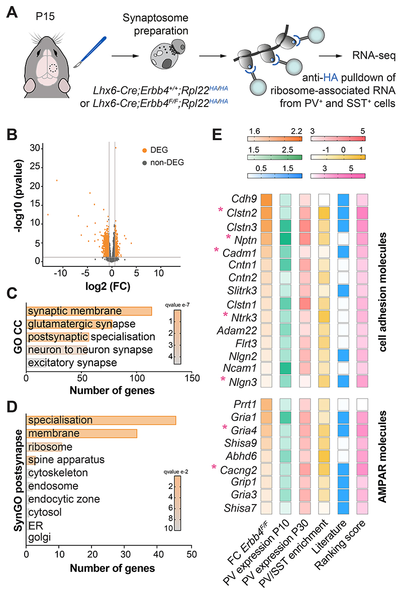
Fig. 3 Synaptic ribosome-associated mRNAs altered in Erbb4 mutants. (A) Schematic of experimental design. (B) Volcano plot displaying significantly differentially expressed ribosome-associated RNAs (DEG, orange) in P15 cortical synaptosomes from homozygous conditional Erbb4 mice compared to controls. Each dot represents one gene. FC: fold-change (FC > 1.5, P < 0.05). (C) Selected Gene Ontology (GO) Cellular Components (CC) terms significantly enriched in the dataset of downregulated genes in homozygous conditional Erbb4 mutants compared to controls. (D) Synaptic Gene Ontology (SynGO) postsynaptic cellular component categories significantly enriched in the dataset of downregulated genes in homozygous conditional Erbb4 mutants compared to controls. (E) Heatmaps showing the selection criteria for 15 “cell adhesion molecules” genes and 9 “AMPA receptors” genes. The asterisks indicate genes selected for validation.
Fig. 3 Synaptic ribosome-associated mRNAs altered in Erbb4 mutants. (A) Schematic of experimental design. (B) Volcano plot displaying significantly differentially expressed ribosome-associated RNAs (DEG, orange) in P15 cortical synaptosomes from homozygous conditional Erbb4 mice compared to controls. Each dot represents one gene. FC: fold-change (FC > 1.5, P < 0.05). (C) Selected Gene Ontology (GO) Cellular Components (CC) terms significantly enriched in the dataset of downregulated genes in homozygous conditional Erbb4 mutants compared to controls. (D) Synaptic Gene Ontology (SynGO) postsynaptic cellular component categories significantly enriched in the dataset of downregulated genes in homozygous conditional Erbb4 mutants compared to controls. (E) Heatmaps showing the selection criteria for 15 “cell adhesion molecules” genes and 9 “AMPA receptors” genes. The asterisks indicate genes selected for validation.CC BY · PubMed Central
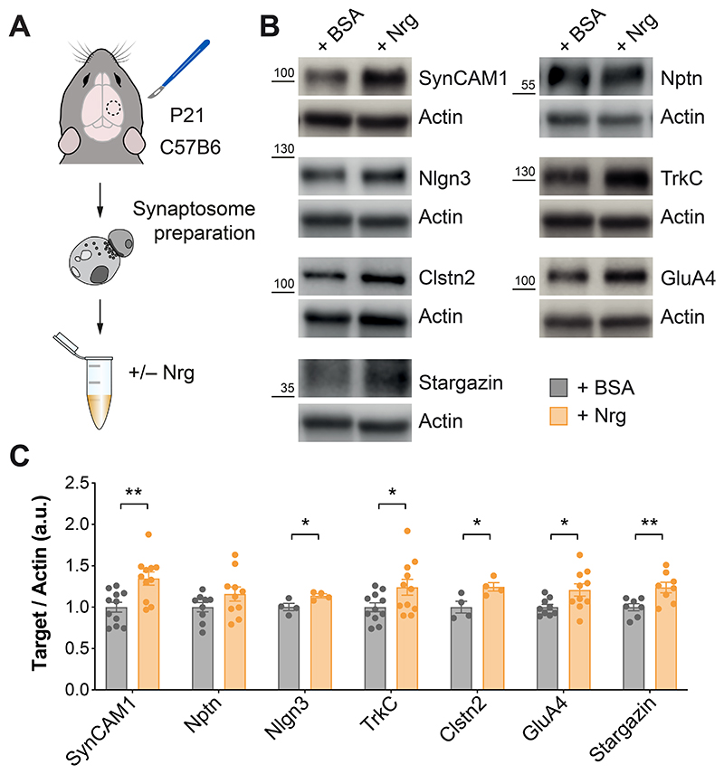
Fig. 4 ErbB4 regulates the local translation of synaptic proteins. (A) Schematic of experimental design. (B) Protein expression of SynCAM1, Nptn, Nlgn3, TrkC, Clstn2, GluA4, Stargazin and actin assessed by Western blot of cortical synaptic fractions treated with neuregulin (Nrg) from P21 C57B6 mice. (C) Quantification of expression levels of SynCAM1, Nptn, Nlgn3. TrkC, Clstn2, GluA4 and Stargazin normalized to actin. One-tailed Student’s unpaired t-tests: *P < 0.05, **P < 0.01 (SynCAM1 and TrkC: +BSA, n =11 synaptosomes: +Nrg, n = 11 synaptosomes; Nptn and GluA4: +BSA, n = 9 synaptosomes, +Nrg, n = 10 synaptosomes; Nlgn3 and Clstn2: +BSA, n = 4 synaptosomes, +Nrg, n = 4 synaptosomes; Stargazin: +BSA, n = 7 synaptosomes, +Nrg, n = 8 synaptosomes). Data are mean ± s.e.m.
Fig. 4 ErbB4 regulates the local translation of synaptic proteins. (A) Schematic of experimental design. (B) Protein expression of SynCAM1, Nptn, Nlgn3, TrkC, Clstn2, GluA4, Stargazin and actin assessed by Western blot of cortical synaptic fractions treated with neuregulin (Nrg) from P21 C57B6 mice. (C) Quantification of expression levels of SynCAM1, Nptn, Nlgn3. TrkC, Clstn2, GluA4 and Stargazin normalized to actin. One-tailed Student’s unpaired t-tests: *P < 0.05, **P < 0.01 (SynCAM1 and TrkC: +BSA, n =11 synaptosomes: +Nrg, n = 11 synaptosomes; Nptn and GluA4: +BSA, n = 9 synaptosomes, +Nrg, n = 10 synaptosomes; Nlgn3 and Clstn2: +BSA, n = 4 synaptosomes, +Nrg, n = 4 synaptosomes; Stargazin: +BSA, n = 7 synaptosomes, +Nrg, n = 8 synaptosomes). Data are mean ± s.e.m.CC BY · PubMed Central
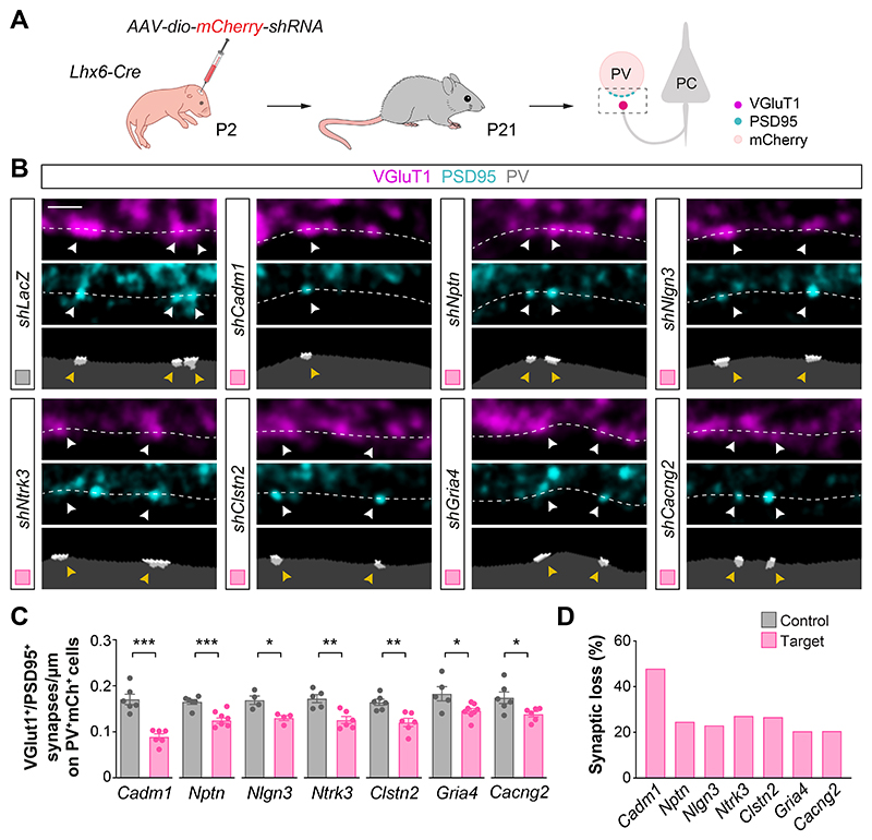
Fig. 5 ErbB4 targets control excitatory synapse formation on PV+ interneurons. (A) Schematic of experimental design. (B) Confocal images (top) and binary images (bottom) illustrating presynaptic VGluT1+ puncta (magenta) and postsynaptic PSD95+ clusters (cyan) in PV+ interneurons (grey) from P21 Lhx6-Cre mice injected with viruses expressing shRNAs targeting the genes of interest or with a control virus (shLacZ). (C) Quantification of the density of VGluT1+PSD95+ synapses contacting PV+ interneurons in knockdown and control mice. Two-tailed Student’s unpaired t-tests: *P < 0.05, **P < 0.01, ***P < 0.001; shCadm1 (n = 122 cells from 6 mice) and control (n = 113 cells from 6 mice); shNptn (n = 131 cells from 6 mice) and control (n = 119 cells from 6 mice); shNlgn3 (n = 86 cells from 4 mice) and control (n = 68 cells from 4 mice); shNtrk3 (n = 126 cells from 6 mice) and control (n = 105 cells from 5 mice); shClstn2 (n = 120 cells from 6 mice) and control (n = 112 cells from 6 mice); shGria4 (n = 121 cells from 8 mice) and control (n = 93 cells from 5 mice); shCacng2 (n = 115 cells from 6 mice) and control (n = 109 cells from 6 mice). (D) Proportion of synaptic loss in PV+ interneurons upon knockdown of ErbB4 downstream targets. Data are mean ± s.e.m. Scale bar, 1 μm.
Fig. 5 ErbB4 targets control excitatory synapse formation on PV+ interneurons. (A) Schematic of experimental design. (B) Confocal images (top) and binary images (bottom) illustrating presynaptic VGluT1+ puncta (magenta) and postsynaptic PSD95+ clusters (cyan) in PV+ interneurons (grey) from P21 Lhx6-Cre mice injected with viruses expressing shRNAs targeting the genes of interest or with a control virus (shLacZ). (C) Quantification of the density of VGluT1+PSD95+ synapses contacting PV+ interneurons in knockdown and control mice. Two-tailed Student’s unpaired t-tests: *P < 0.05, **P < 0.01, ***P < 0.001; shCadm1 (n = 122 cells from 6 mice) and control (n = 113 cells from 6 mice); shNptn (n = 131 cells from 6 mice) and control (n = 119 cells from 6 mice); shNlgn3 (n = 86 cells from 4 mice) and control (n = 68 cells from 4 mice); shNtrk3 (n = 126 cells from 6 mice) and control (n = 105 cells from 5 mice); shClstn2 (n = 120 cells from 6 mice) and control (n = 112 cells from 6 mice); shGria4 (n = 121 cells from 8 mice) and control (n = 93 cells from 5 mice); shCacng2 (n = 115 cells from 6 mice) and control (n = 109 cells from 6 mice). (D) Proportion of synaptic loss in PV+ interneurons upon knockdown of ErbB4 downstream targets. Data are mean ± s.e.m. Scale bar, 1 μm.CC BY · PubMed Central

Verification

Claims the paper makes

The claims and statistics of this paper haven’t been checked yet.

Run the check — free, no account needed. It reads the open-access text and checks the sources it cites.

A few free checks a day · papers already verified open instantly · results are public

Claims other papers make about this one

Each is checked against this paper’s own text: does it say what the citing paper says it does?

Self-check

Percentages, sums and reported differences re-computed from the paper’s own numbers.

The self-check needs the paper’s full text.

Citations

Cites

Show all 13 references

Research works printed but not linked: 62

  1. 10.1126/science.1149381 · DOI · Paper page
  2. 10.1146/annurev.neuro.27.070203.144152 · DOI · Paper page
  3. 10.1016/j.cell.2004.10.004 · DOI
  4. 10.1016/j.neuron.2011.06.019 · DOI
  5. 10.1016/j.cell.2014.06.047 · DOI
  6. 10.1126/science.aau8977 · DOI
  7. 10.1126/science.aav7969 · DOI
  8. 10.1038/nrn.2015.3 · DOI
  9. 10.1146/annurev.neuro.051508.135302 · DOI
  10. 10.1146/annurev-biochem-013118-111042 · DOI
  11. 10.1038/ncb847 · DOI
  12. 10.1038/ncb839 · DOI · Paper page
  13. 10.1016/S0896-6273(01)00551-7 · DOI
  14. 10.1016/S0092-8674(02)00813-9 · DOI
  15. 10.1038/s41586-018-0847-y · DOI
  16. 10.1126/science.273.5280.1402 · DOI
  17. 10.1523/JNEUROSCI.19-01-00168.1999 · DOI
  18. 10.1016/S0896-6273(02)00785-7 · DOI · Paper page
  19. 10.1073/pnas.012605299 · DOI · Paper page
  20. 10.1016/j.neuron.2016.09.040 · DOI
  21. 10.1126/science.aau3644 · DOI · Paper page
  22. 10.7554/eLife.63092 · DOI
  23. 10.1101/gad.995802 · DOI · Paper page
  24. 10.1038/s41467-019-12962-4 · DOI
  25. 10.1038/ncb840 · DOI · Paper page
  26. 10.1523/JNEUROSCI.2538-10.2011 · DOI
  27. 10.1016/j.neuron.2013.07.010 · DOI · Paper page
  28. 10.1038/sj.onc.1202612 · DOI
  29. 10.1523/JNEUROSCI.2454-09.2009 · DOI
  30. 10.15252/embj.201798858 · DOI
  31. 10.7554/eLife.57000 · DOI
  32. 10.1073/pnas.0907143106 · DOI · Paper page
  33. 10.1016/j.neuron.2019.05.002 · DOI · Paper page
  34. 10.1016/j.neuron.2014.06.007 · DOI
  35. 10.1016/j.neuron.2017.06.028 · DOI · Paper page
  36. 10.1146/annurev-genet-030321-054851 · DOI
  37. 10.1038/nn.4471 · DOI
  38. 10.1523/JNEUROSCI.0189-16.2016 · DOI
  39. 10.1016/j.celrep.2018.12.069 · DOI
  40. 10.1126/sciadv.abj0790 · DOI
  41. 10.1126/science.aau9952 · DOI
  42. 10.1016/j.cell.2016.05.029 · DOI
  43. 10.1016/j.celrep.2019.11.025 · DOI
  44. 10.1016/j.neuron.2018.06.004 · DOI
  45. 10.1016/j.cell.2008.10.017 · DOI · Paper page
  46. 10.1016/j.neuron.2019.02.041 · DOI · Paper page
  47. 10.1038/nature10658 · DOI · Paper page
  48. 10.1002/ajmg.a.35911 · DOI
  49. 10.1038/s41588-021-00899-8 · DOI
  50. 10.1146/annurev-genom-083118-015354 · DOI · Paper page
  51. 10.1146/annurev-neuro-080317-061747 · DOI
  52. 10.1016/j.neuron.2015.09.016 · DOI · Paper page
  53. 10.1038/nature12866 · DOI · Paper page
  54. 10.1002/dvg.20271 · DOI
  55. 10.1523/JNEUROSCI.1629-07.2007 · DOI · Paper page
  56. 10.1038/nn.2467 · DOI · Paper page
  57. 10.1016/j.bbr.2003.11.010 · DOI
  58. 10.1093/bioinformatics/bts635 · DOI · Paper page
  59. 10.1016/j.stemcr.2015.10.006 · DOI
  60. 10.1186/s13059-014-0550-8 · DOI · Paper page
  61. 10.1093/nar/gkz401 · DOI · Paper page
  62. 10.1038/s41593-019-0465-5 · DOI

Cited by

Sorted by citations

Metadata

Authors
Bernard, C., Exposito-Alonso, D., Selten, M., Sanalidou, S., Hanusz-Godoy, A., Aguilera, A., Hamid, F., Oozeer, F., Maeso, P., Allison, L., Russell, M., Fleck, R. A., Rico, B., & Marín, O.
Affiliations
  • Centre for Developmental Neurobiology, Institute of Psychiatry, Psychology and Neuroscience, King’s College London, London SE1 1UL, UK.
  • Centre for Ultrastructural Imaging, King’s College London, London SE1 1UL, UK.
Venue
Science
Published
2022-11-25
Volume
378
Issue
6622
ISSN
  • 0036-8075
  • 1095-9203
Cited by
81
References
13
Language
en

About these checks

Registry, retraction, Cites and statistics results are computed, not judged, with one exception: a printed reference that no index lists is read once by a model before it counts as a citation error. Claims are extracted by a model and kept only when their quote appears verbatim in the text; each cited source is then checked, with a model’s help, for whether it says what the claim relies on. Those verdicts can be wrong — read the claim’s quote and the cited source before relying on one.

Statistics and arithmetic are recomputed, not judged.

Confidence bands are shown only for fields calibrated on our annotated test set. None is calibrated yet, so no band is shown.

What counts as a claim ›How we measure confidence ›How we check this ›

Are you an author, or do you see a problem with this page? Request a review