

The DOI matches its Crossref record; 89 of the 90 references it cites were found in a registry or an index, and 1 are not checked yet.
Structural basis of SARS-CoV-2 Omicron immune evasion and receptor engagement
Open accessPublished version of a preprint
Abstract
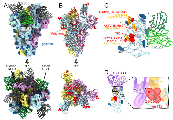
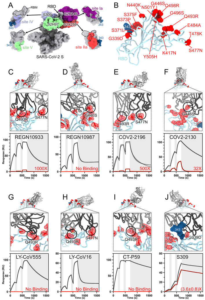
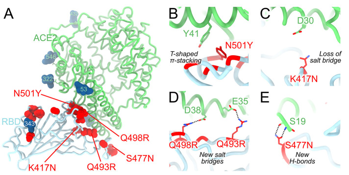
The severe acute respiratory syndrome coronavirus 2 (SARS-CoV-2) Omicron variant of concern evades antibody-mediated immunity that comes from vaccination or infection with earlier variants due to accumulation of numerous spike mutations. To understand the Omicron antigenic shift, we determined cryo–electron microscopy and x-ray crystal structures of the spike protein and the receptor-binding domain bound to the broadly neutralizing sarbecovirus monoclonal antibody (mAb) S309 (the parent mAb of sotrovimab) and to the human ACE2 receptor. We provide a blueprint for understanding the marked reduction of binding of other therapeutic mAbs that leads to dampened neutralizing activity. Remodeling of interactions between the Omicron receptor-binding domain and human ACE2 likely explains the enhanced affinity for the host receptor relative to the ancestral virus.
Overview
The claims and statistics of this paper haven’t been checked yet.
Run the check — free, no account needed. It reads the open-access text and checks the sources it cites.
Integrity signals
Registry
Cites
Statistics
Figures
Verification
Claims the paper makes
The claims and statistics of this paper haven’t been checked yet.
Run the check — free, no account needed. It reads the open-access text and checks the sources it cites.
Claims other papers make about this one
Each is checked against this paper’s own text: does it say what the citing paper says it does?
The cited source says what the claim attributes to it.
In the citing paper
“Several VOCs which have mutated, such as Beta1 and Omicron2, evade humoral responses elicited by vaccines based on ancestral S-protein sequences3.”
From Efficacy of an unmodified bivalent mRNA vaccine against SARS-CoV-2 variants in female small animal models (2023) · cites this paper as [2]
In the citing paper
“Reinfections and vaccine breakthrough infections accounted for the majority of Omicron BA.1/2 infections, likely attributable to a minority of population remaining naïve prior to the Omicron BA.1/2 wave and to Omicron BA.1/2's immune-escape properties 16,21,22 .”
From Rapidly shifting immunologic landscape and severity of SARS-CoV-2 in the Omicron era in South Africa (2023) · cites this paper as [22]
Self-check
Percentages, sums and reported differences re-computed from the paper’s own numbers.
The self-check needs the paper’s full text.
Citations
Cites
UCSF Chimera—A visualization system for exploratory research and analysis
Pettersen, E. F., Goddard, T. D., Huang, C. C., Couch, G. S., Greenblatt, D. M., Meng, E. C., & Ferrin, T. E. (2004) · Journal of Computational Chemistry · doi:10.1002/jcc.20084 · Cited by 45,882
Features and development of Coot
Emsley, P., Lohkamp, B., Scott, W. G., & Cowtan, K. (2010) · Acta Crystallographica Section D Biological Crystallography · doi:10.1107/s0907444910007493 · Cited by 29,915
Phaser crystallographic software
McCoy, A. J., Grosse-Kunstleve, R. W., Adams, P. D., Winn, M. D., Storoni, L. C., & Read, R. J. (2007) · Journal of Applied Crystallography · doi:10.1107/s0021889807021206 · Cited by 20,612
MolProbity : all-atom structure validation for macromolecular crystallography
Chen, V. B., Arendall, W. B., Headd, J. J., Keedy, D. A., Immormino, R. M., Kapral, G. J., Murray, L. W., Richardson, J. S., & Richardson, D. C. (2010) · Acta Crystallographica Section D Biological Crystallography · doi:10.1107/s0907444909042073 · Cited by 14,115
cryoSPARC: algorithms for rapid unsupervised cryo-EM structure determination
Punjani, A., Rubinstein, J. L., Fleet, D. J., & Brubaker, M. A. (2017) · Nature Methods · doi:10.1038/nmeth.4169 · Cited by 11,626
Cryo-EM structure of the 2019-nCoV spike in the prefusion conformation
Wrapp, D., Wang, N., Corbett, K. S., Goldsmith, J. A., Hsieh, C.-L., Abiona, O., Graham, B. S., & McLellan, J. S. (2020) · Science · doi:10.1126/science.abb2507 · Cited by 8,897
Structure, function and antigenicity of the SARS-CoV-2 spike glycoprotein
Walls, A. C., Park, Y.-J., Tortorici, M. A., Wall, A., McGuire, A. T., & Veesler, D. (2020) · doi:10.1101/2020.02.19.956581 · Cited by 8,586 · cited 1×
“However, the region comprising residues 366 to 375, which harbors the S371L/S373P/S375F substitutions, deviates markedly from the conformation observed for the Wuhan-Hu-1 RBD, irrespective of the presence of bound linoleic acid ( 4 , 42 , 43 ).”
Self-citationREFMAC 5 for the refinement of macromolecular crystal structures
Murshudov, G. N., Skubák, P., Lebedev, A. A., Pannu, N. S., Steiner, R. A., Nicholls, R. A., Winn, M., Long, F., & Vagin, A. A. (2011) · Acta Crystallographica Section D Biological Crystallography · doi:10.1107/s0907444911001314 · Cited by 8,415
Liebschner, D., Afonine, P. V., Baker, M. L., Bunkóczi, G., Chen, V. B., Croll, T. I., Hintze, B., Hung, L.-W., Jain, S., McCoy, A. J., et al. (2019) · Acta Crystallographica Section D Structural Biology · doi:10.1107/s2059798319011471 · Cited by 7,953
Self-citationStructure of the SARS-CoV-2 spike receptor-binding domain bound to the ACE2 receptor
Lan, J., Ge, J., Yu, J., Shan, S., Zhou, H., Fan, S., Zhang, Q., Shi, X., Wang, Q., Zhang, L., et al. (2020) · Nature · doi:10.1038/s41586-020-2180-5 · Cited by 5,983 · cited 2×
“The Omicron RBD is structurally similar to the Wuhan-Hu-1 RBD and both structures can be superimposed with an r.m.s.d. of 0.8Å over 183 aligned C ɑ residues (as compared to PDB 6m0j ( 42 )).”
Research works printed but not linked: 2
- 10.1016/j.cell.2020.02.058 · DOI · Paper page
- 10.1126/science.abh1139 · DOI · Paper page
Cited by
Sorted by citations
Markov, P. V., Ghafari, M., Beer, M., Lythgoe, K., Simmonds, P., Stilianakis, N. I., & Katzourakis, A. (2023) · Nature Reviews Microbiology · doi:10.1038/s41579-023-00878-2 · Cited by 1,179
Altered TMPRSS2 usage by SARS-CoV-2 Omicron impacts infectivity and fusogenicity
Meng, B., Abdullahi, A., Ferreira, I. A. T. M., Goonawardane, N., Saito, A., Kimura, I., Yamasoba, D., Gerber, P. P., Fatihi, S., Rathore, S., et al. (2022) · Nature · doi:10.1038/s41586-022-04474-x · Cited by 1,146
Self-citationSARS-CoV-2 Omicron variant: Antibody evasion and cryo-EM structure of spike protein–ACE2 complex
Mannar, D., Saville, J. W., Zhu, X., Srivastava, S. S., Berezuk, A. M., Tuttle, K. S., Marquez, A. C., Sekirov, I., & Subramaniam, S. (2022) · Science · doi:10.1126/science.abn7760 · Cited by 630
SARS-CoV-2 Omicron virus causes attenuated disease in mice and hamsters
Halfmann, P., Iida, S., Iwatsuki‐Horimoto, K., Maemura, T., Kiso, M., Scheaffer, S. M., Darling, T. L., Joshi, A., Loeber, S. J., Singh, G., et al. (2022) · Nature · doi:10.1038/s41586-022-04441-6 · Cited by 664
Self-citationAntibody escape of SARS-CoV-2 Omicron BA.4 and BA.5 from vaccine and BA.1 serum
Tuekprakhon, A., Nutalai, R., Dijokaite-Guraliuc, A., Zhou, D., Ginn, H. M., Selvaraj, M., Liu, C., Mentzer, A. J., Supasa, P., Duyvesteyn, H. M. E., et al. (2022) · Cell · doi:10.1016/j.cell.2022.06.005 · Cited by 687
Metadata
- Authors
- McCallum, M., Czudnochowski, N., Rosen, L. E., Zepeda, S. K., Bowen, J. E., Walls, A. C., Hauser, K., Joshi, A., Stewart, C., Dillen, J. R., Powell, A. E., Croll, T. I., Nix, J., Virgin, H. W., Corti, D., Snell, G., & Veesler, D.
- Affiliations
- University of Washington (US)
- VIR Biotechnology (United States) (US)
- Howard Hughes Medical Institute (US)
- University of Cambridge (GB)
- Lawrence Berkeley National Laboratory (US)
- Washington University in St. Louis (US)
- Vir Biotechnology (Switzerland) (CH)
- Corresponding author
- McCallum, Matthew
- Czudnochowski, Nadine
- Snell, Gyorgy
- Veesler, David
- ORCID
- Venue
- Science
- Published
- 2022-02-25
- Volume
- 375
- Issue
- 6583
- Pages
- 864-868
- ISSN
- 0036-8075 (ISSN-L)
- 1095-9203
- PMC
- PMC9427005
- OpenAlex
- W4220861013
- Cited by
- 535
- References
- 85
- Citations by year
- 2026: 25 · 2025: 56 · 2024: 85 · 2023: 122 · 2022: 246
- FWCI
- 62.11
- Type
- journal article
- Language
- en
- Open access
- hybrid
- doi.org
- Licence
- cc-by
- Locations
- Science (publishedVersion, cc-by)
- bioRxiv (Cold Spring Harbor Laboratory) (acceptedVersion, cc-by-nc-nd)
- PubMed Central (publishedVersion, cc-by)
- PubMed (publishedVersion)
- OSTI OAI (U.S. Department of Energy Office of Scientific and Technical Information) (submittedVersion, cc-by)
- eScholarship (California Digital Library) (submittedVersion)
- Funders
- Howard Hughes Medical Institute (DE-AC02-05CH11231)
- U.S. Department of Energy (-AC02-05CH11231, Contract No. DE-AC02-05-CH11231, 05CH11231, No. DE-AC02-05-CH11231, DE AC02 05CH11231, DE-AC02-)
- Burroughs Wellcome Fund (DE-AC02-05CH11231)
- University of Washington
- Wellcome Trust (Z/17/Z, 209407/Z/17/Z)
- Fast Grants
- National Institutes of Health (S10 OD032290, DE-AC02–05CH11231, 1s10od023476-01, HHSN272201700059C)
- Office of Science (No. DE-AC02- 05-CH11231, DEAC02–05CH11231, AC02-05CH11231, DE-AC02, 05CH11231, Contract No. DE-AC02-05CH11231)
- National Institute of General Medical Sciences (DE-AC02-05CH11231, R01GM120553, T32GM008268, P30GM124169)
- National Institute of Allergy and Infectious Diseases (HHSN272201700059C, DP1AI158186)
- Topics
- Health Sciences › Medicine › Infectious Diseases › SARS-CoV-2 and COVID-19 Research
- Life Sciences › Biochemistry, Genetics and Molecular Biology › Molecular Biology › vaccines and immunoinformatics approaches
- Life Sciences › Agricultural and Biological Sciences › Animal Science and Zoology › Animal Virus Infections Studies
- Keywords
- Omicron variant, spike protein, receptor binding domain, antibody escape, sotrovimab, ACE2 receptor, antigenic shift, spike protein mutations, antibody affinity
- MeSH terms
- Protein Domains, Broadly Neutralizing Antibodies, Receptors, Coronavirus, Angiotensin-Converting Enzyme 2, SARS-CoV-2, Antigenic Drift and Shift, Antibodies, Monoclonal, Antibodies, Viral, Humans, Models, Molecular, Mutation, Protein Binding, Protein Conformation, Crystallography, X-Ray, Amino Acid Substitution, Cryoelectron Microscopy, Protein Interaction Domains and Motifs, Immune Evasion, Spike Glycoprotein, Coronavirus
About these checks
Registry, retraction, Cites and statistics results are computed, not judged, with one exception: a printed reference that no index lists is read once by a model before it counts as a citation error. Claims are extracted by a model and kept only when their quote appears verbatim in the text; each cited source is then checked, with a model’s help, for whether it says what the claim relies on. Those verdicts can be wrong — read the claim’s quote and the cited source before relying on one.
Statistics and arithmetic are recomputed, not judged.
Confidence bands are shown only for fields calibrated on our annotated test set. None is calibrated yet, so no band is shown.
What counts as a claim ›How we measure confidence ›How we check this ›
Are you an author, or do you see a problem with this page? Request a review
