

The DOI matches its Crossref record; 9 of the 9 references it cites were found in a registry or an index.
Drug-induced liver injury: recent advances in diagnosis and risk assessment
Abstract
Idiosyncratic drug-induced liver injury (IDILI) is a rare but potentially severe adverse drug reaction that should be considered in patients who develop laboratory criteria for liver injury secondary to the administration of a potentially hepatotoxic drug. Although currently used liver parameters are sensitive in detecting DILI, they are neither specific nor able to predict the patient's subsequent clinical course. Genetic risk assessment is useful mainly due to its high negative predictive value, with several human leucocyte antigen alleles being associated with DILI. New emerging biomarkers which could be useful in assessing DILI include total keratin18 (K18) and caspase-cleaved keratin18 (ccK18), macrophage colony-stimulating factor receptor 1, high mobility group box 1 and microRNA-122. From the numerous in vitro test systems that are available, monocyte-derived hepatocytes generated from patients with DILI show promise in identifying the DILI-causing agent from among a panel of coprescribed drugs. Several computer-based algorithms are available that rely on cumulative scores of known risk factors such as the administered dose or potential liabilities such as mitochondrial toxicity, inhibition of the bile salt export pump or the formation of reactive metabolites. A novel DILI cluster score is being developed which predicts DILI from multiple complimentary cluster and classification models using absorption–distribution–metabolism–elimination-related as well as physicochemical properties, diverse substructural descriptors and known structural liabilities. The provision of more advanced scientific and regulatory guidance for liver safety assessment will depend on validating the new diagnostic markers in the ongoing DILI registries, biobanks and public–private partnerships.
Overview
The claims and statistics of this paper haven’t been checked yet.
Run the check — free, no account needed. It reads the open-access text and checks the sources it cites.
Integrity signals
Registry
Cites
Statistics
Figures
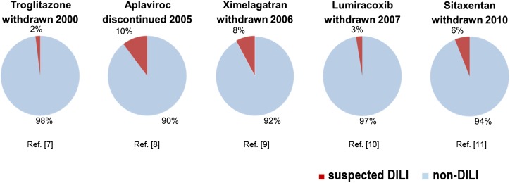
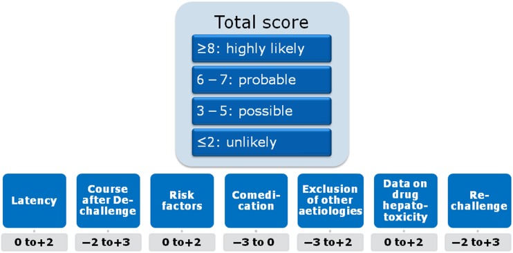
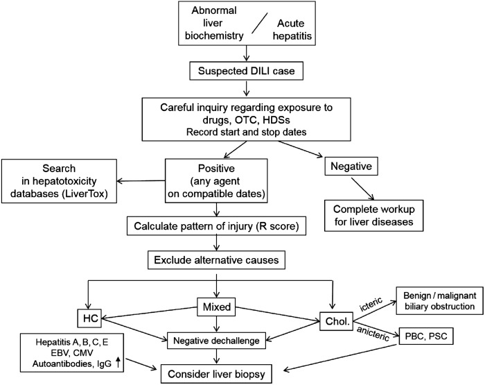
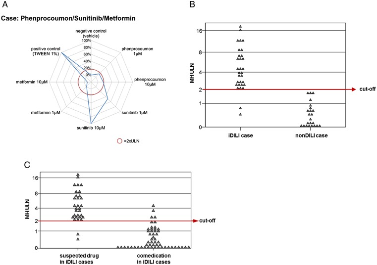
Verification
Claims the paper makes
The claims and statistics of this paper haven’t been checked yet.
Run the check — free, no account needed. It reads the open-access text and checks the sources it cites.
Claims other papers make about this one
Each is checked against this paper’s own text: does it say what the citing paper says it does?
The cited source says what the claim attributes to it.
In the citing paper
“DILI is a major cause of the withdrawal of potentially valuable therapies post-marketing.80”
From Nomenclature, diagnosis and management of drug-induced autoimmune-like hepatitis (DI-ALH): An expert opinion meeting report (2023) · cites this paper as [80]
Self-check
Percentages, sums and reported differences re-computed from the paper’s own numbers.
The self-check needs the paper’s full text.
Citations
Cites
Godoy, P., Hewitt, N. J., Albrecht, U., Andersen, M. E., Ansari, N., Bhattacharya, S., Bode, J. G., Bolleyn, J., Borner, C., Böttger, J., et al. (2013) · Archives of Toxicology · doi:10.1007/s00204-013-1078-5 · Cited by 1,280
Self-citationCirculating microRNAs, potential biomarkers for drug-induced liver injury
Wang, K., Zhang, S., Marzolf, B., Troisch, P., Brightman, A., Hu, Z., Hood, L. E., & Galas, D. J. (2009) · Proceedings of the National Academy of Sciences · doi:10.1073/pnas.0813371106 · Cited by 1,149
Idiosyncratic drug hepatotoxicity
Kaplowitz, N. (2005) · Nature Reviews Drug Discovery · doi:10.1038/nrd1750 · Cited by 1,054
Björnsson, E. S., Bergmann, O. M., Björnsson, H. K., Kvaran, R. B., & Olafsson, S. (2013) · Gastroenterology · doi:10.1053/j.gastro.2013.02.006 · Cited by 865
Drug-Induced Acute Liver Failure: Results of a U.S. Multicenter, Prospective Study
Reuben, A., Koch, D. G., & Lee, W. M. (2010) · Hepatology · doi:10.1002/hep.23937 · Cited by 772
Wysowski, D. K., & Swartz, L. (2005) · Archives of Internal Medicine · doi:10.1001/archinte.165.12.1363 · Cited by 471
Liver injury from herbals and dietary supplements in the U.S. Drug-Induced Liver Injury Network
Navarro, V. J., Barnhart, H., Bonkovsky, H. L., Davern, T., Fontana, R. J., Grant, L., Reddy, R. K., Seeff, L. B., Serrano, J., Sherker, A. H., et al. (2014) · Hepatology · doi:10.1002/hep.27317 · Cited by 418 · cited 1×
“33 34 Due to the lack of a reliable diagnostic in vitro test, there is no objective method beyond expert opinion that assesses causality of a given drug in individual cases.”
Chen, M., Suzuki, A., Thakkar, S., Yu, K., Hu, C., & Tong, W. (2016) · Drug Discovery Today · doi:10.1016/j.drudis.2016.02.015 · Cited by 398
The future of drug safety testing: expanding the view and narrowing the focus
Stevens, J. L., & Baker, T. K. (2009) · Drug Discovery Today · doi:10.1016/j.drudis.2008.11.009 · Cited by 286
Research works printed but not linked: 112
- 10.1016/j.cld.2013.07.001 · DOI
- 10.1097/00042737-200502000-00002 · DOI
- Fontana, 2014, Idiosyncratic drug-induced liver injury is associated with substantial morbidity and mortality within 6 months from onset, Gastroenterology, 147, 96, 10.1053/j.gastro.2014.03.045 · DOI · Paper page
- 10.1053/jhep.2002.34857 · DOI · Paper page
- 10.1002/pds.1209 · DOI
- 10.1128/AAC.00821-07 · DOI
- 10.2165/00002018-200528040-00006 · DOI
- 10.1016/S0140-6736(04)16893-1 · DOI
- 10.1378/chest.07-0767 · DOI
- 10.1001/jama.296.1.87 · DOI
- 10.1007/s11095-013-1007-6 · DOI
- 10.1172/JCI59755 · DOI
- 10.1016/j.cld.2013.07.002 · DOI · Paper page
- 10.1002/hep.23720 · DOI
- 10.1111/liv.12836 · DOI
- Lee, 2016, Hepatotoxicity of targeted therapy for cancer, Expert Opin Drug Metab Toxicol, 12, 789, 10.1080/17425255.2016.1190831 · DOI
- 10.1016/j.lungcan.2015.01.026 · DOI
- 10.1007/s40264-014-0186-3 · DOI
- Weiler, 2015, Drug-induced liver injury: the dawn of biomarkers?, F1000Prime Rep, 7, 34, 10.12703/P7-34 · DOI
- 10.1038/ajg.2014.131 · DOI · Paper page
- Danan, 2015, RUCAM in drug and herb induced liver injury: the update, Int J Mol Sci, 17, E14, 10.3390/ijms17010014 · DOI · Paper page
- 10.1016/j.jhep.2011.02.007 · DOI · Paper page
- Hayashi, 2016, Drug-induced liver injury network causality assessment: criteria and experience in the United States, Int J Mol Sci, 17, 201, 10.3390/ijms17020201 · DOI
- 10.1002/hep.23577 · DOI · Paper page
- 10.1517/14740338.2013.774371 · DOI
- Devarbhavi, 2016, Drug-induced liver injury associated with Stevens-Johnson syndrome/toxic epidermal necrolysis: patient characteristics, causes, and outcome in 36 cases, Hepatology, 63, 993, 10.1002/hep.28270 · DOI
- 10.1016/j.gastro.2005.05.006 · DOI · Paper page
- Chalasani, 2015, Features and Outcomes of 899 Patients With Drug-Induced Liver Injury: The DILIN Prospective Study, Gastroenterology, 148, 1340, 10.1053/j.gastro.2015.03.006 · DOI · Paper page
- Andrade, 2007, Assessment of drug-induced hepatotoxicity in clinical practice: a challenge for gastroenterologists, World J Gastroenterol, 13, 329, 10.3748/wjg.v13.i3.329 · DOI
- 10.1053/j.gastro.2011.07.051 · DOI
- Ortega-Alonso, 2016, Case characterization, clinical features and risk factors in drug-induced liver injury, Int J Mol Sci, 17, E714, 10.3390/ijms17050714 · DOI
- 10.1038/clpt.2011.58 · DOI · Paper page
- Zimmerman, 1968, The spectrum of hepatotoxicity, Perspect Biol Med, 12, 135, 10.1353/pbm.1968.0004 · DOI
- 10.1002/hep.20800 · DOI · Paper page
- 10.1053/j.gastro.2014.03.050 · DOI · Paper page
- de Boer, 2017, Features of autoimmune hepatitis in patients with drug-induced liver injury, Clin Gastroenterol Hepatol, 15, 103, 10.1016/j.cgh.2016.05.043 · DOI · Paper page
- Lucena, 2011, Recurrent drug-induced liver injury (DILI) with different drugs in the Spanish Registry: the dilemma of the relationship to autoimmune hepatitis, J Hepatol, 55, 820, 10.1016/j.jhep.2010.12.041 · DOI · Paper page
- 10.1055/s-0034-1375959 · DOI
- 10.1002/hep.24481 · DOI · Paper page
- 10.1111/cei.12558 · DOI
- Andrade, 2009, Rechallenge in drug-induced liver injury: the attractive hazard, Expert Opin Drug Saf, 8, 709, 10.1517/14740330903397378 · DOI · Paper page
- Senior, 2016, Can rechallenge be done safely after mild or moderate drug-induced liver injury?, Hepatology, 63, 691, 10.1002/hep.28353 · DOI
- Medina-Caliz, 2016, Definition and risk factors for chronicity following acute idiosyncratic drug-induced liver injury, J Hepatol, 65, 532, 10.1016/j.jhep.2016.05.003 · DOI · Paper page
- LeCluyse, 2005, Isolation and culture of primary human hepatocytes, Methods Mol Biol, 290, 207
- 10.2174/138161210791208910 · DOI
- 10.1038/clpt.2011.9 · DOI
- 10.1093/toxsci/kfv117 · DOI
- Atienzar, 2016, Key challenges and opportunities associated with the use of in vitro models to detect human DILI: integrated risk assessment and mitigation plans, Biomed Res Int, 2016, 9737920, 10.1155/2016/9737920 · DOI
- 10.1016/j.tiv.2015.09.019 · DOI · Paper page
- 10.1038/labinvest.2012.158 · DOI
- 10.1124/jpet.114.220798 · DOI
- 10.1016/j.drudis.2008.08.002 · DOI
- 10.1016/j.taap.2008.03.025 · DOI
- 10.1038/labinvest.2012.64 · DOI
- Benesic, 2015, Drug-induced liver injury and individual cell models, Dig Dis, 33, 486, 10.1159/000374094 · DOI
- 10.1136/gutjnl-2015-309528 · DOI · Paper page
- 10.1016/0895-4356(93)90101-6 · DOI · Paper page
- 10.1016/j.drudis.2011.05.007 · DOI · Paper page
- Chen, 2013, High lipophilicity and high daily dose of oral medications are associated with significant risk for drug-induced liver injury, Hepatology, 58, 388, 10.1002/hep.26208 · DOI · Paper page
- 10.1124/dmd.113.056267 · DOI · Paper page
- Chen, 2016, A model to predict severity of drug-induced liver injury in humans, Hepatology, 64, 931, 10.1002/hep.28678 · DOI
- 10.1002/hep.27206 · DOI · Paper page
- 10.1016/S0016-5085(00)70224-1 · DOI
- 10.1093/toxsci/kfq269 · DOI · Paper page
- 10.1124/dmd.111.040758 · DOI
- Woodhead, 2014, Exploring BSEP inhibition-mediated toxicity with a mechanistic model of drug-induced liver injury, Front Pharmacol, 5, 240, 10.3389/fphar.2014.00240 · DOI
- 10.1002/psp4.12053 · DOI
- 10.1038/psp.2014.25 · DOI
- 10.1002/hep.24538 · DOI
- 10.1002/hep.26294 · DOI
- Thulin, 2014, Keratin-18 and microRNA-122 complement alanine aminotransferase as novel safety biomarkers for drug-induced liver injury in two human cohorts, Liver Int, 34, 367, 10.1111/liv.12322 · DOI
- 10.1186/2050-6511-15-42 · DOI
- 10.1038/clpt.2012.101 · DOI
- 10.1016/j.jhep.2011.12.019 · DOI
- 10.1002/hep.24734 · DOI
- Mikus, 2017, Elevated levels of circulating CDH5 and FABP1 in association with human drug-induced liver injury, Liver Int, 37, 132, 10.1111/liv.13174 · DOI
- Pelsers, 2002, Liver fatty acid-binding protein as a sensitive serum marker of acute hepatocellular damage in liver transplant recipients, Clin Chem, 48, 2055, 10.1093/clinchem/48.11.2055 · DOI
- 10.1016/j.arcmed.2012.11.007 · DOI
- 10.1038/clpt.2012.40 · DOI
- 10.1093/toxsci/kft009 · DOI
- Beckett, 1989, Plasma glutathione S-transferase and F protein are more sensitive than alanine aminotransferase as markers of paracetamol (acetaminophen)-induced liver damage, Clin Chem, 35, 2186, 10.1093/clinchem/35.11.2186 · DOI
- 10.1093/toxsci/kfs243 · DOI
- 10.1093/toxsci/kfm246 · DOI
- Arriazu E , Ge X , Leung TM , et al . Signalling via the osteopontin and high mobility group box-1 axis drives the fibrogenic response to liver injury. Gut Published online first: 27 January 2016. doi:10.1136/gutjnl-2015-310752 · DOI · Paper page
- 10.3109/13547500903261354 · DOI
- 10.1517/17425255.2014.920823 · DOI
- Clarke, 2016, Recent advances in biomarkers and therapeutic interventions for hepatic drug safety—false dawn or new horizon?, Expert Opin Drug Saf, 15, 625, 10.1517/14740338.2016.1160057 · DOI · Paper page
- 10.1093/toxsci/kfu195 · DOI
- Schadt, 2016, Bile acids in drug induced liver injury: key players and surrogate markers, Clin Res Hepatol Gastroenterol, 40, 257, 10.1016/j.clinre.2015.12.017 · DOI
- 10.1093/toxsci/kft221 · DOI
- 10.1124/mol.58.3.542 · DOI
- Obermayer-Straub, 2000, Target proteins in human autoimmunity: cytochromes P450 and UDP- glucuronosyltransferases, Can J Gastroenterol, 14, 429, 10.1155/2000/910107 · DOI
- Sutti, 2014, CYP2E1 autoantibodies in liver diseases, Redox Biol, 3, 72, 10.1016/j.redox.2014.11.004 · DOI
- 10.1002/hep.26564 · DOI
- Gao, 2015, Blood pyrrole-protein adducts as a diagnostic and prognostic index in pyrrolizidine alkaloid-hepatic sinusoidal obstruction syndrome, Drug Des Devel Ther, 9, 4861, 10.2147/DDDT.S87858 · DOI · Paper page
- 10.1038/sj.tpj.6500458 · DOI
- 10.1038/ng.379 · DOI
- 10.1038/ng.632 · DOI
- 10.1200/JCO.2010.31.3197 · DOI
- 10.1038/tpj.2015.40 · DOI
- 10.1053/j.gastro.2011.04.001 · DOI · Paper page
- 10.1097/FPC.0b013e3283589a76 · DOI
- Nicoletti, 2016, HLA-DRB1*16: 01-DQB1*05: 02 is a novel genetic risk factor for flupirtine-induced liver injury, Pharmacogenet Genomics, 26, 218, 10.1097/FPC.0000000000000209 · DOI
- Nicoletti P , Aithal GP , Bjornsson ES , et al . Association of liver injury from specific drugs, or groups of drugs, with polymorphisms in HLA and other genes in a genome-wide association study. Gastroenterology 2016. doi:10.1053/j.gastro.2016.12.016 [Epub ahead of print 30 Dec 2016]. doi:10.1053/j.gastro.2016.12.016 · DOI · Paper page
- Urban, 2015, Minocycline hepatotoxicity: clinical characterization and identification of HLA-B 35:02 as a risk factor, Hepatology, 62, 1149A
- Aithal, 2015, Genome-wide association studies in drug-induced liver injury: step change in understanding the pathogenesis, Semin Liver Dis, 35, 421, 10.1055/s-0035-1567829 · DOI
- 10.1016/j.jhep.2010.05.033 · DOI
- 10.1038/ng.3093 · DOI
- Grove, 2015, Human leukocyte antigen genetic risk factors of drug-induced liver toxicology, Expert Opin Drug Metab Toxicol, 11, 395, 10.1517/17425255.2015.992414 · DOI
- Björnsson E , Aithal G . Immune-mediated drug-induced liver injury. In: Gershwin ME , Vierling JM , Manns MP , eds. Liver immunology. Springer International Publishing, 2014:401–12. · DOI
- Aithal, 2010, Preempting and preventing drug-induced liver injury, Nat Genet, 42, 650, 10.1038/ng0810-650 · DOI
- 10.1038/nrgastro.2011.22 · DOI
Cited by
Sorted by citations
EASL Clinical Practice Guidelines: Drug-induced liver injury
Andrade, R. J., Aithal, G. P., Björnsson, E. S., Kaplowitz, N., Kullak-Ublick, G. A., Larrey, D., & Karlsen, T. H. (2019) · Journal of Hepatology · doi:10.1016/j.jhep.2019.02.014 · Cited by 1,276
Self-citationAndrade, R. J., Chalasani, N., Björnsson, E. S., Suzuki, A., Kullak-Ublick, G. A., Watkins, P. B., Devarbhavi, H., Merz, M., Lucena, M. I., Kaplowitz, N., et al. (2019) · Nature Reviews Disease Primers · doi:10.1038/s41572-019-0105-0 · Cited by 924
Self-citationDrug-induced liver injury—types and phenotypes
Hoofnagle, J. H., & Björnsson, E. S. (2019) · New England Journal of Medicine · doi:10.1056/nejmra1816149 · Cited by 652
Reproducing human and cross-species drug toxicities using a liver-chip
Jang, K.-J., Otieno, M. A., Ronxhi, J., Lim, H.-K., Ewart, L., Kodella, K. R., Petropolis, D. B., Kulkarni, G., Rubins, J. E., Conegliano, D., et al. (2019) · Science Translational Medicine · doi:10.1126/scitranslmed.aax5516 · Cited by 511
AASLD practice guidance on drug, herbal, and dietary supplement–induced liver injury
Fontana, R. J., Liou, I., Reuben, A., Suzuki, A., Fiel, M. I., Lee, W., & Navarro, V. (2023) · Hepatology · doi:10.1002/hep.32689 · Cited by 340
Metadata
- Authors
- Kullak-Ublick, G. A., Andrade, R. J., Merz, M., End, P., Benesic, A., Gerbes, A. L., & Aithal, G. P.
- Affiliations
- Department of Clinical Pharmacology and Toxicology, University Hospital Zurich and University of Zurich, Zurich, Switzerland
- Unidad de Gestión Clínica de Aparato Digestivo, Instituto de Investigación Biomédica de Málaga-IBIMA, Hospital Universitario Virgen de la Victoria, Universidad de Málaga, Centro de Investigación Biomédica en Red de Enfermedades Hepáticas y Digestivas (CIBERehd), Málaga, Spain
- Novartis Institutes for BioMedical Research, Novartis Campus, Basel, Switzerland
- Department of Medicine II, Klinikum Grosshadern of the University of Munich (KUM), University of Munich, Munich, Germany
- National Institute for Health Research (NIHR), Nottingham Digestive Diseases Biomedical Research Unit, Nottingham University Hospitals NHS Trust and the University of Nottingham, Nottingham, UK
- ORCID
- Benesic, Andreas
- Venue
- Gut
- Publisher
- BMJ
- Published
- 2017-03-23
- Volume
- 66
- Issue
- 6
- Pages
- 1154-1164
- ISSN
- 0017-5749
- 1468-3288
- PMC
- PMC5532458
- Cited by
- 438
- References
- 9
- Language
- en
- Funders
- Schweizerischer Nationalfonds zur Förderung der Wissenschaftlichen Forschung (320030_144193)
About these checks
Registry, retraction, Cites and statistics results are computed, not judged, with one exception: a printed reference that no index lists is read once by a model before it counts as a citation error. Claims are extracted by a model and kept only when their quote appears verbatim in the text; each cited source is then checked, with a model’s help, for whether it says what the claim relies on. Those verdicts can be wrong — read the claim’s quote and the cited source before relying on one.
Statistics and arithmetic are recomputed, not judged.
Confidence bands are shown only for fields calibrated on our annotated test set. None is calibrated yet, so no band is shown.
What counts as a claim ›How we measure confidence ›How we check this ›
Are you an author, or do you see a problem with this page? Request a review
