

Genetically defined individual reference ranges for tryptase limit unnecessary procedures and unmask myeloid neoplasms
Open accessPublished version of a preprint
The DOI matches its Crossref record; 36 of the 36 references it cites were found in a registry or an index.
Abstract
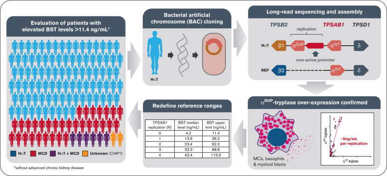
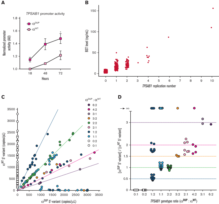
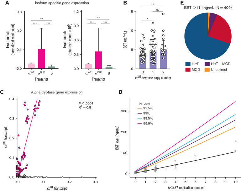
Serum tryptase is a biomarker used to aid in the identification of certain myeloid neoplasms, most notably systemic mastocytosis, where basal serum tryptase (BST) levels >20 ng/mL are a minor criterion for diagnosis. Although clonal myeloid neoplasms are rare, the common cause for elevated BST levels is the genetic trait hereditary α-tryptasemia (HαT) caused by increased germline TPSAB1 copy number. To date, the precise structural variation and mechanism(s) underlying elevated BST in HαT and the general clinical utility of tryptase genotyping, remain undefined. Through cloning, long-read sequencing, and assembling of the human tryptase locus from an individual with HαT, and validating our findings in vitro and in silico, we demonstrate that BST elevations arise from overexpression of replicated TPSAB1 loci encoding canonical α-tryptase protein owing to coinheritance of a linked overactive promoter element. Modeling BST levels based on TPSAB1 replication number, we generate new individualized clinical reference values for the upper limit of normal. Using this personalized laboratory medicine approach, we demonstrate the clinical utility of tryptase genotyping, finding that in the absence of HαT, BST levels >11.4 ng/mL frequently identify indolent clonal mast cell disease. Moreover, substantial BST elevations (eg, >100 ng/mL), which would ordinarily prompt bone marrow biopsy, can result from TPSAB1 replications alone and thus be within normal limits for certain individuals with HαT.
Overview
The claims and statistics of this paper haven’t been checked yet.
Free: it reads the open-access text and checks the first 5 claims that cite a source we can check. Create a free account to check every claim.
Integrity signals
Registry
Cites
Statistics
Figures


Verification
Claims the paper makes
The claims and statistics of this paper haven’t been checked yet.
Free: it reads the open-access text and checks the first 5 claims that cite a source we can check. Create a free account to check every claim.
Claims other papers make about this one
Each is checked against this paper’s own text: does it say what the citing paper says it does?
The cited source says what the claim attributes to it.
In the citing paper
“Another way is by using an online calculator tool (https://bst-calculater.niaid.nih.gov/ (accessed on 13 November 2023)) developed by Chovanek et al. [44].”
From Diagnostic Significance of Tryptase for Suspected Mast Cell Disorders (2023) · cites this paper as [44]
The cited source says what the claim attributes to it.
In the citing paper
“Further studies have elucidated as the number of TPSAB1 copies determined an increase in α-tryptase levels, on turn ultimately leading to elevated BST, each additional copy number adding approximately 9–10 ng/ml to the basal BST levels (12, 13).”
From Defining “Normal” basal serum tryptase levels: a context-dependent approach to improve diagnostics in systemic mastocytosis (2025) · cites this paper as [13]
The cited source says what the claim attributes to it.
In the citing paper
“In addition to this, three other methods of correction have been proposed (5, 13, 23) (Table 2).”
From Defining “Normal” basal serum tryptase levels: a context-dependent approach to improve diagnostics in systemic mastocytosis (2025) · cites this paper as [13]
In the citing paper
“In HαT, increased TPSAB1 copy number encoding α-tryptase results in increased BST levels as a result of promoter-driven overexpression of the replicated gene.37”
From Mast cell activation syndrome: Current understanding and research needs (2024) · cites this paper as [37]
In the citing paper
“HαT is a recently described genetic trait reported among 5% to 7% of Whites.17,37”
From Mast cell activation syndrome: Current understanding and research needs (2024) · cites this paper as [37]
Self-check
Percentages, sums and reported differences re-computed from the paper’s own numbers.
The self-check needs the paper’s full text.
Citations
Cites
Trimmomatic: a flexible trimmer for Illumina sequence data
Bolger, A. M., Lohse, M., & Usadel, B. (2014) · Bioinformatics · doi:10.1093/bioinformatics/btu170 · Cited by 65,548
The Ensembl Variant Effect Predictor
McLaren, W., Gil, L., Hunt, S. E., Riat, H. S., Ritchie, G. R. S., Thormann, A., Flicek, P., & Cunningham, F. (2016) · Genome Biology · doi:10.1186/s13059-016-0974-4 · Cited by 8,099
Sensitive detection of somatic point mutations in impure and heterogeneous cancer samples
Cibulskis, K., Lawrence, M. S., Carter, S. L., Sivachenko, A., Jaffe, D., Sougnez, C., Gabriel, S., Meyerson, M., Lander, E. S., & Getz, G. (2013) · Nature Biotechnology · doi:10.1038/nbt.2514 · Cited by 4,981 · cited 1×
“7.(26) In addition to paired calling, we also performed tumor-only calling with a combination of MuTect2 GATK version 4.”
Strelka2: fast and accurate calling of germline and somatic variants
Kim, S., Scheffler, K., Halpern, A. L., Bekritsky, M. A., Noh, E., Källberg, M., Chen, X., Kim, Y., Beyter, D., Krusche, P., et al. (2018) · Nature Methods · doi:10.1038/s41592-018-0051-x · Cited by 1,632
Diagnostic criteria and classification of mastocytosis: a consensus proposal
Valent, P., Horny, H.-P., Escribano, L., Longley, B. J., Li, C. Y., Schwartz, L. B., Marone, G., Nuñez, R., Akin, C., Sotlar, K., et al. (2001) · Leukemia Research · doi:10.1016/s0145-2126(01)00038-8 · Cited by 1,108
Self-citationVarDict: a novel and versatile variant caller for next-generation sequencing in cancer research
Lai, Z., Markovets, A., Ahdesmaki, M., Chapman, B., Hofmann, O., McEwen, R., Johnson, J., Dougherty, B., Barrett, J. C., & Dry, J. R. (2016) · Nucleic Acids Research · doi:10.1093/nar/gkw227 · Cited by 970 · cited 1×
“0.(27) The latter approach of performing somatic variant detection without including the paired PBMC sample was done to account for the fact that somatic variants in the BM granulocytes and BM mononuclear cells could also be present at low fractions in the PBMC sample, and therefore, be excluded as contaminating germ…”
BackgroundSAMBLASTER: fast duplicate marking and structural variant read extraction
Faust, G. G., & Hall, I. M. (2014) · Bioinformatics · doi:10.1093/bioinformatics/btu314 · Cited by 909
Tryptase levels as an indicator of mast-cell activation in systemic anaphylaxis and mastocytosis
Schwartz, L. B., Metcalfe, D. D., Miller, J. S., Earl, H., & Sullivan, T. (1987) · New England Journal of Medicine · doi:10.1056/nejm198706253162603 · Cited by 784 · cited 1×
“Whereas HaT is the most common heritable cause for elevated BST, the clonal mast cell 112 disorder systemic mastocytosis (SM) is a common acquired cause (10, 12), and BST levels are 113 used routinely as a biomarker to screen for this disorder.”
BackgroundSelf-citationKirshenbaum, A. S., Goff, J. P., Semere, T., Foster, B., Scott, L. M., & Metcalfe, D. D. (1999) · Blood · doi:10.1182/blood.v94.7.2333.419k30_2333_2342 · Cited by 420
Self-citationLyons, J. J., Yu, X., Hughes, J. D., Le, Q. T., Jamil, A., Bai, Y., Ho, N., Zhao, M., Liu, Y., O'Connell, M. P., et al. (2016) · Nature Genetics · doi:10.1038/ng.3696 · Cited by 394 · cited 7×
“220 Using these haplotypes, we next re-examined genome sequence reads from 183 individuals 221 without increased TPSAB1 copy number, and from 32 affected individuals from 10 families with 222 HaT (5).”
BackgroundMethodInfluentialSelf-citation
Research works printed but not linked: 1
- 1989, Molecular Cloning: A Laboratory Manual
Cited by
Human germline heterozygous gain-of-function STAT6 variants cause severe allergic disease
Sharma, M., Leung, D., Momenilandi, M., Jones, L. C. W., Pacillo, L., James, A. E., Murrell, J. R., Delafontaine, S., Maimaris, J., Vaseghi-Shanjani, M., et al. (2023) · Journal of Experimental Medicine · doi:10.1084/jem.20221755 · Cited by 125
Self-citationIncorporating Tryptase Genotyping Into the Workup and Diagnosis of Mast Cell Diseases and Reactions
Lyons, J. J., Greiner, G., Hoermann, G., & Metcalfe, D. D. (2022) · The Journal of Allergy and Clinical Immunology: In Practice · doi:10.1016/j.jaip.2022.05.003 · Cited by 47
Self-citationMast cell activation syndrome: Current understanding and research needs
Castells, M., Giannetti, M. P., Hamilton, M. J., Novak, P., Pozdnyakova, O., Nicoloro-SantaBarbara, J., Jennings, S. V., Francomano, C., Kim, B., Glover, S. C., et al. (2024) · Journal of Allergy and Clinical Immunology · doi:10.1016/j.jaci.2024.05.025 · Cited by 45
Self-citationClonal mast cell disorders and hereditary α-tryptasemia as risk factors for anaphylaxis
Kačar, M., Rijavec, M., Šelb, J., & Korošec, P. (2023) · Clinical & Experimental Allergy · doi:10.1111/cea.14264 · Cited by 37
Self-citationKorošec, P., Sturm, G. J., Lyons, J. J., Marolt, T. P., Svetina, M., Košnik, M., Zidarn, M., Kačar, M., Frelih, N., Lalek, N., et al. (2024) · Allergy · doi:10.1111/all.16084 · Cited by 46
Self-citation
Metadata
- Authors
- Chovanec, J., Tunc, I., Hughes, J., Halstead, J., Mateja, A., Liu, Y., O’Connell, M. P., Kim, J., Park, Y. H., Wang, Q., Le, Q., Pirooznia, M., Trivedi, N. N., Bai, Y., Yin, Y., Hsu, A. P., McElwee, J., Lassiter, S., Nelson, C., Bandoh, J., DiMaggio, T., Šelb, J., Rijavec, M., Carter, M. C., Komarow, H. D., Sabato, V., Steinberg, J., Hafer, K. M., Feuille, E., Hourigan, C. S., Lack, J., Khoury, P., Maric, I., Zanotti, R., Bonadonna, P., Schwartz, L. B., Milner, J. D., Glover, S. C., Ebo, D. G., Korošec, P., Caughey, G. H., Brittain, E. H., Busby, B., Metcalfe, D. D., & Lyons, J. J.
- Affiliations
- National Institutes of Health (US)
- Foundation Medicine (United States) (US)
- Birmingham Women’s and Children’s NHS Foundation Trust (GB)
- Frederick National Laboratory for Cancer Research (US)
- 1Translational Allergic Immunopathology Unit, Laboratory of Allergic Diseases, National Institute of Allergy and Infectious Diseases (NIAID), National Institutes of Health (NIH), Bethesda, MD
- Virginia Commonwealth University (US)
- San Francisco VA Medical Center (US)
- 12Nimbus Therapeutics, Cambridge, MA
- University Clinic of Pulmonary and Allergic Diseases Golnik (SI)
- University of Antwerp (BE)
- Medical College of Wisconsin (US)
- Stanford Medicine (US)
- Cornell University (US)
- 23Hematology Service, Department of Laboratory Medicine, Clinical Center, NIH, Bethesda, MD
- University of Verona (IT)
- Azienda Ospedaliera Universitaria Integrata Verona (IT)
- Columbia University (US)
- University of Mississippi Medical Center (US)
- Corresponding author
- Lyons, Jonathan J.
- ORCID
- Chovanec, Jack
- Tunc, Ilker
- Mateja, Allyson
- Liu, Yihui
- O’Connell, Michael P.
- Park, Young Hwan
- Le, Quang
- Pirooznia, Mehdi
- Bai, Yun
- Yin, Yuzhi
- Hsu, Amy P.
- McElwee, Joshua
- Nelson, Celeste
- DiMaggio, Thomas
- Šelb, Julij
- Rijavec, Matija
- Carter, Melody C.
- Komarow, Hirsh D.
- Sabato, Vito
- Steinberg, Joshua
- Feuille, Elizabeth
- Hourigan, Christopher S.
- Lack, Justin
- Khoury, Paneez
- Zanotti, Roberta
- Bonadonna, Patrizia
- Schwartz, Lawrence B.
- Milner, Joshua D.
- Glover, Sarah C.
- Ebo, Didier G.
- Korošec, Peter
- Caughey, George H.
- Brittain, Erica H.
- Busby, Ben
- Metcalfe, Dean D.
- Lyons, Jonathan J.
- Venue
- Blood Advances
- Publisher
- American Society of Hematology
- Published
- 2023-04-28
- Volume
- 7
- Issue
- 9
- Pages
- 1796-1810
- ISSN
- 2473-9529 (ISSN-L)
- 2473-9537
- PMC
- PMC10164828
- OpenAlex
- W4297520278
- Cited by
- 62
- References
- 36
- Citations by year
- 2026: 11 · 2025: 24 · 2024: 13 · 2023: 11
- FWCI
- 4.68
- Type
- journal article
- Language
- en
- Open access
- gold
- doi.org
- Licence
- cc-by-nc-nd
- Locations
- Blood Advances (publishedVersion, cc-by-nc-nd)
- medRxiv (acceptedVersion)
- PubMed Central (publishedVersion, cc-by-nc-nd)
- PubMed (publishedVersion)
- Funders
- U.S. Department of Health and Human Services (HHSN261201500003I)
- Fonds Wetenschappelijk Onderzoek (1804518N, 1800614N, FWO: 1800614N)
- Vlaamse regering
- National Institutes of Health (75N910D00024, HHSN261201500003I)
- National Heart, Lung, and Blood Institute
- National Cancer Institute (75N910D00024, HHSN261201500003I, HHSN261201500003C)
- National Institute of Allergy and Infectious Diseases (HHSN261201500003I, 75N910D00024)
- National Institute of Dental and Craniofacial Research (R21DE028378)
- U.S. National Library of Medicine
- NHLBI Division of Intramural Research
- Topics
- Life Sciences › Immunology and Microbiology › Immunology › Mast cells and histamine
- Life Sciences › Biochemistry, Genetics and Molecular Biology › Genetics › Genomics and Rare Diseases
- Health Sciences › Medicine › Hematology › Acute Myeloid Leukemia Research
- Keywords
- serum tryptase, hereditary alpha-tryptasemia, upper limit of normal, systemic mastocytosis, myeloid neoplasms
- MeSH terms
- Mast Cell Activation Syndrome, Humans, Mast Cells, Mastocytosis, Myeloproliferative Disorders, Reference Values, Unnecessary Procedures, Tryptases
About these checks
Registry, retraction, Cites and statistics results are computed, not judged, with one exception: a printed reference that no index lists is read once by a model before it counts as a citation error. Claims are extracted by a model and kept only when their quote appears verbatim in the text; each cited source is then checked, with a model’s help, for whether it says what the claim relies on. Those verdicts can be wrong — read the claim’s quote and the cited source before relying on one.
Statistics and arithmetic are recomputed, not judged.
Confidence bands are shown only for fields calibrated on our annotated test set. None is calibrated yet, so no band is shown.
What counts as a claim ›How we measure confidence ›How we check this ›
Are you an author, or do you see a problem with this page? Request a review
How to cite
Cite CoChat for the checks on this page, and the paper for its own findings.
- This page
- CoChat. (2026). Citation check: Genetically defined individual reference ranges for tryptase limit unnecessary procedures and unmask myeloid neoplasms. https://app.cochat.ai/papers/10.1182/bloodadvances.2022007936
- The paper
- Chovanec, J., Tunc, I., Hughes, J., Halstead, J., Mateja, A., Liu, Y., O’Connell, M. P., Kim, J., Park, Y. H., Wang, Q., Le, Q., Pirooznia, M., Trivedi, N. N., Bai, Y., Yin, Y., Hsu, A. P., McElwee, J., Lassiter, S., Nelson, C., . . . Lyons, J. J. (2023). Genetically defined individual reference ranges for tryptase limit unnecessary procedures and unmask myeloid neoplasms. Blood Advances. https://doi.org/10.1182/bloodadvances.2022007936
Data sources
Bibliographic data: Semantic Scholar, OpenAlex, Crossref.
Contains information from the Semantic Scholar Academic Graph Datasets, made available under the ODC Attribution License (ODC-BY 1.0).
Semantic Scholar’s data is described in: Kinney, R., et al. (2023). The Semantic Scholar Open Data Platform. arXiv:2301.10140.
