

The DOI matches its Crossref record; 48 of the 48 references it cites were found in a registry or an index.
Microvesicles secreted by macrophages shuttle invasion-potentiating microRNAs into breast cancer cells
Open accessAbstract
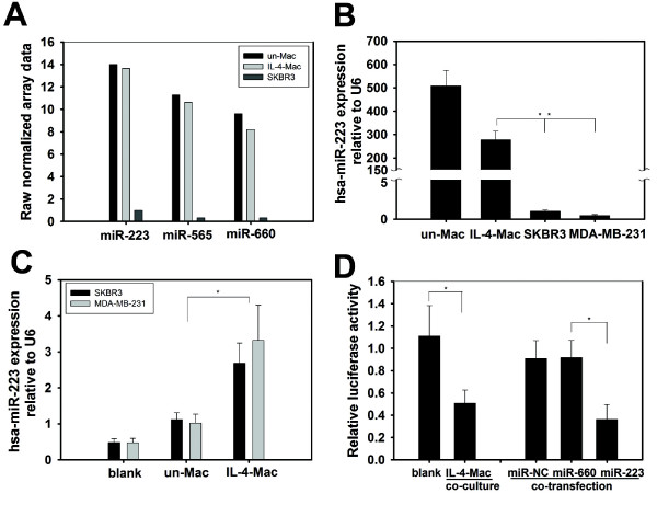
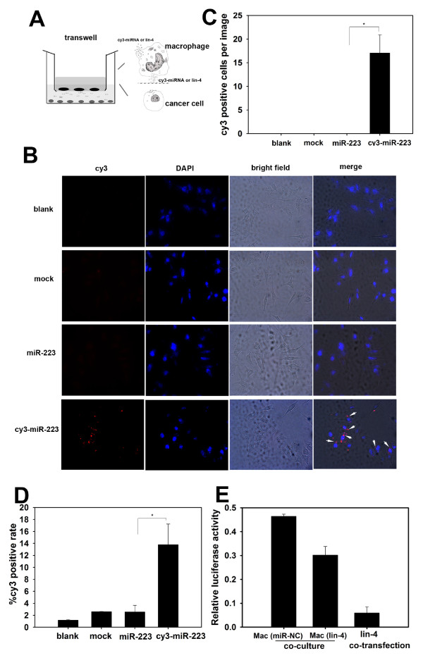
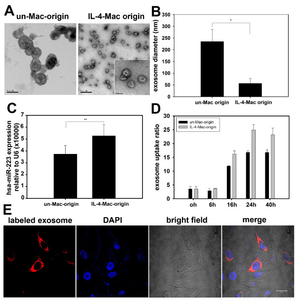
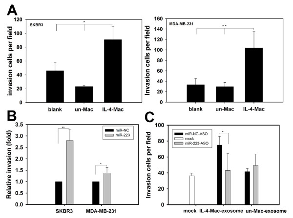
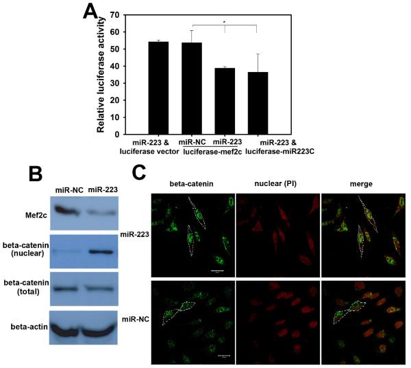
BACKGROUND: Tumor-associated macrophages (TAMs) are alternatively activated cells induced by interleukin-4 (IL-4)-releasing CD4+ T cells. TAMs promote breast cancer invasion and metastasis; however, the mechanisms underlying these interactions between macrophages and tumor cells that lead to cancer metastasis remain elusive. Previous studies have found microRNAs (miRNAs) circulating in the peripheral blood and have identified microvesicles, or exosomes, as mediators of cell-cell communication. Therefore, one alternative mechanism for the promotion of breast cancer cell invasion by TAMs may be through macrophage-secreted exosomes, which would deliver invasion-potentiating miRNAs to breast cancer cells. RESULTS: We utilized a co-culture system with IL-4-activated macrophages and breast cancer cells to verify that miRNAs are transported from macrophages to breast cancer cells. The shuttling of fluorescently-labeled exogenous miRNAs from IL-4-activated macrophages to co-cultivated breast cancer cells without direct cell-cell contact was observed. miR-223, a miRNA specific for IL-4-activated macrophages, was detected within the exosomes released by macrophages and was significantly elevated in the co-cultivated SKBR3 and MDA-MB-231 cells. The invasiveness of the co-cultivated breast cancer cells decreased when the IL-4-activated macrophages were treated with a miR-223 antisense oligonucleotide (ASO) that would inhibit miR-223 expression. Furthermore, results from a functional assay revealed that miR-223 promoted the invasion of breast cancer cells via the Mef2c-β-catenin pathway. CONCLUSIONS: We conclude that macrophages regulate the invasiveness of breast cancer cells through exosome-mediated delivery of oncogenic miRNAs. Our data provide insight into the mechanisms underlying the metastasis-promoting interactions between macrophages and breast cancer cells.
Overview
The claims and statistics of this paper haven’t been checked yet.
Run the check — free, no account needed. It reads the open-access text and checks the sources it cites.
Integrity signals
Registry
Cites
Statistics
Figures
Verification
Claims the paper makes
The claims and statistics of this paper haven’t been checked yet.
Run the check — free, no account needed. It reads the open-access text and checks the sources it cites.
Claims other papers make about this one
Each is checked against this paper’s own text: does it say what the citing paper says it does?
In the citing paper
“Secretion of miRNAs via exosomes have also been reported to be regulated by signaling molecules, such as interleukin-4 (IL4) (132) and Docosahexaenoic acid (DHA) (133).”
From Overview of MicroRNA Biogenesis, Mechanisms of Actions, and Circulation (2018) · cites this paper as [132]
In the citing paper
“IL4-activated macrophages were found to secrete exosomes carrying oncogenic miRNAs to promote invasiveness of breast cancer cells (132).”
From Overview of MicroRNA Biogenesis, Mechanisms of Actions, and Circulation (2018) · cites this paper as [132]
Self-check
Percentages, sums and reported differences re-computed from the paper’s own numbers.
The self-check needs the paper’s full text.
Citations
Cites
1 retracted
RETRACTED: MicroRNA signatures of tumor-derived exosomes as diagnostic biomarkers of ovarian cancer
Taylor, D. D., & Gerçel-Taylor, Ç. (2008) · Gynecologic Oncology · doi:10.1016/j.ygyno.2008.04.033 · Cited by 2,333
RetractedValadi, H., Ekström, K., Bossios, A., Sjöstrand, M., Lee, J. J., & Lötvall, J. O. (2007) · Nature Cell Biology · doi:10.1038/ncb1596 · Cited by 12,834
The functions of animal microRNAs
Ambros, V. (2004) · Nature · doi:10.1038/nature02871 · Cited by 10,728
Circulating microRNAs as stable blood-based markers for cancer detection
Mitchell, P. S., Parkin, R. K., Kroh, E. M., Fritz, B. R., Wyman, S. K., Pogosova-Agadjanyan, E. L., Peterson, A., Noteboom, J., O'Briant, K. C., Allen, A., et al. (2008) · Proceedings of the National Academy of Sciences · doi:10.1073/pnas.0804549105 · Cited by 7,969
Microenvironmental regulation of metastasis
Joyce, J. A., & Pollard, J. W. (2009) · Nature Reviews Cancer · doi:10.1038/nrc2618 · Cited by 3,684
B lymphocytes secrete antigen-presenting vesicles.
Raposo, G., Nijman, H. W., Stoorvogel, W., Liejendekker, R., Harding, C. V., Melief, C. J., & Geuze, H. J. (1996) · The Journal of experimental medicine · doi:10.1084/jem.183.3.1161 · Cited by 3,546
Tumour-educated macrophages promote tumour progression and metastasis
Pollard, J. W. (2004) · Nature reviews. Cancer · doi:10.1038/nrc1256 · Cited by 3,413
Macrophage activation and polarization
Martinez, F. O. (2008) · Frontiers in Bioscience · doi:10.2741/2692 · Cited by 3,160
Bingle, L., Brown, N. J., & Lewis, C. E. (2002) · The Journal of Pathology · doi:10.1002/path.1027 · Cited by 2,022
DeNardo, D. G., Barreto, J., Andreu, P., Vásquez, L. J., Tawfik, D., Kolhatkar, N. S., & Coussens, L. M. (2009) · Cancer Cell · doi:10.1016/j.ccr.2009.06.018 · Cited by 1,391
Ratajczak, J., Wysoczynski, M., Hayek, F., Janowska-Wieczorek, A., & Ratajczak, M. Z. (2006) · Leukemia · doi:10.1038/sj.leu.2404296 · Cited by 1,340
Regulation of progenitor cell proliferation and granulocyte function by microRNA-223
Johnnidis, J. B., Harris, M. H., Wheeler, R. T., Stehling-Sun, S., Lam, M. H., Kirak, O., Brummelkamp, T. R., Fleming, M. D., & Camargo, F. D. (2008) · Nature · doi:10.1038/nature06607 · Cited by 1,208
Exosomes/microvesicles as a mechanism of cell-to-cell communication
Camussi, G., Deregibus, M. C., Bruno, S., Cantaluppi, V., & Biancone, L. (2010) · Kidney International · doi:10.1038/ki.2010.278 · Cited by 1,200
Association of macrophage infiltration with angiogenesis and prognosis in invasive breast carcinoma.
Leek, R., Lewis, C. E., Whitehouse, R. M., Greenall, M. J., Clarke, J., & Harris, A. L. (1996) · PubMed · Cited by 1,187
Yan, L., Huang, X.-F., Shao, Q., Huang, M., Deng, L., Wu, Q., Zeng, Y.-X., & Shao, J. (2008) · RNA · doi:10.1261/rna.1034808 · Cited by 1,159
Exosome: from internal vesicle of the multivesicular body to intercellular signaling device
Denzer, K., Kleijmeer, M. J., Heijnen, H. F. G., Stoorvogel, W., & Geuze, H. J. (2000) · Journal of Cell Science · doi:10.1242/jcs.113.19.3365 · Cited by 1,067
Molecular Characterization of Dendritic Cell-Derived Exosomes
Théry, C., Régnault, A., Garin, J., Wolfers, J., Zitvogel, L., Ricciardi‐Castagnoli, P., Raposo, G., & Amigorena, S. (1999) · The Journal of Cell Biology · doi:10.1083/jcb.147.3.599 · Cited by 1,065
The inflammatory micro-environment in tumor progression: the role of tumor-associated macrophages.
Allavena, P., Sica, A., Solinas, G., Porta, C., & Mantovani, A. (2008) · Critical Reviews in Oncology/Hematology · doi:10.1016/j.critrevonc.2007.07.004 · Cited by 1,044
Fazi, F., Rosa, A., Fatica, A., Gelmetti, V., De Marchis, M. L., Nervi, C., & Bozzoni, I. (2005) · Cell · doi:10.1016/j.cell.2005.09.023 · Cited by 894
TCR Activation of Human T Cells Induces the Production of Exosomes Bearing the TCR/CD3/ζ Complex
Blanchard, N., Lankar, D., Faure, F., Regnault, A., Dumont, C., Raposo, G., & Hivroz, C. (2002) · The Journal of Immunology · doi:10.4049/jimmunol.168.7.3235 · Cited by 660
Genomic instability in laminopathy-based premature aging
Liu, B., Wang, J., Chan, K. M., Tjia, W. M., Deng, W., Guan, X., Huang, J.-D., Li, K. M., Chau, P. Y., Chen, D. J., et al. (2005) · Nature Medicine · doi:10.1038/nm1266 · Cited by 650
Self-citationMicro-RNA profiling in kidney and bladder cancers
Gottardo, F., Liu, C. G., Ferracin, M., Calin, G. A., Fassan, M., Bassi, P., Sevignani, C., Byrne, D., Negrini, M., Pagano, F. C., et al. (2007) · Urologic Oncology Seminars and Original Investigations · doi:10.1016/j.urolonc.2007.01.019 · Cited by 601
Induction of myeloid‐derived suppressor cells by tumor exosomes
Xiang, X., Poliakov, A., Liu, C., Liu, Y., Deng, Z., Wang, J., Cheng, Z., Shah, S. V., Wang, G., Zhang, L., et al. (2008) · International Journal of Cancer · doi:10.1002/ijc.24249 · Cited by 597
Self-citationOhshima, K., Inoue, K., Fujiwara, A., Hatakeyama, K., Kanto, K., Watanabe, Y., Muramatsu, K., Fukuda, Y., Ogura, S., Yamaguchi, K., et al. (2010) · PLoS ONE · doi:10.1371/journal.pone.0013247 · Cited by 593
Xu, J., Wu, C., Che, X., Wang, L., Yu, D., Zhang, T., Huang, L., Li, H., Tan, W., Wang, C., et al. (2011) · Molecular Carcinogenesis · doi:10.1002/mc.20712 · Cited by 567
Targeting tumor-associated macrophages as a novel strategy against breast cancer
Luo, Y. (2006) · Journal of Clinical Investigation · doi:10.1172/jci27648 · Cited by 551
Luo, S.-S., Ishibashi, O., Ishikawa, G., Ishikawa, T., Katayama, A., Mishima, T., Takizawa, T., Shigihara, T., Goto, T., Izumi, A., et al. (2009) · Biology of Reproduction · doi:10.1095/biolreprod.108.075481 · Cited by 489
Tumor-Associated Macrophages Press the Angiogenic Switch in Breast Cancer
Lin, E. Y., & Pollard, J. W. (2007) · Cancer Research · doi:10.1158/0008-5472.can-07-0912 · Cited by 463
Nanostructural and Transcriptomic Analyses of Human Saliva Derived Exosomes
Palanisamy, V., Sharma, S., Deshpande, A., Zhou, H., Gimzewski, J. K., & Wong, D. T. (2010) · PLoS ONE · doi:10.1371/journal.pone.0008577 · Cited by 363
Mears, R., Craven, R. A., Hanrahan, S. J., Totty, N., Upton, C., Young, S. L., Patel, P. M., Selby, P. J., & Banks, R. E. (2004) · PROTEOMICS · doi:10.1002/pmic.200400876 · Cited by 335
BRF (1976) · Anesthesiology · doi:10.1097/00000542-197608000-00021 · Cited by 323
Loke, P., Nair, M. G., Parkinson, J., Guiliano, D. B., Blaxter, M., & Allen, J. E. (2002) · BMC Immunology · doi:10.1186/1471-2172-3-7 · Cited by 322
The Macrophage Growth Factor CSF-1 in Mammary Gland Development and Tumor Progression
Lin, E. Y., Gouon–Evans, V., Nguyen, A., & Pollard, J. W. (2002) · Journal of Mammary Gland Biology and Neoplasia · doi:10.1023/a:1020399802795 · Cited by 291
An optimized isolation and labeling platform for accurate microRNA expression profiling
SHINGARA, J., KEIGER, K., SHELTON, J., LAOSINCHAI-WOLF, W., POWERS, P., CONRAD, R., BROWN, D., & LABOURIER, E. (2005) · RNA · doi:10.1261/rna.2610405 · Cited by 260
Esser, J., Gehrmann, U., D’Alexandri, F. L., Hidalgo-Estévez, A. M., Wheelock, C. E., Scheynius, A., Gabrielsson, S., & Rådmark, O. (2010) · Journal of Allergy and Clinical Immunology · doi:10.1016/j.jaci.2010.06.039 · Cited by 235
Inflammation-mediated promotion of invasion and metastasis
Solinas, G., Marchesi, F., Garlanda, C., Mantovani, A., & Allavena, P. (2010) · Cancer and Metastasis Reviews · doi:10.1007/s10555-010-9227-2 · Cited by 205
Alternative activation of tumor-associated macrophages by IL-4
Wang, H., & Joyce, J. A. (2010) · Cell Cycle · doi:10.4161/cc.9.24.14322 · Cited by 180
Vallhov, H., Gutzeit, C., Johansson, S. M., Nagy, N., Paul, M., Li, Q., Friend, S. L., George, T. C., Klein, E., Scheynius, A., et al. (2010) · The Journal of Immunology · doi:10.4049/jimmunol.1001145 · Cited by 141
Rosell, R., Wei, J., & Tarón, M. (2009) · Clinical Lung Cancer · doi:10.3816/clc.2009.n.001 · Cited by 133
αT-Catenin: a novel tissue-specific β-catenin-binding protein mediating strong cell-cell adhesion
Janssens, B., Goossens, S., Staes, K., Gilbert, B., van Hengel, J., Colpaert, C., Bruyneel, E. A., Mareel, M. M. K., & Roy, F. M. V. (2001) · Journal of Cell Science · doi:10.1242/jcs.114.17.3177 · Cited by 133
Gourzonès, C., Gelin, A., Bombik, I., Klibi, J., Vérillaud, B., Guigay, J., Lang, P., Temam, S., Schneider, V., Amiel, C., et al. (2010) · Virology Journal · doi:10.1186/1743-422x-7-271 · Cited by 127
Luo, Y., Zhou, H., Krueger, J. S., Kaplan, C., Liao, D., Markowitz, D., Liu, C., Chen, T., Chuang, T. H., Xiang, R., et al. (2009) · Oncogene · doi:10.1038/onc.2009.308 · Cited by 94
O’Brien, J., & Schedin, P. S. (2009) · Journal of Mammary Gland Biology and Neoplasia · doi:10.1007/s10911-009-9118-8 · Cited by 73
Exosomes Secreted by Bacterially Infected Macrophages Are Proinflammatory
O'Neill, H. C., & Quah, B. (2008) · Science Signaling · doi:10.1126/stke.16pe8 · Cited by 57
Vanpoucke, G. (2004) · Nucleic Acids Research · doi:10.1093/nar/gkh727 · Cited by 52
Joimel, U., Gest, C., Soria, J., Pritchard, L.-L., Alexandre, J., Laurent, M., Blot, E., Cazin, L., Vannier, J., Varin, R., et al. (2010) · BMC Cancer · doi:10.1186/1471-2407-10-375 · Cited by 44
The potential role of PDGF, IGF-1, TGF-beta expression in idiopathic pulmonary fibrosis.
Cao, B., Guo, Z., Zhu, Y., & Xu, W. (2000) · PubMed · Cited by 43
More insights into the immunosuppressive potential of tumor exosomes
Huber, V., Filipazzi, P., Iero, M., Fais, S., & Rivoltini, L. (2008) · Journal of Translational Medicine · doi:10.1186/1479-5876-6-63 · Cited by 36
Cited by
Sorted by citations
Overview of MicroRNA Biogenesis, Mechanisms of Actions, and Circulation
O'Brien, J., Hayder, H., Zayed, Y., & Peng, C. (2018) · Frontiers in Endocrinology · doi:10.3389/fendo.2018.00402 · Cited by 5,287
Extracellular vesicles: biology and emerging therapeutic opportunities
EL Andaloussi, S., Mäger, I., Breakefield, X. O., & Wood, M. J. A. (2013) · Nature Reviews Drug Discovery · doi:10.1038/nrd3978 · Cited by 3,571
MicroRNAs and other non-coding RNAs as targets for anticancer drug development
Ling, H. Y., Fabbri, M., & Călin, G. A. (2013) · Nature Reviews Drug Discovery · doi:10.1038/nrd4140 · Cited by 1,489
Exosomes in cancer development, metastasis, and drug resistance: a comprehensive review
Azmi, A. S., Bao, B., & Sarkar, F. H. (2013) · Cancer and Metastasis Reviews · doi:10.1007/s10555-013-9441-9 · Cited by 1,187
Macrophages in Tumor Microenvironments and the Progression of Tumors
Hao, N.-B., Lü, M.-H., Fan, Y.-H., Cao, Y.-L., Zhang, Z.-R., & Yang, S.-M. (2012) · Clinical and Developmental Immunology · doi:10.1155/2012/948098 · Cited by 1,003
Metadata
- Authors
- Yang, M., Chen, J., Su, F., Yu, B., Su, F., Lin, L., Liu, Y., Huang, J.-D., & Song, E.
- Affiliations
- Sun Yat-sen University (CN)
- University of Hong Kong (HK)
- Corresponding author
- Huang, Jian-Dong
- Song, Erwei
- Venue
- Molecular Cancer
- Published
- 2011-09-22
- Volume
- 10
- Issue
- 1
- Pages
- 117-117
- ISSN
- 1476-4598 (ISSN-L)
- PMC
- PMC3190352
- OpenAlex
- W2059266855
- Cited by
- 693
- References
- 48
- Citations by year
- 2026: 11 · 2025: 15 · 2024: 34 · 2023: 43 · 2022: 45 · 2021: 60 · 2020: 66 · 2019: 48 · 2018: 45 · 2017: 50 · 2016: 54 · 2015: 78 · 2014: 54 · 2013: 45 · 2012: 41
- FWCI
- 18.55
- Type
- journal article
- Language
- en
- Open access
- gold
- doi.org
- Licence
- cc-by
- Locations
- Molecular Cancer (publishedVersion, cc-by)
- PubMed Central (publishedVersion, cc-by)
- PubMed (publishedVersion)
- DOAJ (DOAJ: Directory of Open Access Journals) (submittedVersion)
- The HKU Scholars Hub (University of Hong Kong) (submittedVersion, cc-by-nc-nd)
- Funders
- National Natural Science Foundation of China (30972785, 2009CB521706, 2010CB912800, 30921140312, 30830110, KLB09001, 2011CB504203)
- Sun Yat-sen University (KLB09001, 2011CB504203, 2010CB912800)
- Topics
- Life Sciences › Biochemistry, Genetics and Molecular Biology › Molecular Biology › Extracellular vesicles in disease
- Life Sciences › Immunology and Microbiology › Immunology › Immune cells in cancer
- Life Sciences › Biochemistry, Genetics and Molecular Biology › Cancer Research › MicroRNA in disease regulation
- Keywords
- tumor-associated macrophages, microvesicles, exosomes, microRNA, breast cancer invasion, miR-223, IL-4, metastasis, antisense oligonucleotides
- MeSH terms
- Breast Neoplasms, Cell Communication, Female, Humans, Macrophages, Neoplasm Invasiveness, Interleukin-4, Genes, Reporter, Myogenic Regulatory Factors, Coculture Techniques, Cell Culture Techniques, 3' Untranslated Regions, MADS Domain Proteins, RNA Interference, MicroRNAs, Cell Line, Tumor, Luciferases, Renilla, beta Catenin, Exosomes, MEF2 Transcription Factors
About these checks
Registry, retraction, Cites and statistics results are computed, not judged, with one exception: a printed reference that no index lists is read once by a model before it counts as a citation error. Claims are extracted by a model and kept only when their quote appears verbatim in the text; each cited source is then checked, with a model’s help, for whether it says what the claim relies on. Those verdicts can be wrong — read the claim’s quote and the cited source before relying on one.
Statistics and arithmetic are recomputed, not judged.
Confidence bands are shown only for fields calibrated on our annotated test set. None is calibrated yet, so no band is shown.
What counts as a claim ›How we measure confidence ›How we check this ›
Are you an author, or do you see a problem with this page? Request a review
