

The DOI matches its Crossref record; 167 of the 167 references it cites were found in a registry or an index.
A new look at cerebrospinal fluid circulation
Open accessAbstract
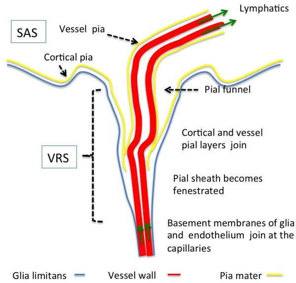
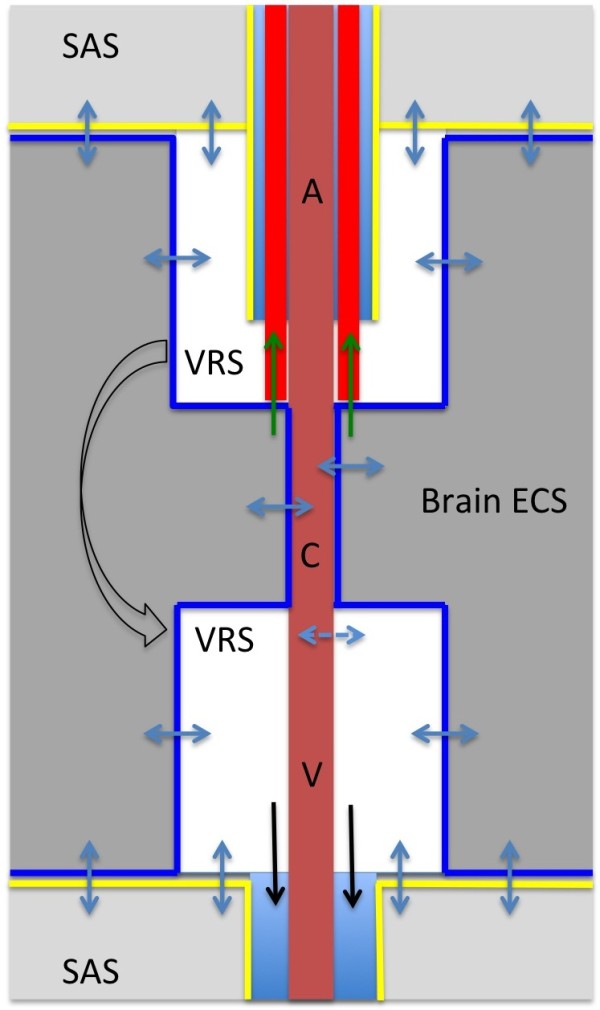
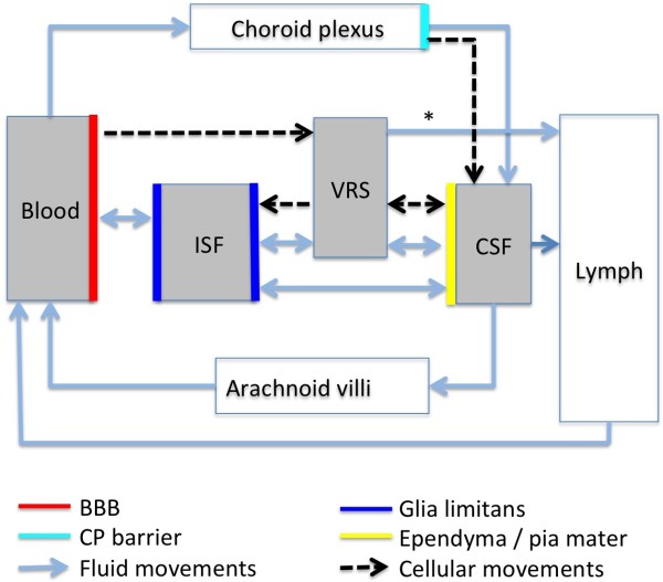
According to the traditional understanding of cerebrospinal fluid (CSF) physiology, the majority of CSF is produced by the choroid plexus, circulates through the ventricles, the cisterns, and the subarachnoid space to be absorbed into the blood by the arachnoid villi. This review surveys key developments leading to the traditional concept. Challenging this concept are novel insights utilizing molecular and cellular biology as well as neuroimaging, which indicate that CSF physiology may be much more complex than previously believed. The CSF circulation comprises not only a directed flow of CSF, but in addition a pulsatile to and fro movement throughout the entire brain with local fluid exchange between blood, interstitial fluid, and CSF. Astrocytes, aquaporins, and other membrane transporters are key elements in brain water and CSF homeostasis. A continuous bidirectional fluid exchange at the blood brain barrier produces flow rates, which exceed the choroidal CSF production rate by far. The CSF circulation around blood vessels penetrating from the subarachnoid space into the Virchow Robin spaces provides both a drainage pathway for the clearance of waste molecules from the brain and a site for the interaction of the systemic immune system with that of the brain. Important physiological functions, for example the regeneration of the brain during sleep, may depend on CSF circulation.
Overview
The claims and statistics of this paper haven’t been checked yet.
Run the check — free, no account needed. It reads the open-access text and checks the sources it cites.
Integrity signals
Registry
Cites
Statistics
Figures
Verification
Claims the paper makes
The claims and statistics of this paper haven’t been checked yet.
Run the check — free, no account needed. It reads the open-access text and checks the sources it cites.
Claims other papers make about this one
Each is checked against this paper’s own text: does it say what the citing paper says it does?
The cited source says what the claim attributes to it.
In the citing paper
“Uptake into arachnoid villi, as proposed more than one hundred years ago, seems to be of minor importance in humans, as reviewed by Brinker et al. (50) and as indicated in our recent study (13).”
From Brain-wide glymphatic enhancement and clearance in humans assessed with MRI. (2018) · cites this paper as [50]
Self-check
Percentages, sums and reported differences re-computed from the paper’s own numbers.
The self-check needs the paper’s full text.
Citations
Cites
Iliff, J. J., Wang, M., Liao, Y., Plogg, B. A., Peng, W., Gundersen, G. A., Benveniste, H., Vates, G. E., Deane, R., Goldman, S. A., et al. (2012) · Science Translational Medicine · doi:10.1126/scitranslmed.3003748 · Cited by 5,824
Sleep Drives Metabolite Clearance from the Adult Brain
Xie, L., Kang, H., Xu, Q., Chen, M. J., Liao, Y., Thiyagarajan, M., O’Donnell, J., Christensen, D. J., Nicholson, C., Iliff, J. J., et al. (2013) · Science · doi:10.1126/science.1241224 · Cited by 5,411
Structure and function of the blood–brain barrier
Abbott, N. J., Patabendige, A. A. K., Dolman, D. E. M., Yusof, S. R., & Begley, D. J. (2010) · Neurobiology of Disease · doi:10.1016/j.nbd.2009.07.030 · Cited by 5,185
Daneman, R., & Prat, A. (2015) · Cold Spring Harbor Perspectives in Biology · doi:10.1101/cshperspect.a020412 · Cited by 3,437
Aquaporin-4 deletion in mice reduces brain edema after acute water intoxication and ischemic stroke
Manley, G. T., Fujimura, M., Ma, T., Noshita, N., Filiz, F., Bollen, A. W., Chan, P. H., & Verkman, A. S. (2000) · Nature Medicine · doi:10.1038/72256 · Cited by 1,501
Diffusion in Brain Extracellular Space
Syková, E., & Nicholson, C. (2008) · Physiological Reviews · doi:10.1152/physrev.00027.2007 · Cited by 1,481
Nielsen, S. M., Nagelhus, E. A., Amiry-Moghaddam, M. R., Bourque, C. W., Agre, P. C., & Ottersen, O. P. (1997) · Journal of Neuroscience · doi:10.1523/jneurosci.17-01-00171.1997 · Cited by 1,440
Brain-wide pathway for waste clearance captured by contrast-enhanced MRI
Iliff, J. J., Lee, H., Yu, M., Feng, T., Logan, J., Nedergaard, M., & Benveniste, H. (2013) · Journal of Clinical Investigation · doi:10.1172/jci67677 · Cited by 1,148
Blood–brain barrier structure and function and the challenges for CNS drug delivery
Abbott, N. J. (2013) · Journal of Inherited Metabolic Disease · doi:10.1007/s10545-013-9608-0 · Cited by 951
Mathiisen, T. M., Lehre, K. P., Danbolt, N. C., & Ottersen, O. (2010) · Glia · doi:10.1002/glia.20990 · Cited by 893
Research works printed but not linked: 1
- 10.1186/1743-8454-5-10 · DOI · Paper page
Cited by
Sorted by citations
Items 401–500 of 821
Strbačko, I., Radoš, M., Jurjević, I., Orešković, D., & Klarica, M. (2025) · Frontiers in Human Neuroscience · doi:10.3389/fnhum.2024.1463740 · Cited by 13
Zhang, L., Hussain, Z., & Ren, Z. (2019) · Current Drug Targets · doi:10.2174/1389450120666190214121342 · Cited by 13
Orozco, G. A., Smith, J. H., & García, J. J. (2020) · Proceedings of the Institution of Mechanical Engineers Part H Journal of Engineering in Medicine · doi:10.1177/0954411920937220 · Cited by 13
Yatsushiro, S., Sunohara, S., Tokushima, T., Takizawa, K., Matsumae, M., Atsumi, H., Horie, T., Kajihara, N., & Kuroda, K. (2021) · Magnetic Resonance in Medical Sciences · doi:10.2463/mrms.mp.2020-0085 · Cited by 13
Cardiovascular risk factors in Chiari malformation and idiopathic intracranial hypertension
Frič, R., Pripp, A. H., & Eide, P. K. (2017) · Brain and Behavior · doi:10.1002/brb3.677 · Cited by 13
A novel view on Alzheimer’s disease pathogenesis: modern conceptof amyloid clearance
Lobzin, V. Y., Kolmakova, K. A., & Emelin, А. Y. (2018) · V M BEKHTEREV REVIEW OF PSYCHIATRY AND MEDICAL PSYCHOLOGY · doi:10.31363/2313-7053-2018-2-22-28 · Cited by 13
Shear-induced amyloid formation of IDPs in the brain
Trumbore, C. N. (2019) · Progress in molecular biology and translational science · doi:10.1016/bs.pmbts.2019.05.008 · Cited by 13
Yang, G., Xu, M. L., Chen, W., Xu, Q., Shi, H., & Hu, Y. (2023) · Frontiers in Neurology · doi:10.3389/fneur.2023.1139048 · Cited by 12
Magnetic resonance imaging analysis of human skull diploic venous anatomy
Alarfaj, A., Sankar, T., Bhargava, R., Tyler, J., Walji, A. H., Fox, R., Sagga, A., Ishaque, A. H., & Aronyk, K. E. (2021) · Surgical Neurology International · doi:10.25259/sni_532_2020 · Cited by 12
Glymphatic System in Ocular Diseases: Evaluation of MRI Findings
Manava, P., Eckrich, C., Luciani, F., Schmidbauer, J. M., Lell, M., & Detmar, K. (2022) · American Journal of Neuroradiology · doi:10.3174/ajnr.a7552 · Cited by 12
Blood-Based Biomarkers for the Identification of Sports-Related Concussion
Anto‐Ocrah, M., Jones, C. M. C., Diacovo, D., & Bazarian, J. J. (2017) · Neurologic Clinics · doi:10.1016/j.ncl.2017.03.008 · Cited by 12
Duru, S., Oria, M., Arévalo, S., Rodó, C., Correa, L., S, F. V., Sanchez-Margallo, F. M., & Peiro, J. L. (2019) · Child s Nervous System · doi:10.1007/s00381-019-04096-1 · Cited by 12
Amount of Brain Edema Correlates With Neurologic Recovery in Pediatric Cerebral Malaria
Kampondeni, S. D., Seydel, K. B., Zhang, B., Small, D. S., Birbeck, G. L., Hammond, C. A., Chilingulo, C., Taylor, T. E., & Potchen, M. J. (2020) · The Pediatric Infectious Disease Journal · doi:10.1097/inf.0000000000002573 · Cited by 12
Role of Interfacial Conditions on Blast Overpressure Propagation Into the Brain
Chen, Y., O’Shaughnessy, T. J., Kamimori, G. H., Horner, D. M., Egnoto, M. J., & Bagchi, A. (2020) · Frontiers in Neurology · doi:10.3389/fneur.2020.00323 · Cited by 12
Lumbar drains: Practical understanding and application for the otolaryngologist
Livingston, A. J., Laing, B., Zwagerman, N. T., & Harris, M. S. (2020) · American Journal of Otolaryngology · doi:10.1016/j.amjoto.2020.102740 · Cited by 12
Toma, A. K. (2015) · Surgery (Oxford) · doi:10.1016/j.mpsur.2015.05.006 · Cited by 12
Xu, H., Miyajima, M., Nakajima, M., Ogino, I., Kawamura, K., Akiba, C., Kamohara, C., Sakamoto, K., Karagiozov, K. L., Nakamura, E., et al. (2022) · Fluids and Barriers of the CNS · doi:10.1186/s12987-022-00341-z · Cited by 12
Morphometry and morphology of rostral cranial fossa in brachycephalic dogs – CT studies
Sokołowski, W., Barszcz, K., Kupczyńska, M., Czopowicz, M., Czubaj, N., Kinda, W., & Kiełbowicz, Z. (2020) · PLoS ONE · doi:10.1371/journal.pone.0240091 · Cited by 11
Riew, T., Hwang, J.-W., Jin, X., Kim, H. L., & Lee, M. (2022) · Frontiers in Molecular Neuroscience · doi:10.3389/fnmol.2022.1033271 · Cited by 11
Wang, Y., Yang, M., Zeng, X., Wang, S., Zhang, W., Wang, W., Du, Y., Ding, J., & Ding, X. (2025) · Frontiers in Neurology · doi:10.3389/fneur.2025.1605889 · Cited by 11
Lametschwandtner, A., & Minnich, B. (2018) · Journal of Morphology · doi:10.1002/jmor.20824 · Cited by 11
Anatomy, Head and Neck, Subarachnoid Space
Shafique, S., & Rayi, A. (2020) · StatPearls · Cited by 11
Seiner, A. J., Burla, G. K. R., Shrestha, D. S., Bowen, M., Horvath, J. D., & Martin, B. A. (2022) · Frontiers in Neuroimaging · doi:10.3389/fnimg.2022.879098 · Cited by 11
Cerebrospinal Fluid Protein Concentrations in Hydrocephalus
Wilhelmy, F., Krause, M., Schob, S., Merkenschlager, A., Wachowiak, R., Härtig, W., Meixensberger, J., Gburek‐Augustat, J., & Wende, T. (2023) · Children · doi:10.3390/children10040644 · Cited by 11
Candanedo, C., Doron, O., Hemphill, J. C., de Noriega, F. R., Manley, G. T., Patal, R., & Rosenthal, G. (2018) · Neurocritical Care · doi:10.1007/s12028-018-0612-y · Cited by 11
Neuropharmacokinetics: a bridging tool between CNS drug development and therapeutic outcome
Chaturvedi, S., Rashid, M., Malik, M. Y., Agarwal, A. K., Singh, S. K., Gayen, J. R., & Wahajuddin, M. (2019) · Drug Discovery Today · doi:10.1016/j.drudis.2019.02.007 · Cited by 11
An Electrical Model of Hydrocephalus Shunt Incorporating the CSF Dynamics
Baghbani, R. (2019) · Scientific Reports · doi:10.1038/s41598-019-46328-z · Cited by 11
Longatti, P., Fiorindi, A., Peruzzo, P., Basaldella, L., & Susin, F. M. (2019) · Journal of neurosurgery · doi:10.3171/2019.5.jns19276 · Cited by 11
Podgoršak, A., Trimmel, N. E., Oertel, M. F., Qvarlander, S., Arras, M., Eklund, A., Weisskopf, M., & Daners, M. S. (2022) · Fluids and Barriers of the CNS · doi:10.1186/s12987-021-00300-0 · Cited by 11
VICTR : Venous transit time imaging by changes in T1 relaxation
Shi, W., Jiang, D., Hu, Z., Yedavalli, V. S., Ge, Y., Moghekar, A., & Lu, H. (2024) · Magnetic Resonance in Medicine · doi:10.1002/mrm.30051 · Cited by 10
Serum GFAP and NfL levels in migraine: A registry for migraine (REFORM) study
Gozalov, E., Karlsson, W. K., Christensen, R. H., Al‐Khazali, H. M., Hansen, M. B., Olsen, D. A., Søndergaard, H. B., Sellebjerg, F. T., Madsen, J. S., Ashina, M., et al. (2025) · Cephalalgia · doi:10.1177/03331024251338178 · Cited by 10
Phatale, V., Khairnar, P., Shukla, S., Puri, N., Sahane, P., & Srivastava, S. (2025) · International Journal of Pharmaceutics · doi:10.1016/j.ijpharm.2025.126077 · Cited by 11
McCully, C. L., Rodgers, L. T., Garica, R. C., Thomas, M. L., Peer, C. J., Figg, W. D., Barnard, D. E., & Warren, K. E. (2020) · Comparative Medicine · doi:10.30802/aalas-cm-99-990010 · Cited by 10
Liu, S., & Shah, D. K. (2022) · Drug Metabolism and Disposition · doi:10.1124/dmd.121.000460 · Cited by 10
Interleukin 6/gp130 axis promotes neural invasion in pancreatic cancer
Suzuki, H., Mitsunaga, S., Ikeda, M., Aoyama, T., Yoshizawa, K., Yamaguchi, M., Suzuki, M., Narita, M., Kawasaki, T., & Ochiai, A. (2022) · Cancer Medicine · doi:10.1002/cam4.4823 · Cited by 10
CSF Dynamics: Implications for Hydrocephalus and Glymphatic Clearance
Bissenas, A., Fleeting, C. R., Patel, D., Al‐Bahou, R., Patel, A., Nguyen, A. D., Woolridge, M. G., Angelle, C., & Lucke-Wold, B. P. (2022) · Current Research in Medical Sciences · doi:10.56397/crms.2022.12.04 · Cited by 10
Experimental Spinal Stenosis in Cats: New Insight in Mechanisms of Hydrocephalus Development
Klarica, M., Jukić, T., Miše, B., Kudelić, N., Radoš, M., & Orešković, D. (2015) · Brain Pathology · doi:10.1111/bpa.12337 · Cited by 10
Trumbore, C. N. (2017) · Journal of Alzheimer s Disease · doi:10.3233/jad-170259 · Cited by 10
Kuroda, T., Honma, M., Mori, Y., Futamura, A., Sugimoto, A., Kasai, H., Yano, S., Hieda, S., Kasuga, K., Ikeuchi, T., et al. (2021) · Journal of Alzheimer s Disease · doi:10.3233/jad-215187 · Cited by 10
Acute Hydrocephalus Resulting from Neuromyelitis Optica: A Case Report and Review of the Literature
Close, L. N., Zanaty, M., Kirby, P., & Dlouhy, B. J. (2019) · World Neurosurgery · doi:10.1016/j.wneu.2019.05.177 · Cited by 10
Roles of the Choroid Plexus in Aging
Cauwenberghe, C. V., Gorlé, N., & Vandenbroucke, R. E. (2020) · Physiology in health and disease · doi:10.1007/978-1-0716-0536-3_9 · Cited by 10
News from the editors of Fluids and Barriers of the CNS
Drewes, L. R., Jones, H. C., & Keep, R. F. (2014) · Fluids and Barriers of the CNS · doi:10.1186/2045-8118-11-13 · Cited by 10
This was the year that was: brain barriers and brain fluid research in 2019
Keep, R. F., Jones, H. C., & Drewes, L. R. (2020) · Fluids and Barriers of the CNS · doi:10.1186/s12987-020-00181-9 · Cited by 10
Dong, Z., Wang, F., Strom, A., Eckstein, K., Bachrátá, B., Robinson, S., Rosen, B. R., Wald, L. L., Lewis, L. D., & Polimeni, J. R. (2025) · bioRxiv (Cold Spring Harbor Laboratory) · doi:10.1101/2025.03.22.644745 · Cited by 11
Adult Hydrocephalus and the Glymphatic System
Eide, P. K. (2025) · Neurosurgery Clinics of North America · doi:10.1016/j.nec.2024.11.002 · Cited by 9
What provokes a disappearing arachnoid cyst? – Case study and literature review
Haddad, G., Alam, R., Atweh, L. A., & Hourani, M. H. (2021) · Clinical Imaging · doi:10.1016/j.clinimag.2021.11.001 · Cited by 9
Transient Receptor Potential Vanilloid 4: a Double-Edged Sword in the Central Nervous System
Zeng, M., Kong, S., Chen, T.-X., & Peng, B. (2022) · Molecular Neurobiology · doi:10.1007/s12035-022-03141-6 · Cited by 9
Lü, J., Zhu, Y., Parkington, H. C., Hussein, M. H., Zhao, J., Bergen, P. J., Rudd, D. A., Deane, M. A., Oberrauch, S., Cornthwaite‐Duncan, L., et al. (2022) · Molecular Neurobiology · doi:10.1007/s12035-022-03140-7 · Cited by 9
Kumar, S., Kabat, M., Basak, S., Babiarz, J. P., Francois Berthiaume, B., & Grumet, M. (2022) · Biomolecules · doi:10.3390/biom12121803 · Cited by 9
Silvero-Isidre, A., Morínigo-Guayuán, S., Meza-Ojeda, A., Mongelós-Cardozo, M., Centurión-Wenninger, C., Figueredo-Thiel, S., Sanchez, D. F., & Acosta, N. (2017) · Parasitology Research · doi:10.1007/s00436-017-5693-6 · Cited by 9
Neuroimaging of Small Vessel Disease in Late-Life Depression
Farhat, N., Theiss, R., Santini, T., Ibrahim, T. S., & Aizenstein, H. J. (2019) · Advances in experimental medicine and biology · doi:10.1007/978-981-32-9721-0_5 · Cited by 9
Hydrocephalus Revisited: New Insights into Dynamics of Neurofluids on Macro- and Microscales
Ludwig, H. C., Bock, H. C., Gärtner, J., Schiller, S. A., Frahm, J., & Dreha‐Kulaczewski, S. F. (2021) · Neuropediatrics · doi:10.1055/s-0041-1731981 · Cited by 9
Heidarzadeh, M., Sokullu, E., Saghati, S., Karimipour, M., & Rahbarghazi, R. (2022) · Molecular Neurobiology · doi:10.1007/s12035-022-02853-z · Cited by 9
Scollato, A., Gallina, P., & Lorenzo, N. D. (2015) · Acta Neurologica Scandinavica · doi:10.1111/ane.12419 · Cited by 9
The prevalence of cardiovascular disease in non-communicating hydrocephalus
Eide, P. K., & Pripp, A. H. (2016) · Clinical Neurology and Neurosurgery · doi:10.1016/j.clineuro.2016.07.024 · Cited by 9
Choroid plexus trophic factors in the developing and adult brain
Arnaud, K., & Nardo, A. A. D. (2016) · Frontiers in Biology · doi:10.1007/s11515-016-1401-7 · Cited by 9
Ultrastructure of the brain waste-clearance pathwayPreprint
Grubb, S. (2023) · bioRxiv (Cold Spring Harbor Laboratory) · doi:10.1101/2023.04.14.536712 · Cited by 9
Chen, Y., Hong, H., Nazeri, A., Markus, H. S., & Luo, X. (2024) · Fluids and Barriers of the CNS · doi:10.1186/s12987-024-00559-z · Cited by 8
Brandhonneur, N., Noury, F., Bruyère, A., Saint‐Jalmes, H., & Corre, P. L. (2016) · European Journal of Pharmaceutics and Biopharmaceutics · doi:10.1016/j.ejpb.2016.04.012 · Cited by 8
CLINICAL PATHOPHYSIOLOGY OF CEREBRAL EDEMA (PART 1)
Задворнов, А. А., Golomidov, A. V., & Grigoryev, E. V. (2017) · Messenger of Anesthesiology and Resuscitation · doi:10.21292/2078-5658-2017-14-3-44-50 · Cited by 8
Cerebrospinal fluid flushing as a means of neuroprotection
Dufwenberg, M. A., Garfinkel, A., Greenhill, M. J., Garewal, A., & Larson, M. C. (2023) · Frontiers in Neuroscience · doi:10.3389/fnins.2023.1288790 · Cited by 8
Lumbar Intrathecal Injection in Adult and Neonatal Mice
Stavrou, M., Georgiou, E., & Kleopa, K. A. (2024) · Current Protocols · doi:10.1002/cpz1.1091 · Cited by 8
Drug delivery in leptomeningeal disease: Navigating barriers and beyond
Arshad, N., Biswas, N., Gill, J. M., Kesari, S., & Ashili, S. (2024) · Drug Delivery · doi:10.1080/10717544.2024.2375521 · Cited by 8
Glymphatics for the Neurosurgeon
Treffy, R., Eraky, A. M., Hussain, O., & Hedayat, H. (2023) · Neurosurgery Open · doi:10.1227/neuprac.0000000000000051 · Cited by 8
Role of frequency-dependent and capacitive tissue properties in spinal cord stimulation models
Khadka, N., Wang, B., & Bikson, M. (2025) · Journal of Neural Engineering · doi:10.1088/1741-2552/add76e · Cited by 8
Intravenous Fluid Bolus Prior to Neonatal and Infant Lumbar Puncture
Rankin, J., Wang, V. J., Goodarzian, F., & Lai, H. (2016) · Archives of Pediatrics and Adolescent Medicine · doi:10.1001/jamapediatrics.2015.4636 · Cited by 8
Hydrocephalus: Generalities and Clinical Presentations
Rocco, C. D., & Frassanito, P. (2019) · Springer eBooks · doi:10.1007/978-3-319-31512-6_11-1 · Cited by 8
Nanobubble technology to treat spinal cord ischemic injury
Naganuma, M., Saiki, Y., Kanda, K., Akiyama, M., Adachi, O., Horii, A., & Saiki, Y. (2020) · JTCVS Open · doi:10.1016/j.xjon.2020.07.005 · Cited by 8
Case Series of Rapid Surgical Interventions in Fulminant Intracranial Hypertension
Ploof, J., Aylward, S. C., Jordan, C. O., & Drapeau, A. I. (2021) · Journal of Child Neurology · doi:10.1177/08830738211026798 · Cited by 8
Autologous Gradient Formation under Differential Interstitial Fluid Flow Environments
Stine, C. A., & Munson, J. M. (2022) · Biophysica · doi:10.3390/biophysica2010003 · Cited by 9
Brain Fluid Channels for Metabolite Removal
Maloveská, M., Humeník, F., Vikartovská, Z., Hudáková, N., Almášiová, V., Krešáková, L., & Čı́žková, D. (2022) · Physiological Research · doi:10.33549/physiolres.934802 · Cited by 8
Blood–brain barrier disruption during spontaneous canine visceral leishmaniasis
de Melo, G. D., Grano, F. G., Silva, J. E. S., Kremer, B. E., de Lima, V. M. F., & Machado, G. F. (2015) · Parasite Immunology · doi:10.1111/pim.12285 · Cited by 8
Metzger, F., Mischek, D., & Stoffers, F. (2017) · Frontiers in Neuroscience · doi:10.3389/fnins.2017.00241 · Cited by 8
Garland, J., McCarthy, S., Hensby-Bennett, S., Philcox, W., O’Regan, T., Rousseau, G., Palmière, C., Elstub, H., Cala, A. D., Clifton, L., et al. (2019) · American Journal of Forensic Medicine & Pathology · doi:10.1097/paf.0000000000000488 · Cited by 8
Is stagnant cerebrospinal fluid involved in the pathophysiology of normal tension glaucoma
Killer, H. E. (2020) · Progress in brain research · doi:10.1016/bs.pbr.2020.06.004 · Cited by 8
Fluidic Considerations of Measuring Intracranial Pressure Using an Open External Ventricular Drain
Beidler, P. G., Novokhodko, A., Prolo, L. M., Browd, S. R., & Lutz, B. R. (2021) · Cureus · doi:10.7759/cureus.15324 · Cited by 8
Yatsushiro, S., Sunohara, S., Matsumae, M., Atsumi, H., Horie, T., Kajihara, N., & Kuroda, K. (2022) · Magnetic Resonance in Medical Sciences · doi:10.2463/mrms.mp.2021-0126 · Cited by 8
Quarleri, J. F., & Delpino, M. V. (2022) · GeroScience · doi:10.1007/s11357-022-00528-0 · Cited by 9
The effects of blood and blood products on the arachnoid cell
Hansen, E. A., Romanova, L. G., Janson, C. G., & Lam, C. H. (2017) · Experimental Brain Research · doi:10.1007/s00221-017-4927-2 · Cited by 8
Bito, Y., Ochi, H., Shirase, R., Yokohama, W., Harada, K., & Kudo, K. (2023) · Magnetic Resonance in Medical Sciences · doi:10.2463/mrms.mp.2023-0081 · Cited by 7
Spencer, A. P., Vilaça, A., Xavier, M., Santos, R., Ionescu, A., Gómez-Lázaro, M., Leiro, V., Perlson, E., Guimarães, S. C., Maoz, B. M., et al. (2025) · Advanced Healthcare Materials · doi:10.1002/adhm.202500107 · Cited by 7
Re-evaluation of foramen magnum decompression with dura left open for Chiari I malformation
Mitsuyama, T., Aihara, Y., Taira, T., Eguchi, S., Chiba, K., Okada, Y., & Kawamata, T. (2017) · Interdisciplinary Neurosurgery · doi:10.1016/j.inat.2017.09.011 · Cited by 7
The possible impact of cervical stenosis on cephalad neuronal dysfunction
Khachatryan, T., & Robinson, J. S. (2018) · Medical Hypotheses · doi:10.1016/j.mehy.2018.06.008 · Cited by 7
Должиков, А. А., Бобынцев, И. И., Белых, А. Е., Shevchenko, O. A., Победа, А. С., Dolzhikova, I. N., & Bibik, P. I. (2020) · Человек и его здоровье · doi:10.21626/vestnik/2020-1/06 · Cited by 7
Onderwater, G. L. J., van Dongen, R. M., Zielman, R., Terwindt, G. M., & Ferrari, M. D. (2017) · Handbook of clinical neurology · doi:10.1016/b978-0-12-804279-3.00016-2 · Cited by 8
Application of Metabolomics in Childhood Leukemia Diagnostics
Kozioł, A., & Pupek, M. (2022) · Archivum Immunologiae et Therapiae Experimentalis · doi:10.1007/s00005-022-00665-6 · Cited by 7
Yang, M., Sun, S.-Y., Lv, H.-G., Wang, W., Li, H.-X., Sun, J., & Zhang, Z. (2023) · ACS Omega · doi:10.1021/acsomega.3c01296 · Cited by 7
RetractedWesselman, H. M., Arceri, L., Nguyen, T. K., Lara, C. M., & Wingert, R. A. (2023) · FEBS Journal · doi:10.1111/febs.17012 · Cited by 7
Bert, R. J., Hayek, S. M., & Yaksh, T. L. (2017) · Anesthesia & Analgesia · doi:10.1213/ane.0000000000002071 · Cited by 7
Novielli‐Kuntz, N. M., Press, E. R., Barr, K., Prado, M. A. M., & Laird, D. W. (2021) · Disease Models & Mechanisms · doi:10.1242/dmm.046235 · Cited by 7
Raybaud, C. (2018) · Springer eBooks · doi:10.1007/978-3-319-31889-9_44-1 · Cited by 7
Physiology and Constituents of CSF
Tumani, H. (2015) · Springer eBooks · doi:10.1007/978-3-319-01225-4_3 · Cited by 7
Advances in brain barriers and brain fluid research and news from Fluids and Barriers of the CNS
Drewes, L. R., Jones, H. C., & Keep, R. F. (2015) · Fluids and Barriers of the CNS · doi:10.1186/s12987-016-0026-1 · Cited by 7
Boulghobra, A., & Bonose, M. (2022) · Critical Reviews in Analytical Chemistry · doi:10.1080/10408347.2022.2151833 · Cited by 6
Modeling dynamic inflow effects in fMRI to quantify cerebrospinal fluid flow
Ashenagar, B., Gomez, D. E. P., & Lewis, L. D. (2025) · Imaging Neuroscience · doi:10.1162/imag.a.9 · Cited by 6
Circadian influences on central nervous system barriers and the glymphatic system
Elliott, B. D., Kisamore, C. O., Nelson, R. J., DeVries, A. C., & Walker, W. H. (2025) · Frontiers in Physiology · doi:10.3389/fphys.2025.1622236 · Cited by 6
Yang, Y., Jia, H., & Liu, H. (2026) · Frontiers in Neuroscience · doi:10.3389/fnins.2026.1775240 · Cited by 6
Quantitative assessment of flow between cerebrospinal and interstitial fluid compartments in humans
Wåhlin, A., Behndig, S., Ryst, J. E. D., Näslund, V. V., Lindström, D. D., Axelsson, J., Björnfot, C., Bylund, M., Garpebring, A., Holmlund, P., et al. (2026) · Proceedings of the National Academy of Sciences · doi:10.1073/pnas.2526239123 · Cited by 6
Álvarez‐Sánchez, L., Ferré‐González, L., Peña‐Bautista, C., Balaguer, Á., Amengual, J. L., Baquero, M., Cubas-Núñez, L., Casanova, B., & Cháfer‐Pericás, C. (2025) · European Journal of Clinical Investigation · doi:10.1111/eci.70034 · Cited by 6
Bubeníková, A., Procházka, V., Vacínek, D., Haratek, K., Skalický, P., Laczó, M., Laczó, J., Vlasák, A., Leško, R., Mládek, A., et al. (2025) · Journal of Neurology Neurosurgery & Psychiatry · doi:10.1136/jnnp-2025-336076 · Cited by 6
Metadata
- Authors
- Brinker, T., Stopa, E., Morrison, J., & Klinge, P.
- Affiliations
- Brown University (US)
- Corresponding author
- Brinker, Thomas
- ORCID
- Klinge, Petra
- Venue
- Fluids and Barriers of the CNS
- Published
- 2014
- Volume
- 11
- Issue
- 1
- Pages
- 10
- ISSN
- 2045-8118 (ISSN-L)
- PMC
- PMC4016637
- OpenAlex
- W2157256907
- Cited by
- 819
- References
- 167
- Citations by year
- 2026: 32 · 2025: 63 · 2024: 66 · 2023: 72 · 2022: 105 · 2021: 72 · 2020: 70 · 2019: 91 · 2018: 74 · 2017: 68 · 2016: 48 · 2015: 42 · 2014: 13
- FWCI
- 6.51
- Type
- journal article
- Language
- en
- Open access
- gold
- doi.org
- Licence
- cc-by
- Locations
- Fluids and Barriers of the CNS (publishedVersion, cc-by)
- Europe PMC (PubMed Central) (publishedVersion, cc-by)
- PubMed Central (publishedVersion, cc-by)
- PubMed (publishedVersion)
- Topics
- Health Sciences › Medicine › Clinical Neurology › Cerebrospinal fluid and hydrocephalus
- Life Sciences › Biochemistry, Genetics and Molecular Biology › Molecular Biology › Ion Transport and Channel Regulation
- Life Sciences › Neuroscience › Neurology › Barrier Structure and Function Studies
- Keywords
- cerebrospinal fluid flow, choroid plexus, arachnoid villi, CSF absorption, interstitial fluid, blood-CSF barrier, astrocytes, aquaporins, brain water homeostasis, perivascular spaces, cerebrospinal fluid drainage
About these checks
Registry, retraction, Cites and statistics results are computed, not judged, with one exception: a printed reference that no index lists is read once by a model before it counts as a citation error. Claims are extracted by a model and kept only when their quote appears verbatim in the text; each cited source is then checked, with a model’s help, for whether it says what the claim relies on. Those verdicts can be wrong — read the claim’s quote and the cited source before relying on one.
Statistics and arithmetic are recomputed, not judged.
Confidence bands are shown only for fields calibrated on our annotated test set. None is calibrated yet, so no band is shown.
What counts as a claim ›How we measure confidence ›How we check this ›
Are you an author, or do you see a problem with this page? Request a review
